. 2018 Dec 6;13(12):e0208316.
doi: 10.1371/journal.pone.0208316. eCollection 2018.

Global analysis of erythroid cells redox status reveals the involvement of Prdx1 and Prdx2 in the severity of beta thalassemia

Affiliations

Global analysis of erythroid cells redox status reveals the involvement of Prdx1 and Prdx2 in the severity of beta thalassemia

Karen S Romanello et al. PLoS One. .

Abstract

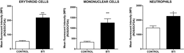
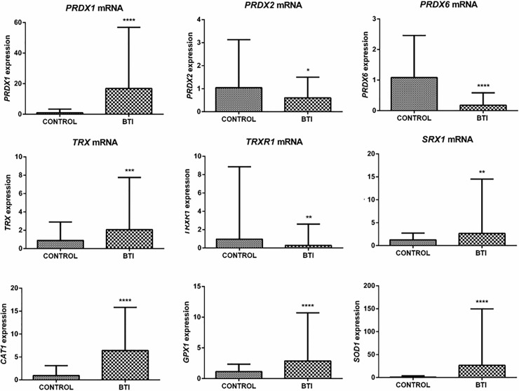
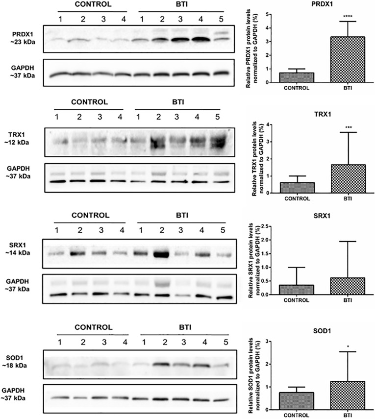
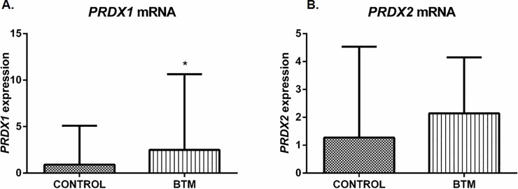
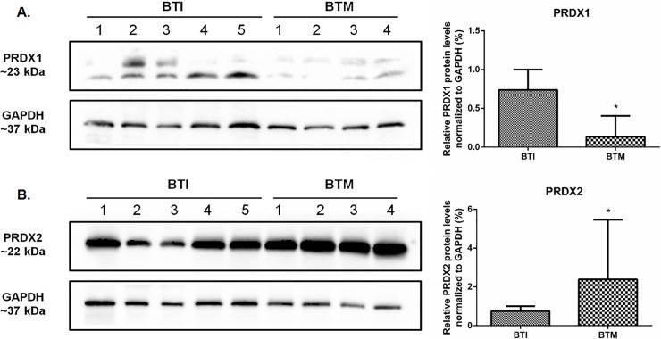
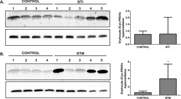
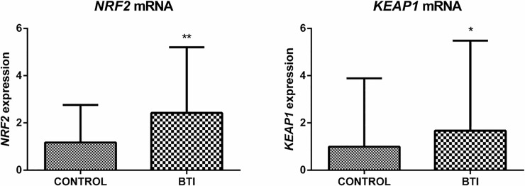
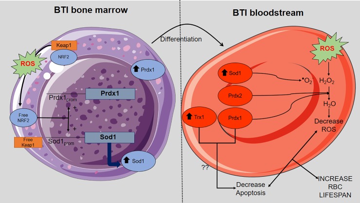
β-thalassemia is a worldwide distributed monogenic red cell disorder, characterized by an absent or reduced beta globin chain synthesis. The unbalance of alpha-gamma chain and the presence of pathological free iron promote severe oxidative damage, playing crucial a role in erythrocyte hemolysis, exacerbating ineffective erythropoiesis and decreasing the lifespan of red blood cells (RBC). Catalase, glutathione peroxidase and peroxiredoxins act together to protect RBCs from hydrogen peroxide insult. Among them, peroxiredoxins stand out for their overall abundance and reactivity. In RBCs, Prdx2 is the third most abundant protein, although Prdxs 1 and 6 isoforms are also found in lower amounts. Despite the importance of these enzymes, Prdx1 and Prdx2 may have their peroxidase activity inactivated by hyperoxidation at high hydroperoxide concentrations, which also promotes the molecular chaperone activity of these proteins. Some studies have demonstrated the importance of Prdx1 and Prdx2 for the development and maintenance of erythrocytes in hemolytic anemia. Now, we performed a global analysis comparatively evaluating the expression profile of several antioxidant enzymes and their physiological reducing agents in patients with beta thalassemia intermedia (BTI) and healthy individuals. Furthermore, increased levels of ROS were observed not only in RBC, but also in neutrophils and mononuclear cells of BTI patients. The level of transcripts and the protein content of Prx1 were increased in reticulocyte and RBCs of BTI patients and the protein content was also found to be higher when compared to beta thalassemia major (BTM), suggesting that this peroxidase could cooperate with Prx2 in the removal of H2O2. Furthermore, Prdx2 production is highly increased in RBCs of BTM patients that present high amounts of hyperoxidized species. A significant increase in the content of Trx1, Srx1 and Sod1 in RBCs of BTI patients suggested protective roles for these enzymes in BTI patients. Finally, the upregulation of Nrf2 and Keap1 transcription factors found in BTI patients may be involved in the regulation of the antioxidant enzymes analyzed in this work.


Conflict of interest statement

The authors have declared that no competing interests exist.

Figures

Fig 1
Fig 1. ROS production was increased in different cell types of patients with β Thalassemia intermedia.
Analysis of DCFDA fluorescence intensity, corresponding to the level of ROS production in erythroid, mononuclear and neutrophils cells. The results are expressed as mean (±SEM) fluorescence intensity (MFI) emitted by the analyzed cells of control and BTI patients. Statistical difference: (*) p < 0.05 and (***) p < 0.001.
Fig 2
Fig 2. Transcription analysis of redox gene expression responsible for the production of antioxidant enzymes in reticulocytes of control individuals and BTI patients.
RT-qPCR was carried out using the primers for each gene described in S1 Table. mRNA abundance for each gene was normalized to Bac, except for TrxR1, where Hprt1 was used as the endogenous reference. Results are presented as mean with standard error (± SEM). Statistical significance: (*p < 0.05), (**p < 0.001), (***p < 0.0001).
Fig 3
Fig 3. Representative western blot of Prdx1, Trx1, Srx1 and Sod1 expression in the erythrocyte cell lysate of beta-thalassemia intermedia patients compared to healthy individuals.
Protein levels were measured in mature cell erythrocyte lysate from 10 patients and 8 healthy subjects. Samples were separated in a 12% (w/v) reducing SDS–PAGE using 50 μg of total protein from each sample. The intensity of the bands was measured using GAPDH as the endogenous reference. Quantitative analyzes were performed by densitometry using ImageJ Software [35]. The results are presented as mean and standard error (± SEM), and are representative of two independent experiments (*p< 0.05), (***p< 0.0001).
Fig 4
Fig 4. Transcription analysis of Prdx1 and Prdx2 gene expression in reticulocytes of BTI and BTM patients.
RT-qPCR was carried out using the primers for each gene as described in S1 Table. mRNA abundance for each gene was normalized to Bac as endogenous reference. Results are presented as mean and standard error (± SEM). Statistical significance: *p < 0.05.
Fig 5
Fig 5. An opposite level of Prdx1 and Prdx2 was observed between beta thalassemia intermedia and major patients.
Protein levels were measured using 50 μg of total protein from each sample running in a 12% (w/v) reducing SDS–PAGE. The intensity of the bands was measured using GAPDH as endogenous reference. Quantitative analyzes were performed by densitometry using ImageJ Software [35]. Results are presented as mean and standard error (± SEM), and are representative of two independent experiments. Statistical significance: *p < 0.05.
Fig 6
Fig 6. Hyperoxidation of 2Cys-Prdx is altered in patients BTM.
Representative western blot for hyperoxidation state of 2CysPrdx. Erythroid cells of BTI (A) and BTM (B) patients were lysed in the presence of 200 uM of Nethylmaleimide (NEM) to prevent further sample oxidation. Hyperoxidation levels were measured using 50 μg of total protein from each sample running in a 12% (w/v) reducing SDS–PAGE. The intensity of the bands was measured using GAPDH as the endogenous control. Quantitative analyzes were performed by densitometry using ImageJ Software [35]. Results are presented as mean and standard error (± SEM). Statistical significance: *p < 0.05.
Fig 7
Fig 7. Analysis of the gene expression of Nrf2 and Keap1 in reticulocytes of BTI patients (n = 15) and healthy individuals (n = 16).
RT-qPCR analyzes showed an increase in the gene expression of Nrf2 (approx. 3 fold change) and Keap1 (approx. 2 fold change) in the reticulocytes of the BTI group when compared to the control group. Primers used for these analyzes are described in S1 Table. mRNA abundance for each gene was normalized to Bac as the endogenous reference. Results are presented as mean and standard error (± SEM). Statistical significance: *p < 0.05, **p < 0.001.
Fig 8
Fig 8. Model of interaction between Prdx1 and Prdx2 in the detoxification of hydroperoxides of BTI RBCs.
An increase in ROS production during the development of RBCs results in Keap1 oxidation and consequent liberation of free Nrf2, contributing to the upregulation of Prdx1 and Sod1. In the bloodstream the upregulation of Prdx1 can act together with Sod1 and Prdx2 in the detoxification of ROS. In addition, Prdx1 associated with Trx1 leads to decrease in apoptosis. Altogether, these processes contribute to increase BTI RBCs lifespan.

References

    1. Olivieri O, De Franceschi L, Capellini MD, Girelli D, Corrocher R, Brugnara C. Oxidative damage and erythrocyte membrane transport abnormalities in thalassemias. Blood. 1994;84(1):315–20. Epub 1994/07/01. . - PubMed
    1. Schrier SL, Centis F, Verneris M, Ma L, Angelucci E. The role of oxidant injury in the pathophysiology of human thalassemias. Redox Rep. 2003;8(5):241–5. Epub 2004/02/14. 10.1179/135100003225002835 . - DOI - PubMed
    1. Weatherall DJ. Disorders of globin synthesis: The Talassemias Williams Hematology. 7 New York: McGraw-Hill Medical; 2006.
    1. Cao A, Galanello R. Beta-thalassemia. Genet Med. 2010;12(2):61–76. Epub 2010/01/26. 10.1097/GIM.0b013e3181cd68ed . - DOI - PubMed
    1. Urbinati F, Madigan C, Malik P. Pathophysiology and therapy for haemoglobinopathies. Part II: thalassaemias. Expert Rev Mol Med. 2006;8(10):1–26. Epub 2006/05/11. 10.1017/S1462399406010805 . - DOI - PubMed

Publication types

MeSH terms