

Dermoscopy, with and without visual inspection, for diagnosing melanoma in adults
- PMID: 30521682
- PMCID: PMC6517096
- DOI: 10.1002/14651858.CD011902.pub2
Dermoscopy, with and without visual inspection, for diagnosing melanoma in adults
Abstract
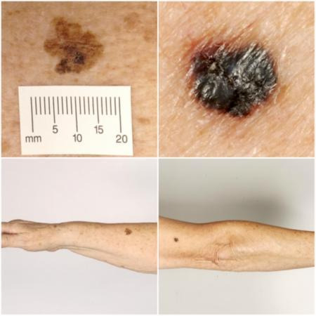
Background: Melanoma has one of the fastest rising incidence rates of any cancer. It accounts for a small percentage of skin cancer cases but is responsible for the majority of skin cancer deaths. Although history-taking and visual inspection of a suspicious lesion by a clinician are usually the first in a series of 'tests' to diagnose skin cancer, dermoscopy has become an important tool to assist diagnosis by specialist clinicians and is increasingly used in primary care settings. Dermoscopy is a magnification technique using visible light that allows more detailed examination of the skin compared to examination by the naked eye alone. Establishing the additive value of dermoscopy over and above visual inspection alone across a range of observers and settings is critical to understanding its contribution for the diagnosis of melanoma and to future understanding of the potential role of the growing number of other high-resolution image analysis techniques.
Objectives: To determine the diagnostic accuracy of dermoscopy alone, or when added to visual inspection of a skin lesion, for the detection of cutaneous invasive melanoma and atypical intraepidermal melanocytic variants in adults. We separated studies according to whether the diagnosis was recorded face-to-face (in-person), or based on remote (image-based), assessment.
Search methods: We undertook a comprehensive search of the following databases from inception up to August 2016: CENTRAL; MEDLINE; Embase; CINAHL; CPCI; Zetoc; Science Citation Index; US National Institutes of Health Ongoing Trials Register; NIHR Clinical Research Network Portfolio Database; and the World Health Organization International Clinical Trials Registry Platform. We studied reference lists and published systematic review articles.
Selection criteria: Studies of any design that evaluated dermoscopy in adults with lesions suspicious for melanoma, compared with a reference standard of either histological confirmation or clinical follow-up. Data on the accuracy of visual inspection, to allow comparisons of tests, was included only if reported in the included studies of dermoscopy.
Data collection and analysis: Two review authors independently extracted all data using a standardised data extraction and quality assessment form (based on QUADAS-2). We contacted authors of included studies where information related to the target condition or diagnostic threshold were missing. We estimated accuracy using hierarchical summary receiver operating characteristic (SROC),methods. Analysis of studies allowing direct comparison between tests was undertaken. To facilitate interpretation of results, we computed values of sensitivity at the point on the SROC curve with 80% fixed specificity and values of specificity with 80% fixed sensitivity. We investigated the impact of in-person test interpretation; use of a purposely developed algorithm to assist diagnosis; observer expertise; and dermoscopy training.
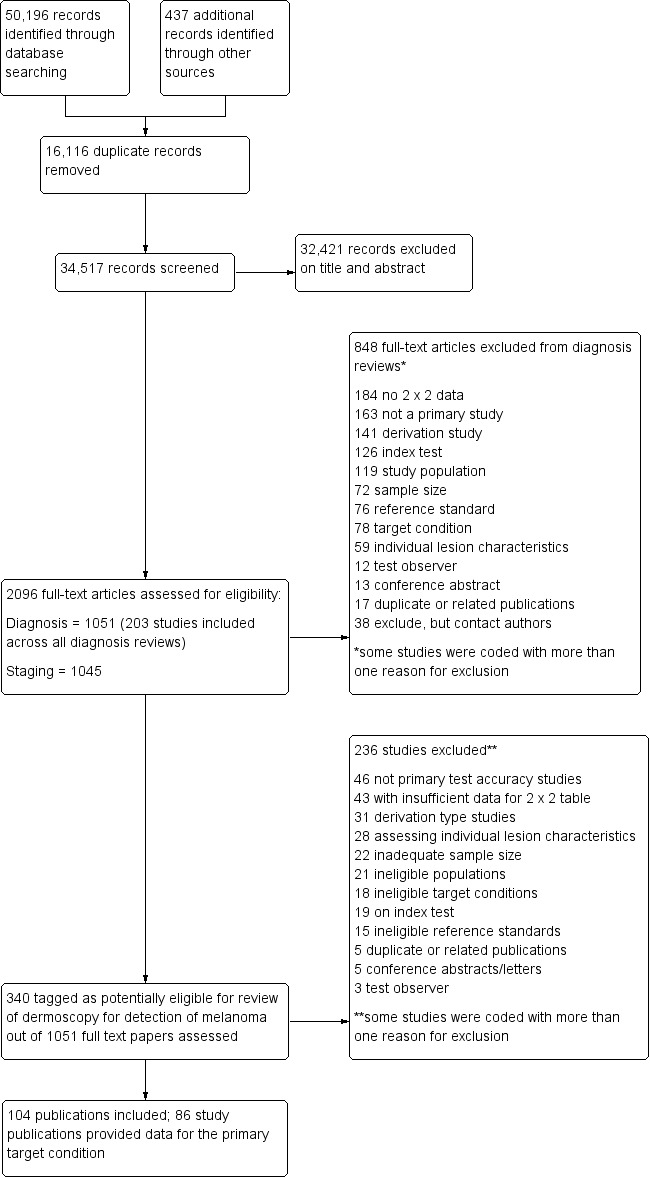
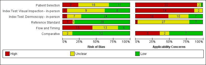
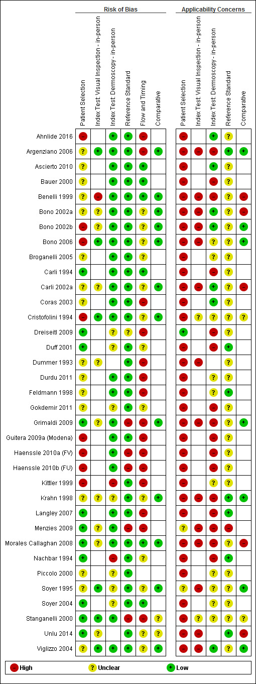
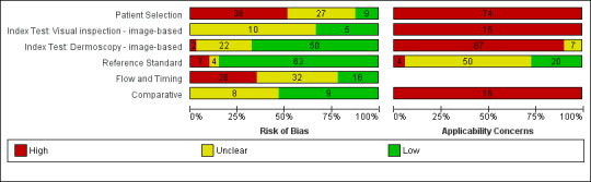
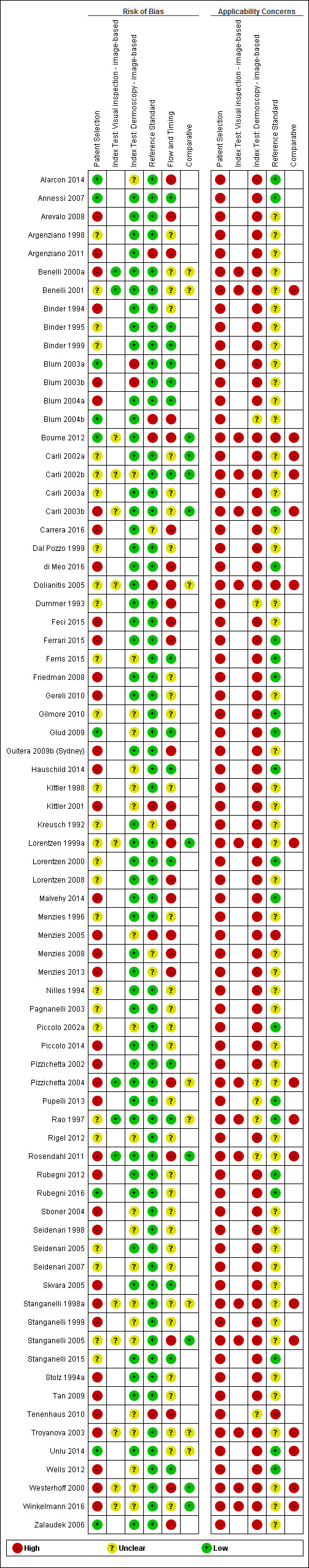
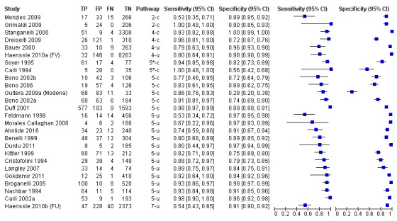
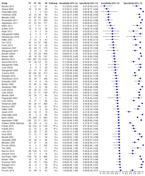
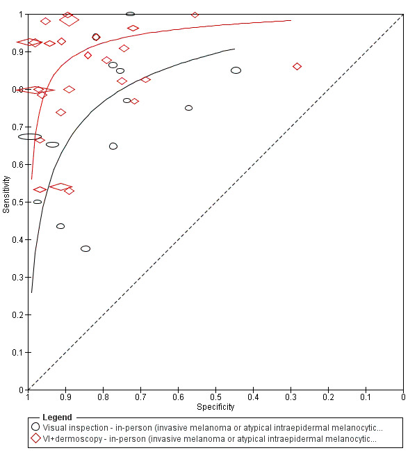
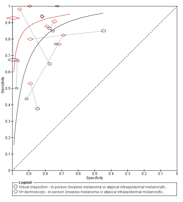
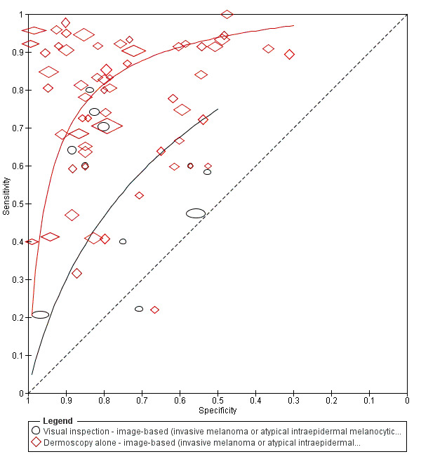
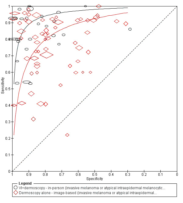
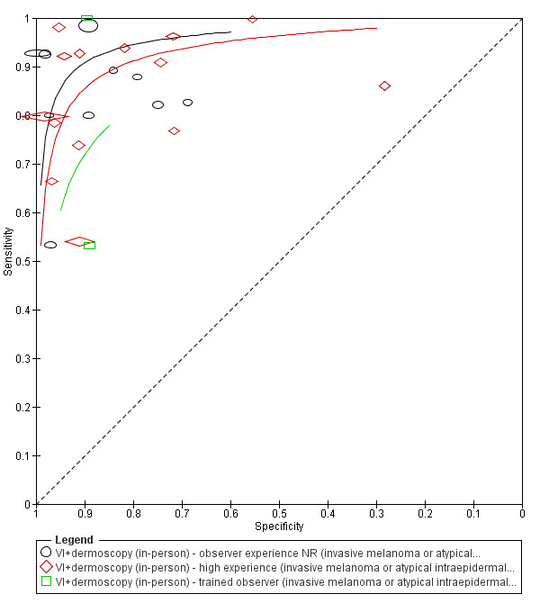
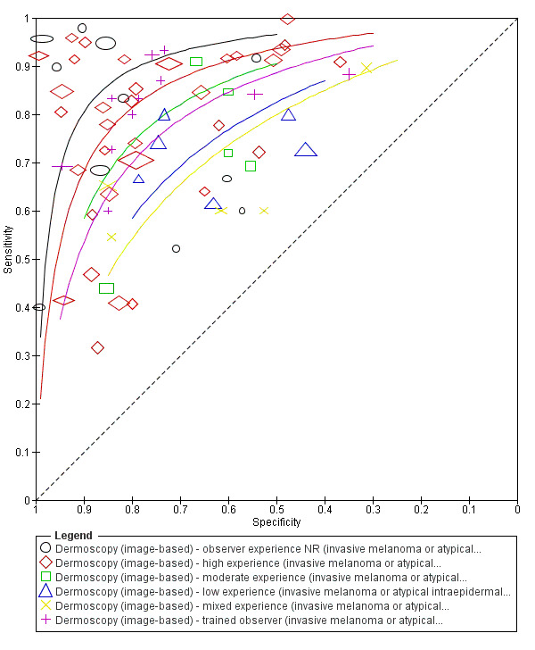
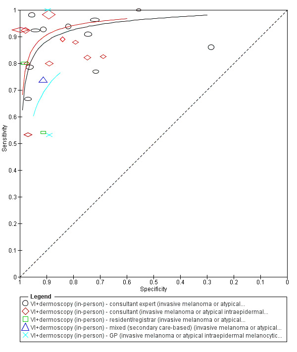
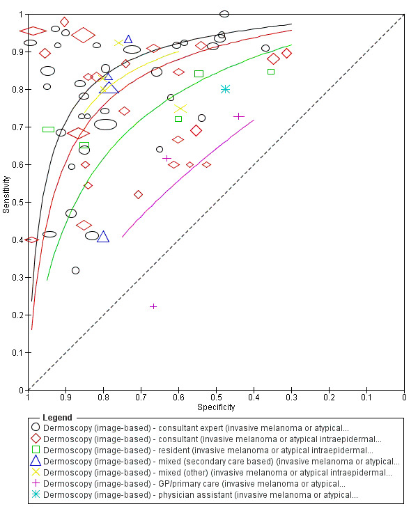
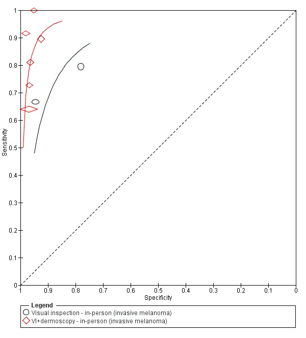
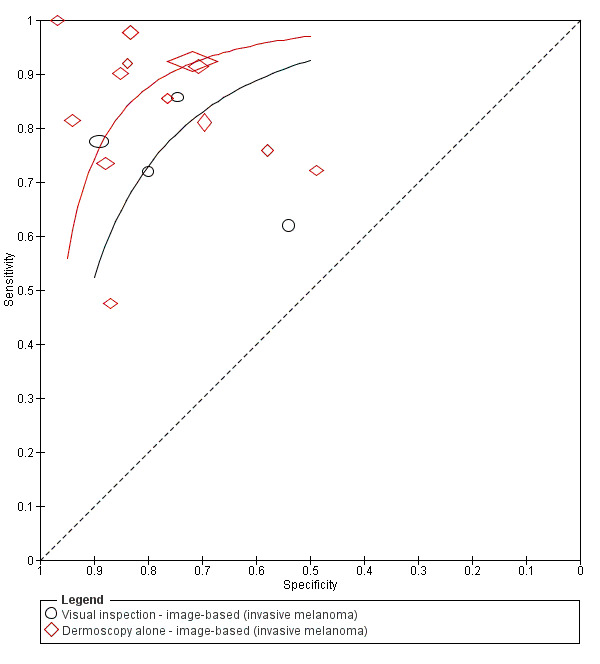
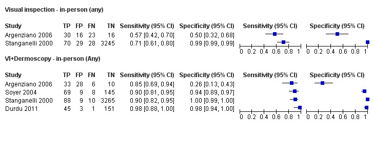
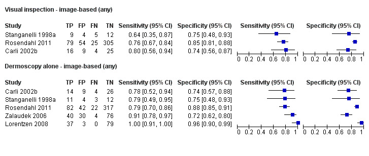
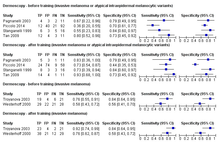
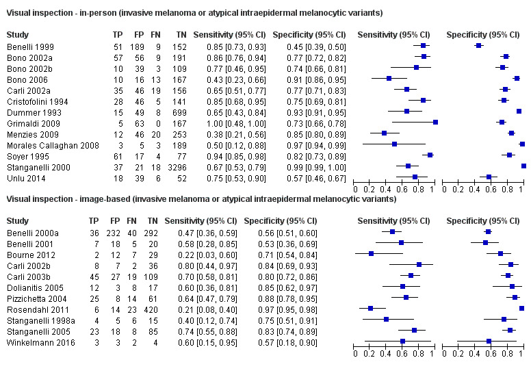
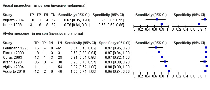
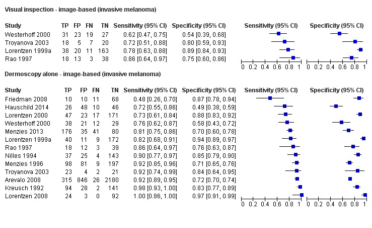
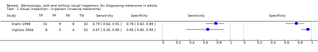
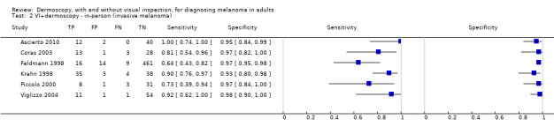
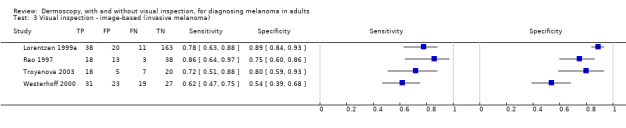
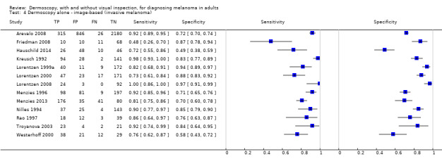
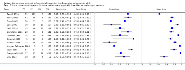
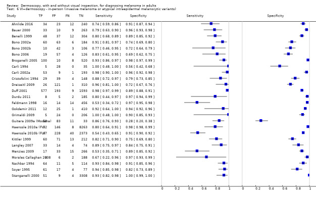
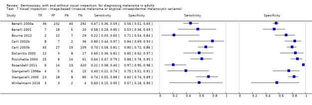
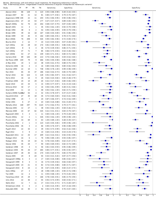
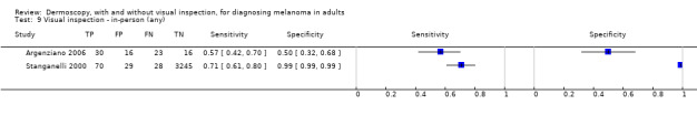
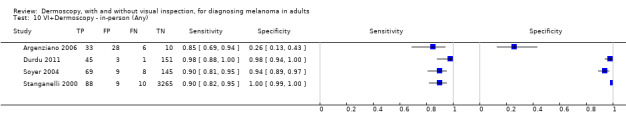
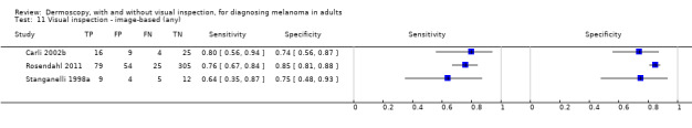
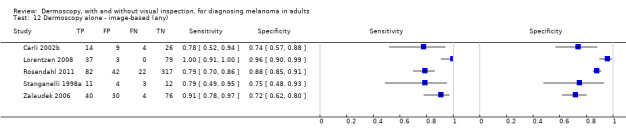
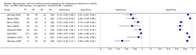
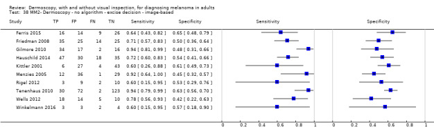
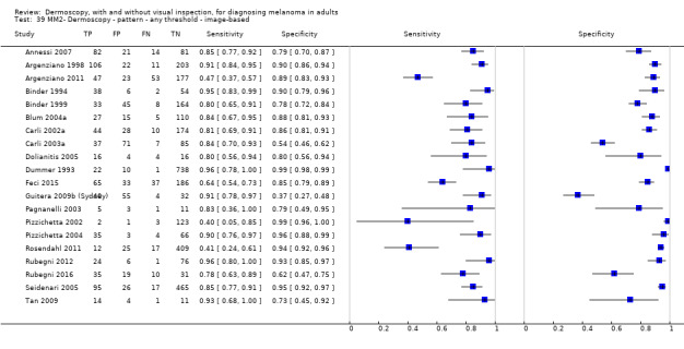
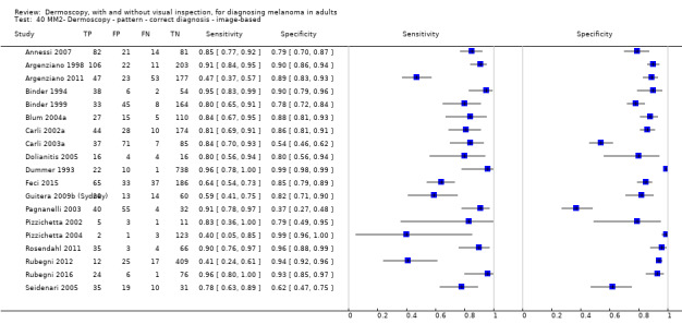
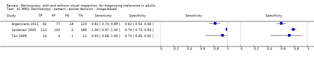
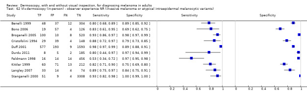
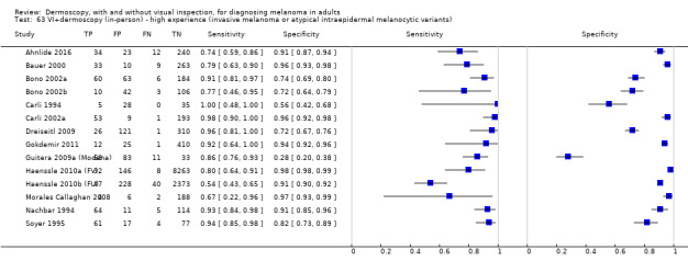
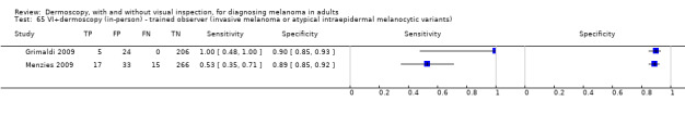
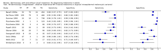
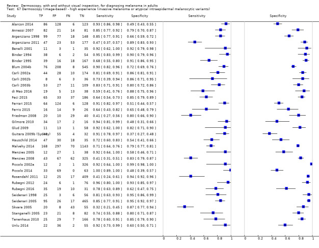
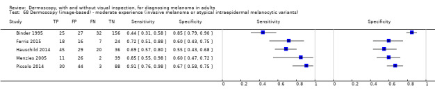
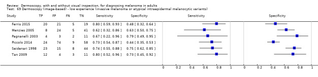
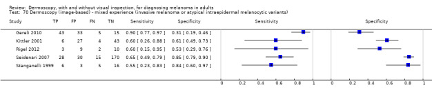
Main results: We included a total of 104 study publications reporting on 103 study cohorts with 42,788 lesions (including 5700 cases), providing 354 datasets for dermoscopy. The risk of bias was mainly low for the index test and reference standard domains and mainly high or unclear for participant selection and participant flow. Concerns regarding the applicability of study findings were largely scored as 'high' concern in three of four domains assessed. Selective participant recruitment, lack of reproducibility of diagnostic thresholds and lack of detail on observer expertise were particularly problematic.The accuracy of dermoscopy for the detection of invasive melanoma or atypical intraepidermal melanocytic variants was reported in 86 datasets; 26 for evaluations conducted in person (dermoscopy added to visual inspection), and 60 for image-based evaluations (diagnosis based on interpretation of dermoscopic images). Analyses of studies by prior testing revealed no obvious effect on accuracy; analyses were hampered by the lack of studies in primary care, lack of relevant information and the restricted inclusion of lesions selected for biopsy or excision. Accuracy was higher for in-person diagnosis compared to image-based evaluations (relative diagnostic odds ratio (RDOR) 4.6, 95% confidence interval (CI) 2.4 to 9.0; P < 0.001).We compared accuracy for (a), in-person evaluations of dermoscopy (26 evaluations; 23,169 lesions and 1664 melanomas),versus visual inspection alone (13 evaluations; 6740 lesions and 459 melanomas), and for (b), image-based evaluations of dermoscopy (60 evaluations; 13,475 lesions and 2851 melanomas),versus image-based visual inspection (11 evaluations; 1740 lesions and 305 melanomas). For both comparisons, meta-analysis found dermoscopy to be more accurate than visual inspection alone, with RDORs of (a), 4.7 (95% CI 3.0 to 7.5; P < 0.001), and (b), 5.6 (95% CI 3.7 to 8.5; P < 0.001). For a), the predicted difference in sensitivity at a fixed specificity of 80% was 16% (95% CI 8% to 23%; 92% for dermoscopy + visual inspection versus 76% for visual inspection), and predicted difference in specificity at a fixed sensitivity of 80% was 20% (95% CI 7% to 33%; 95% for dermoscopy + visual inspection versus 75% for visual inspection). For b) the predicted differences in sensitivity was 34% (95% CI 24% to 46%; 81% for dermoscopy versus 47% for visual inspection), at a fixed specificity of 80%, and predicted difference in specificity was 40% (95% CI 27% to 57%; 82% for dermoscopy versus 42% for visual inspection), at a fixed sensitivity of 80%.Using the median prevalence of disease in each set of studies ((a), 12% for in-person and (b), 24% for image-based), for a hypothetical population of 1000 lesions, an increase in sensitivity of (a), 16% (in-person), and (b), 34% (image-based), from using dermoscopy at a fixed specificity of 80% equates to a reduction in the number of melanomas missed of (a), 19 and (b), 81 with (a), 176 and (b), 152 false positive results. An increase in specificity of (a), 20% (in-person), and (b), 40% (image-based), at a fixed sensitivity of 80% equates to a reduction in the number of unnecessary excisions from using dermoscopy of (a), 176 and (b), 304 with (a), 24 and (b), 48 melanomas missed.The use of a named or published algorithm to assist dermoscopy interpretation (as opposed to no reported algorithm or reported use of pattern analysis), had no significant impact on accuracy either for in-person (RDOR 1.4, 95% CI 0.34 to 5.6; P = 0.17), or image-based (RDOR 1.4, 95% CI 0.60 to 3.3; P = 0.22), evaluations. This result was supported by subgroup analysis according to algorithm used. We observed higher accuracy for observers reported as having high experience and for those classed as 'expert consultants' in comparison to those considered to have less experience in dermoscopy, particularly for image-based evaluations. Evidence for the effect of dermoscopy training on test accuracy was very limited but suggested associated improvements in sensitivity.
Authors' conclusions: Despite the observed limitations in the evidence base, dermoscopy is a valuable tool to support the visual inspection of a suspicious skin lesion for the detection of melanoma and atypical intraepidermal melanocytic variants, particularly in referred populations and in the hands of experienced users. Data to support its use in primary care are limited, however, it may assist in triaging suspicious lesions for urgent referral when employed by suitably trained clinicians. Formal algorithms may be of most use for dermoscopy training purposes and for less expert observers, however reliable data comparing approaches using dermoscopy in person are lacking.
Conflict of interest statement
Jacqueline Dinnes: nothing to declare. Jonathan J Deeks: nothing to declare. Naomi Chuchu: nothing to declare. Lavinia Ferrante di Ruffano: nothing to declare. Rubeta N Matin: my institution received a grant for a Barco NV commercially sponsored study to evaluate digital dermoscopy in the skin cancer clinic. My institution also received Oxfordshire Health Services Research Charitable Funds for carrying out a study of feasibility of using the Skin Cancer Quality of Life Impact Tool (SCQOLIT) in non melanoma skin cancer. I have received payment from Public Health England for the "Be Clear on Cancer Skin Cancer" report and royalties for the
Figures


Update of
References
References to studies included in this review
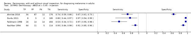
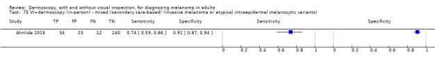
Ahnlide 2016 {published data only}
-
- Ahnlide I, Bjellerup M, Nilsson F, Nielsen K. Validity of ABCD Rule of Dermoscopy in Clinical Practice. Acta Dermato‐Venereologica 2016;96(3):367‐72. [ER4:25012370; PUBMED: 26351008] - PubMed
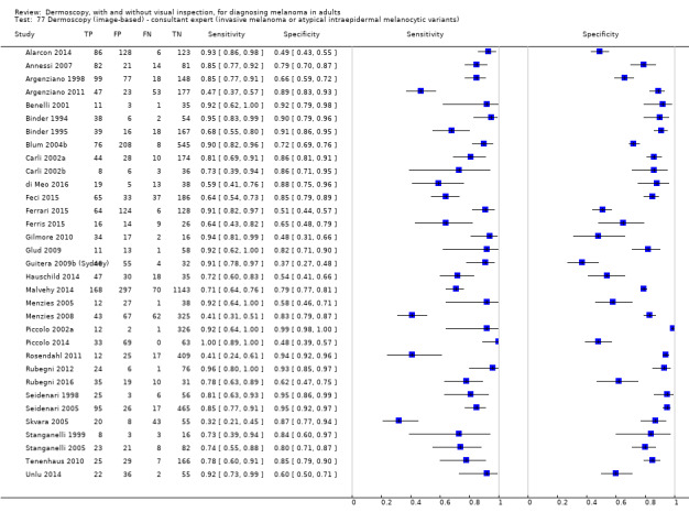
Alarcon 2014 {published data only}
Annessi 2007 {published data only}
-
- Annessi G, Bono R, Sampogna F, Faraggiana T, Abeni D. Sensitivity, specificity, and diagnostic accuracy of three dermoscopic algorithmic methods in the diagnosis of doubtful melanocytic lesions: the importance of light brown structureless areas in differentiating atypical melanocytic nevi from thin melanomas. Journal of the American Academy of Dermatology 2007;56(5):759‐67. [ER4:15465846; PUBMED: 17316894] - PubMed
Arevalo 2008 {published data only}
-
- Arevalo A, Altamura D, Avramidis M, Blum A, Menzies S. The significance of eccentric and central hyperpigmentation, multifocal hyper/hypopigmentation, and the multicomponent pattern in melanocytic lesions lacking specific dermoscopic features of melanoma. Archives of Dermatology 2008;144(11):1440‐4. [ER4:19728335; PUBMED: 19015418] - PubMed
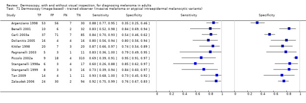
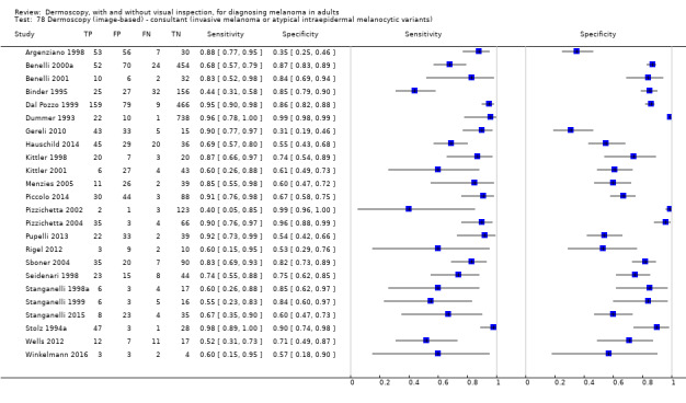
Argenziano 1998 {published data only}
-
- Argenziano G, Fabbrocini G, Carli P, Giorgi V, Sammarco E, Delfino M. Epiluminescence microscopy for the diagnosis of doubtful melanocytic skin lesions. Comparison of the ABCD rule of dermatoscopy and a new 7‐point checklist based on pattern analysis. Archives of Dermatology 1998;134(12):1563‐70. [ER4:15465850; PUBMED: 9875194] - PubMed
Argenziano 2006 {published data only}
-
- Argenziano G, Puig S, Zalaudek I, Sera F, Corona R, Alsina M, et al. Dermoscopy improves accuracy of primary care physicians to triage lesions suggestive of skin cancer. Journal of Clinical Oncology 2006;24(12):1877‐82. [ER4:17940973; PUBMED: 16622262] - PubMed
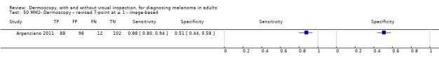
Argenziano 2011 {published data only}
-
- Argenziano G, Catricala C, Ardigo M, Buccini P, Simone P, Eibenschutz L, et al. Seven‐point checklist of dermoscopy revisited. British Journal of Dermatology 2011;164(4):785‐90. [ER4:15465848; PUBMED: 21175563] - PubMed
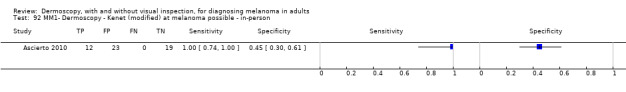
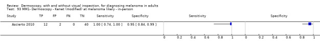
Ascierto 2010 {published data only}
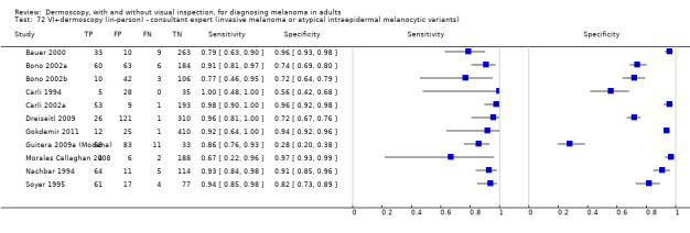
Bauer 2000 {published data only}
-
- Bauer P, Cristofolini P, Boi S, Burroni M, Dell'Eva G, Micciolo R, et al. Digital epiluminescence microscopy: usefulness in the differential diagnosis of cutaneous pigmentary lesions. A statistical comparison between visual and computer inspection. Melanoma Research 2000;10(4):345‐9. [ER4:15465861; PUBMED: 10985668] - PubMed
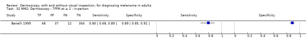
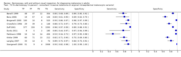
Benelli 1999 {published data only}
-
- Benelli C, Roscetti E, Pozzo VD, Gasparini G, Cavicchini S. The dermoscopic versus the clinical diagnosis of melanoma. European Journal of Dermatology 1999;9(6):470‐6. [ER4:18375029; PUBMED: 10491506] - PubMed
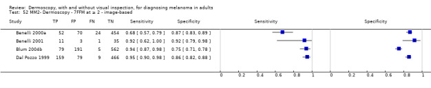
Benelli 2000a {published data only}
-
- Benelli C, Roscetti E, Dal Pozzo V. The dermoscopic (7FFM) versus the clinical (ABCDE) diagnosis of small diameter melanoma. European Journal of Dermatology 2000;10(4):282‐7. [PUBMED: 10846255] - PubMed
Benelli 2001 {published data only}
-
- Benelli C, Roscetti E, Dal Pozzo V. Reproducibility of the clinical criteria (ABCDE rule) and dermatoscopic features (7FFM) for the diagnosis of malignant melanoma. European Journal of Dermatology 2001;11(3):234‐9. [ER4:18375028; PUBMED: 11358731] - PubMed
Binder 1994 {published data only}
-
- Binder M, Steiner A, Schwarz M, Knollmayer S, Wolff K, Pehamberger H. Application of an artificial neural network in epiluminescence microscopy pattern analysis of pigmented skin lesions: a pilot study. British Journal of Dermatology 1994;130(4):460‐5. [ER4:18375032; PUBMED: 8186110] - PubMed
Binder 1995 {published data only}
-
- Binder M, Schwarz M, Winkler A, Steiner A, Kaider A, Wolff K, et al. Epiluminescence microscopy. A useful tool for the diagnosis of pigmented skin lesions for formally trained dermatologists. Archives of Dermatology 1995;131(3):286‐91. [ER4:18375031; PUBMED: 7887657] - PubMed
Binder 1999 {published data only}
-
- Binder M, Kittler H, Steiner A, Dawid M, Pehamberger H, Wolff K. Reevaluation of the ABCD rule for epiluminescence microscopy. Journal of the American Academy of Dermatology 1999;40(2 Pt 1):171‐6. [ER4:15465864; PUBMED: 10025741] - PubMed
Blum 2003a {published data only}
-
- Blum A, Rassner G, Garbe C. Modified ABC‐point list of dermoscopy: a simplified and highly accurate dermoscopic algorithm for the diagnosis of cutaneous melanocytic lesions. Journal of the American Academy of Dermatology 2003;48(5):672‐8. [ER4:15465867; PUBMED: 12734495] - PubMed
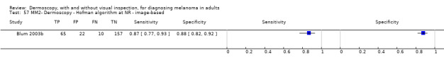
Blum 2003b {published data only}
-
- Blum A, Soyer HP, Garbe C, Kerl H, Rassner G, Hofmann‐Wellenhof R. The dermoscopic classification of atypical melanocytic naevi (Clark naevi) is useful to discriminate benign from malignant melanocytic lesions. British Journal of Dermatology 2003;149(6):1159‐64. [ER4:15465868; PUBMED: 14674892] - PubMed
Blum 2004a {published data only}
-
- Blum A, Hofmann‐Wellenhof R, Luedtke H, Ellwanger U, Steins A, Roehm S, et al. Value of the clinical history for different users of dermoscopy compared with results of digital image analysis. Journal of the European Academy of Dermatology & Venereology 2004;18(6):665‐9. [ER4:15465865; PUBMED: 15482291] - PubMed
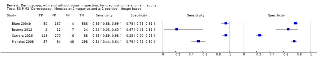
Blum 2004b {published data only}
-
- Blum A, Luedtke H, Ellwanger U, Schwabe R, Rassner G, Garbe C. Digital image analysis for diagnosis of cutaneous melanoma. Development of a highly effective computer algorithm based on analysis of 837 melanocytic lesions. British Journal of Dermatology 2004;151(5):1029‐38. [ER4:15465866; PUBMED: 15541081] - PubMed
Bono 2002a {published data only}
-
- Bono A, Bartoli C, Cascinelli N, Lualdi M, Maurichi A, Moglia D, et al. Melanoma detection. A prospective study comparing diagnosis with the naked eye, dermatoscopy and telespectrophotometry. Dermatology 2002;205(4):362‐6. [ER4:15465870; PUBMED: 12444332] - PubMed
Bono 2002b {published data only}
-
- Bono A, Bartoli C, Baldi M, Tomatis S, Bifulco C, Santinami M. Clinical and dermatoscopic diagnosis of small pigmented skin lesions. European Journal of Dermatology 2002;12(6):573‐6. [ER4:18375034; PUBMED: 12459531] - PubMed
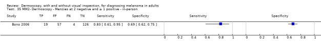
Bono 2006 {published data only}
-
- Bono A, Tolomio E, Trincone S, Bartoli C, Tomatis S, Carbone A, et al. Micro‐melanoma detection: a clinical study on 206 consecutive cases of pigmented skin lesions with a diameter < or = 3 mm. British Journal of Dermatology 2006;155(3):570‐3. [ER4:15465872; PUBMED: 16911283] - PubMed
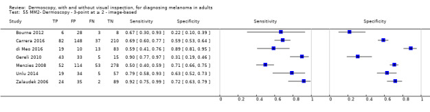
Bourne 2012 {published data only}
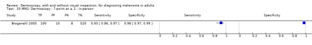
Broganelli 2005 {published data only}
-
- Broganelli P, Chiaretta A, Sacerdote C, Pippione M. The epiluminescence microscopy in the ambulatory clinical practice: diagnostic accuracy and usefulness of videodermatoscopic monitoring [L'epiluminescenza nella pratica clinica ambulatoriale: accuratezza diagnostica ed utilita del monitoraggio videodermatoscopico]. Giornale Italiano di Dermatologia e Venereologia 2005;140(1):15‐25. [ER4:18375073]
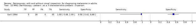
Carli 1994 {published data only}
-
- Carli P, Giorgi V, Donati E, Pestelli E, Giannotti B. Epiluminescence microscopy reduces the risk of removing clinically atypical, but histologically common, melanocytic lesions [La microscopia a epiluminescenza (Elm) riduce il rischio di asportare lesioni melanocitarie clinicamente sospette ma istologicamente comuni]. Giornale Italiano di Dermatologia e Venereologia 1994;129(12):599‐605. [ER4:18375075]
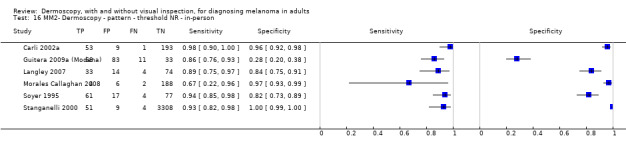
Carli 2002a {published data only}
-
- Carli P, Giorgi V, Argenziano G, Palli D, Giannotti B. Pre‐operative diagnosis of pigmented skin lesions: in vivo dermoscopy performs better than dermoscopy on photographic images. Journal of the European Academy of Dermatology & Venereology 2002;16(4):339‐46. [ER4:15465882; PUBMED: 12224689] - PubMed
Carli 2002b {published data only}
-
- Carli P, Giorgi V, Salvini C, Mannone F, Chiarugi A. The gold standard for photographing pigmented skin lesions for diagnostic purposes: contact versus distant imaging. Skin Research & Technology 2002;8(4):255‐9. [ER4:15465888; PUBMED: 12423545] - PubMed
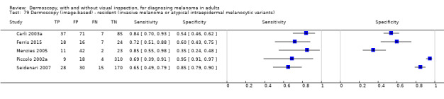
Carli 2003a {published data only}
-
- Carli P, Quercioli E, Sestini S, Stante M, Ricci L, Brunasso G, et al. Pattern analysis, not simplified algorithms, is the most reliable method for teaching dermoscopy for melanoma diagnosis to residents in dermatology. British Journal of Dermatology 2003;148(5):981‐4. [ER4:15465890; PUBMED: 12786829] - PubMed
Carli 2003b {published data only}
-
- Carli P, Giorgi V, Chiarugi A, Nardini P, Mannone F, Stante M, et al. Effect of lesion size on the diagnostic performance of dermoscopy in melanoma detection. Dermatology 2003;206(4):292‐6. [ER4:15465883; PUBMED: 12771468] - PubMed
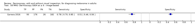
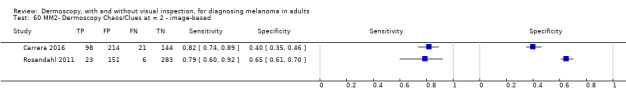
Carrera 2016 {published data only}
-
- Carrera C, Marchetti MA, Dusza SW, Argenziano G, Braun RP, Halpern AC, et al. Validity and reliability of dermoscopic criteria used to differentiate nevi from melanoma a web‐based international dermoscopy society study. JAMA Dermatology 2016;152(7):798‐806. [ER4:25233595; PUBMED: 27074267] - PMC - PubMed
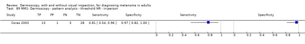
Coras 2003 {published data only}
-
- Coras B, Glaessl A, Kinateder J, Klovekorn W, Braun R, Lepski U, et al. Teledermatoscopy in daily routine‐‐results of the first 100 cases. Current Problems in Dermatology 2003;32:207‐12. [PUBMED: 12472014] - PubMed
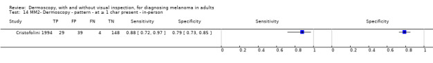
Cristofolini 1994 {published data only}
-
- Cristofolini M, Zumiani G, Bauer P, Cristofolini P, Boi S, Micciolo R. Dermatoscopy: usefulness in the differential diagnosis of cutaneous pigmentary lesions. Melanoma Research 1994;4(6):391‐4. [ER4:15465898; PUBMED: 7703719] - PubMed
Dal Pozzo 1999 {published data only}
-
- Dal Pozzo V, Benelli C, Roscetti E. The seven features for melanoma: a new dermoscopic algorithm for the diagnosis of malignant melanoma. European Journal of Dermatology 1999;9(4):303‐8. [ER4:18375041; PUBMED: 10356410] - PubMed
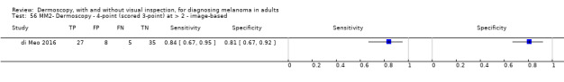
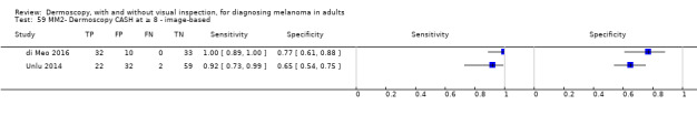
di Meo 2016 {published data only}
-
- Meo N, Stinco G, Bonin S, Gatti A, Trevisini S, Damiani G, et al. CASH algorithm versus 3‐point checklist and its modified version in evaluation of melanocytic pigmented skin lesions: the 4‐point checklist. Journal of Dermatology 2016;43(6):682‐5. [ER4:25012343; PUBMED: 26589251] - PubMed
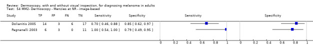
Dolianitis 2005 {published data only}
-
- Dolianitis C, Kelly J, Wolfe R, Simpson P. Comparative performance of 4 dermoscopic algorithms by nonexperts for the diagnosis of melanocytic lesions. Archives of Dermatology 2005;141(8):1008‐14. [ER4:15465906; PUBMED: 16103330] - PubMed
Dreiseitl 2009 {published data only}
-
- Dreiseitl S, Binder M, Hable K, Kittler H. Computer versus human diagnosis of melanoma: evaluation of the feasibility of an automated diagnostic system in a prospective clinical trial. Melanoma Research 2009;19(3):180‐4. [ER4:15465907; PUBMED: 19369900] - PubMed
Duff 2001 {published data only}
-
- Duff CG, Melsom D, Rigby HS, Kenealy JM, Townsend PL. A 6 year prospective analysis of the diagnosis of malignant melanoma in a pigmented‐lesion clinic: even the experts miss malignant melanomas, but not often. British Journal of Plastic Surgery 2001;54(4):317‐21. [DOI: ; ER4:20569450; PUBMED: 11355986] - PubMed
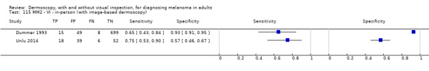
Dummer 1993 {published data only}
-
- Dummer W, Doehnel KA, Remy W. Videomicroscopy in differential diagnosis of skin tumors and secondary prevention of malignant melanoma. Hautarzt 1993;44(12):772‐6. [ER4:18375044; PUBMED: 8113040] - PubMed
Durdu 2011 {published data only}
-
- Durdu M, Baba M, Seckin D. Dermatoscopy versus Tzanck smear test: a comparison of the value of two tests in the diagnosis of pigmented skin lesions. Journal of the American Academy of Dermatology 2011;65(5):972‐82. [ER4:15465910; PUBMED: 21565420] - PubMed
Feci 2015 {published data only}
-
- Feci L, Cevenini G, Nami N, Fagiolini A, Perotti R, Miracco C, et al. Influence of ambient stressors and time constraints on diagnostic accuracy of borderline pigmented skin lesions. Dermatology 2015;231(3):269‐73. [ER4:25012339; PUBMED: 26375805] - PubMed
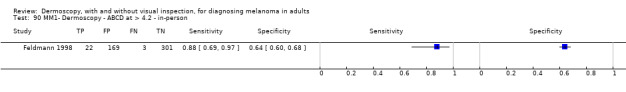
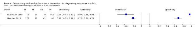
Feldmann 1998 {published data only}
-
- Feldmann R, Fellenz C, Gschnait F. The ABCD rule in dermatoscopy: analysis of 500 melanocytic lesions. Hautarzt 1998;49(6):473‐6. [ER4:15465916; PUBMED: 9675574] - PubMed
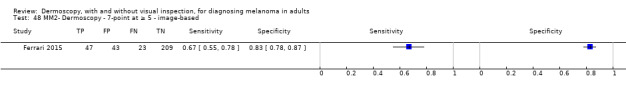
Ferrari 2015 {published data only}
-
- Ferrari B, Pupelli G, Farnetani F, Carvalho NT, Longo C, Reggiani C, et al. Dermoscopic difficult lesions: an objective evaluation of reflectance confocal microscopy impact for accurate diagnosis. Journal of the European Academy of Dermatology and Venereology 2015;29(6):1135‐40. [DOI: 10.1111/jdv.12769; ER4:20569458; PUBMED: 25303304] - DOI - PubMed
Ferris 2015 {published data only}
-
- Ferris LK, Harkes JA, Gilbert B, Winger DG, Golubets K, Akilov O, et al. Computer‐aided classification of melanocytic lesions using dermoscopic images. Journal of the American Academy of Dermatology 2015;73(5):769‐76. [ER4:25012337; PUBMED: 26386631] - PubMed
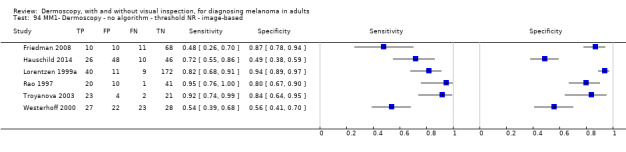
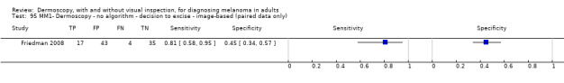
Friedman 2008 {published data only}
-
- Friedman RJ, Gutkowicz‐Krusin D, Farber MJ, Warycha M, Schneider‐Kels L, Papastathis N, et al. The diagnostic performance of expert dermoscopists vs a computer‐vision system on small‐diameter melanomas. Archives of Dermatology 2008;144(4):476‐82. [ER4:15465921; PUBMED: 18427041] - PubMed
Gereli 2010 {published data only}
-
- Gereli MC, Onsun N, Atilganoglu U, Demirkesen C. Comparison of two dermoscopic techniques in the diagnosis of clinically atypical pigmented skin lesions and melanoma: seven‐point and three‐point checklists. International Journal of Dermatology 2010;49(1):33‐8. [ER4:15465929; PUBMED: 20465608] - PubMed
Gilmore 2010 {published data only}
-
- Gilmore S, Hofmann‐Wellenhof R, Soyer HP. A support vector machine for decision support in melanoma recognition. Experimental Dermatology 2010;19(9):830‐5. [ER4:15465935; PUBMED: 20629732] - PubMed
Glud 2009 {published data only}
-
- Glud M, Gniadecki R, Drzewiecki KT. Spectrophotometric intracutaneous analysis versus dermoscopy for the diagnosis of pigmented skin lesions: prospective, double‐blind study in a secondary reference centre. Melanoma Research 2009;19(3):176‐9. [ER4:18375045; PUBMED: 19319002] - PubMed
Gokdemir 2011 {published data only}
-
- Gokdemir A, Guler OM, Bek Y, Aydin F, Senturk N, Canturk T, et al. Dermoscopic and histopathological correlation in melanocytic and non‐melanocytic lesions [Melanositik ve non‐melanositik lezyonlarda dermoskopik ve histopatolojik tani korelasyonu]. Turkiye Klinikleri Dermatoloji 2011;21(1):7‐16. [EMBASE: 361807346; ER4:18375084]
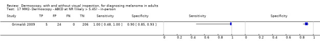
Grimaldi 2009 {published data only}
-
- Grimaldi L, Silvestri A, Brandi C, Nisi G, Brafa A, Calabro M, et al. Digital epiluminescence dermoscopy for pigmented cutaneous lesions, primary care physicians, and telediagnosis: a useful tool?. Journal of Plastic, Reconstructive & Aesthetic Surgery 2009;62(8):1054‐8. [ER4:15465940; PUBMED: 18547883] - PubMed
Guitera 2009a (Modena) {published data only}
-
- Guitera P, Pellacani G, Longo C, Seidenari S, Avramidis M, Menzies SW. In vivo reflectance confocal microscopy enhances secondary evaluation of melanocytic lesions. Journal of Investigative Dermatology 2009;129(1):131‐8. [PUBMED: 18633444] - PubMed
Guitera 2009b (Sydney) {published data only}
-
- Guitera P, Pellacani G, Longo C, Seidenari S, Avramidis M, Menzies SW. In vivo reflectance confocal microscopy enhances secondary evaluation of melanocytic lesions. Journal of Investigative Dermatology 2009;129(1):131‐8. [ER4:15465945; PUBMED: 18633444] - PubMed
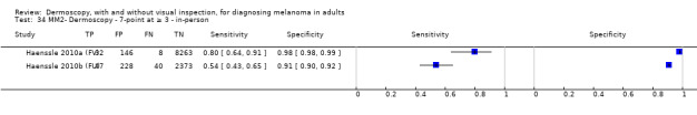
Haenssle 2010a (FV) {published data only}
-
- Haenssle HA, Korpas B, Hansen‐Hagge C, Buhl T, Kaune KM, Rosenberger A, et al. Seven‐point checklist for dermatoscopy: performance during 10 years of prospective surveillance of patients at increased melanoma risk. Journal of the American Academy of Dermatology 2010;62(5):785‐93. [PUBMED: 20226567] - PubMed
Haenssle 2010b (FU) {published data only}
-
- Haenssle HA, Korpas B, Hansen‐Hagge C, Buhl T, Kaune KM, Rosenberger A, et al. Seven‐point checklist for dermatoscopy: performance during 10 years of prospective surveillance of patients at increased melanoma risk. Journal of the American Academy of Dermatology 2010;62(5):785‐93. [PUBMED: 20226567] - PubMed
Hauschild 2014 {published data only}
-
- Hauschild A, Chen SC, Weichenthal M, Blum A, King HC, Goldsmith J, et al. To excise or not: impact of MelaFind on German dermatologists' decisions to biopsy atypical lesions. Journal der Deutschen Dermatologischen Gesellschaft 2014;12(7):606‐14. [ER4:17941085; PUBMED: 24944011] - PubMed
Kittler 1998 {published data only}
-
- Kittler H, Seltenheim M, Pehamberger H, Wolff K, Binder M. Diagnostic informativeness of compressed digital epiluminescence microscopy images of pigmented skin lesions compared with photographs. Melanoma Research 1998;8(3):255‐60. [ER4:17941060; PUBMED: 9664147] - PubMed
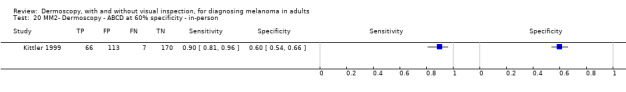
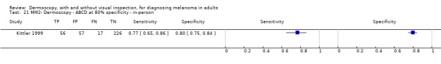
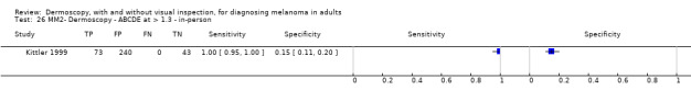
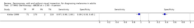
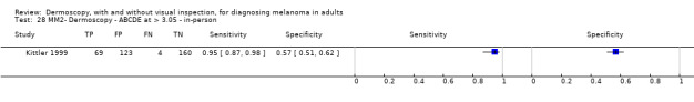
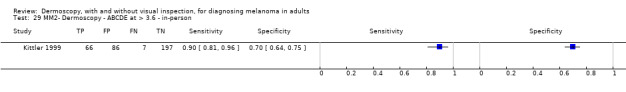
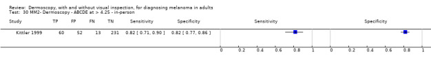
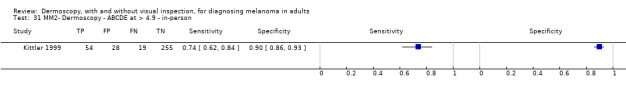
Kittler 1999 {published data only}
-
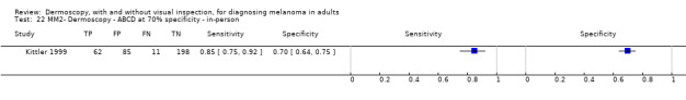
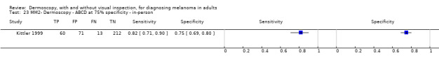
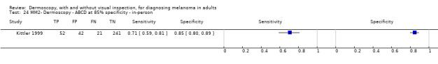
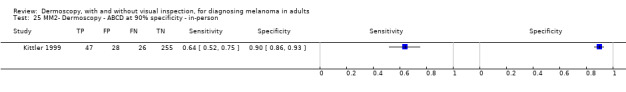
- Kittler H, Seltenheim M, Dawid M, Pehamberger H, Wolff K, Binder M. Morphologic changes of pigmented skin lesions: a useful extension of the ABCD rule for dermatoscopy. Journal of the American Academy of Dermatology 1999;40(4):558‐62. [ER4:15465976; PUBMED: 10188673] - PubMed
Kittler 2001 {published data only}
-
- Kittler H, Binder M. Risks and benefits of sequential imaging of melanocytic skin lesions in patients with multiple atypical nevi. Archives of Dermatology 2001;137(12):1590‐5. [ER4:20569472; PUBMED: 11735709] - PubMed
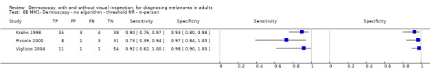
Krahn 1998 {published data only}
-
- Krahn G, Gottlober P, Sander C, Peter RU. Dermatoscopy and high frequency sonography: two useful non‐invasive methods to increase preoperative diagnostic accuracy in pigmented skin lesions. Pigment Cell Research 1998;11(3):151‐4. [ER4:15465981; PUBMED: 9730322] - PubMed
Kreusch 1992 {published data only}
-
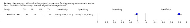
- Kreusch J, Rassner G, Trahn C, Pietsch‐Breitfeld B, Henke D, Selbmann HK. Epiluminescent microscopy: a score of morphological features to identify malignant melanoma. Pigment Cell Research 1992;Suppl 2:295‐8. [PUBMED: 1409432] - PubMed
Langley 2007 {published data only}
-
- Langley RG, Walsh N, Sutherland AE, Propperova I, Delaney L, Morris SF, et al. The diagnostic accuracy of in vivo confocal scanning laser microscopy compared to dermoscopy of benign and malignant melanocytic lesions: a prospective study. Dermatology 2007;215(4):365‐72. [ER4:15465985; PUBMED: 17912001] - PubMed
Lorentzen 1999a {published data only}
-
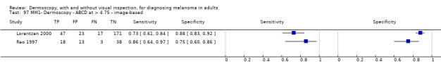
- Lorentzen H, Weismann K, Petersen CS, Larsen FG, Secher L, Skodt V. Clinical and dermatoscopic diagnosis of malignant melanoma. Assessed by expert and non‐expert groups. Acta Dermato‐Venereologica 1999;79(4):301‐4. [ER4:17941062; PUBMED: 10429989] - PubMed
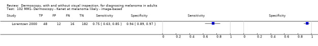
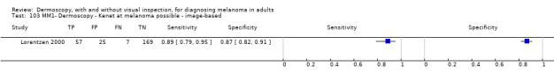
Lorentzen 2000 {published data only}
-
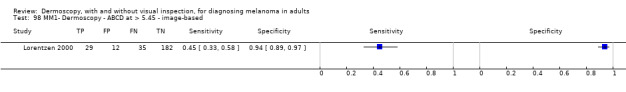
- Lorentzen H, Weismann K, Kenet RO, Secher L, Larsen FG. Comparison of dermatoscopic ABCD rule and risk stratification in the diagnosis of malignant melanoma. Acta Dermato‐Venereologica 2000;80(2):122‐6. [ER4:17941061; PUBMED: 10877133] - PubMed
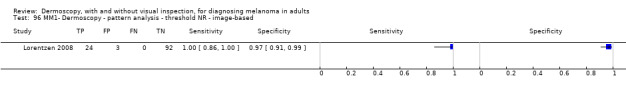
Lorentzen 2008 {published data only}
-
- Lorentzen HF, Eefsen RL, Weismann K. Comparison of classical dermatoscopy and acrylic globe magnifier dermatoscopy. Acta Dermato‐Venereologica 2008;88(2):139‐42. [ER4:15465993; PUBMED: 18311441] - PubMed
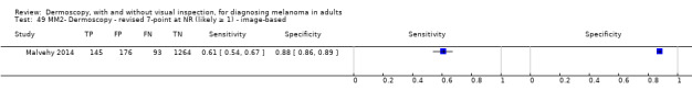
Malvehy 2014 {published data only}
-
- Malvehy J, Hauschild A, Curiel‐Lewandrowski C, Mohr P, Hofmann‐Wellenhof R, Motley R, et al. Clinical performance of the Nevisense system in cutaneous melanoma detection: an international, multicentre, prospective and blinded clinical trial on efficacy and safety. British Journal of Dermatology 2014;171(5):1099‐107. [PUBMED: 24841846] - PMC - PubMed
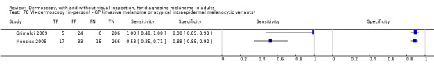
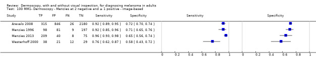
Menzies 1996 {published data only}
-
- Menzies SW, Ingvar C, Crotty KA, McCarthy WH. Frequency and morphologic characteristics of invasive melanomas lacking specific surface microscopic features. Archives of Dermatology 1996;132(10):1178‐82. [ER4:21450627; PUBMED: 8859028] - PubMed
Menzies 2005 {published data only}
-
- Menzies SW, Bischof L, Talbot H, Gutenev A, Avramidis M, Wong L, et al. The performance of SolarScan: an automated dermoscopy image analysis instrument for the diagnosis of primary melanoma. Archives of Dermatology 2005;141(11):1388‐96. [ER4:20569478; PUBMED: 16301386] - PubMed
Menzies 2008 {published data only}
-
- Menzies SW, Kreusch J, Byth K, Pizzichetta MA, Marghoob A, Braun R, et al. Dermoscopic evaluation of amelanotic and hypomelanotic melanoma. Archives of Dermatology 2008;144(9):1120‐7. [PUBMED: 18794455] - PubMed
Menzies 2009 {published data only}
-
- Menzies SW, Emery J, Staples M, Davies S, McAvoy B, Fletcher J, et al. Impact of dermoscopy and short‐term sequential digital dermoscopy imaging for the management of pigmented lesions in primary care: a sequential intervention trial. British Journal of Dermatology 2009;161(6):1270‐7. [DOI: 10.1111/j.1365-2133.2009.09374.x; ER4:15466005; PUBMED: 19747359] - DOI - PubMed
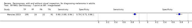
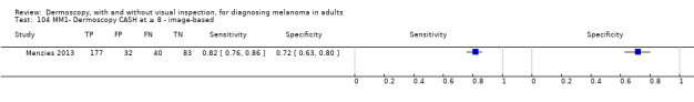
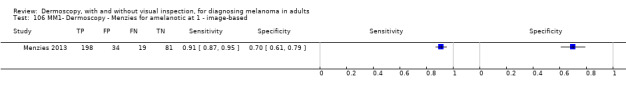
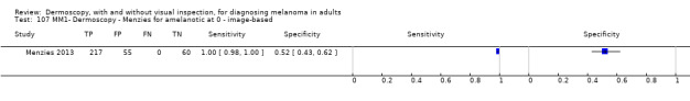
Menzies 2013 {published data only}
-
- Menzies SW, Moloney FJ, Byth K, Avramidis M, Argenziano G, Zalaudek I, et al. Dermoscopic evaluation of nodular melanoma. JAMA Dermatology 2013;149(6):699‐709. [PUBMED: 23553375] - PubMed
Morales Callaghan 2008 {published data only}
-
- Morales‐Callaghan AM, Castrodeza‐Sanz J, Martinez‐Garcia G, Peral‐Martinez I, Miranda‐Romero A. Correlation between clinical, dermatoscopic, and histopathologic variables in atypical melanocytic nevi. Actas Dermo‐Sifiliograficas 2008;99(5):380‐9. [ER4:17941068; PUBMED: 18501170] - PubMed
Nachbar 1994 {published data only}
-
- Nachbar F, Stolz W, Merkle T, Cognetta AB, Vogt T, Landthaler M, et al. The ABCD rule of dermatoscopy. High prospective value in the diagnosis of doubtful melanocytic skin lesions. Journal of the American Academy of Dermatology 1994;30(4):551‐9. [ER4:15466022; PUBMED: 8157780] - PubMed
Nilles 1994 {published data only}
-
- Nilles M, Boedeker RH, Schill WB. Surface microscopy of naevi and melanomas‐‐clues to melanoma. British Journal of Dermatology 1994;130(3):349‐55. [ER4:18375123; PUBMED: 8148277] - PubMed
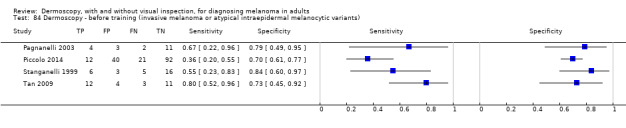
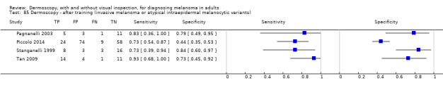
Pagnanelli 2003 {published data only}
-
- Pagnanelli G, Soyer HP, Argenziano G, Talamini R, Barbati R, Bianchi L, et al. Diagnosis of pigmented skin lesions by dermoscopy: web‐based training improves diagnostic performance of non‐experts. British Journal of Dermatology 2003;148(4):698‐702. [ER4:15466036; PUBMED: 12752126] - PubMed
Piccolo 2000 {published data only}
-
- Piccolo D, Smolle J, Argenziano G, Wolf IH, Braun R, Cerroni L, et al. Teledermoscopy‐‐results of a multicentre study on 43 pigmented skin lesions. Journal of Telemedicine & Telecare 2000;6(3):132‐7. [PUBMED: 10912329] - PubMed
Piccolo 2002a {published data only}
-
- Piccolo D, Ferrari A, Peris K, Diadone R, Ruggeri B, Chimenti S. Dermoscopic diagnosis by a trained clinician vs. a clinician with minimal dermoscopy training vs. computer‐aided diagnosis of 341 pigmented skin lesions: a comparative study. British Journal of Dermatology 2002;147(3):481‐6. [ER4:15466057; PUBMED: 12207587] - PubMed
Piccolo 2014 {published data only}
-
- Piccolo D, Crisman G, Schoinas S, Altamura D, Peris K. Computer‐automated ABCD versus dermatologists with different degrees of experience in dermoscopy. European Journal of Dermatology 2014;24(4):477‐81. [ER4:17941089; PUBMED: 24721784] - PubMed
Pizzichetta 2002 {published data only}
-
- Pizzichetta MA, Talamini R, Piccolo D, Trevisan G, Veronesi A, Carbone A, et al. Interobserver agreement of the dermoscopic diagnosis of 129 small melanocytic skin lesions. Tumori 2002;88(3):234‐8. [ER4:18375049; PUBMED: 12195762] - PubMed
Pizzichetta 2004 {published data only}
-
- Pizzichetta MA, Talamini R, Stanganelli I, Puddu P, Bono R, Argenziano G, et al. Amelanotic/hypomelanotic melanoma: clinical and dermoscopic features. British Journal of Dermatology 2004;150(6):1117‐24. [ER4:15466066; PUBMED: 15214897] - PubMed
Pupelli 2013 {published data only}
-
- Pupelli G, Longo C, Veneziano L, Cesinaro AM, Ferrara G, Piana S, et al. Small‐diameter melanocytic lesions: morphological analysis by means of in vivo confocal microscopy. British Journal of Dermatology 2013;168(5):1027‐33. [ER4:15466070; PUBMED: 23301553] - PubMed
Rao 1997 {published data only}
-
- Rao BK, Marghoob AA, Stolz W, Kopf AW, Slade J, Wasti Q, et al. Can early malignant melanoma be differentiated from atypical melanocytic nevi by in vivo techniques? Part I. Clinical and dermoscopic characteristics. Skin Research and Technology 1997;3(1):8‐14. [ER4:17941048; PUBMED: 27333167] - PubMed
Rigel 2012 {published data only}
-
- Rigel DS, Roy M, Yoo J, Cockerell CJ, Robinson JK, White R. Impact of guidance from a computer‐aided multispectral digital skin lesion analysis device on decision to biopsy lesions clinically suggestive of melanoma. Archives of Dermatology 2012;148(4):541‐3. [ER4:15466080; PUBMED: 22351788] - PubMed
Rosendahl 2011 {published data only}
-
- Rosendahl C, Tschandl P, Cameron A, Kittler H. Diagnostic accuracy of dermatoscopy for melanocytic and nonmelanocytic pigmented lesions. Journal of the American Academy of Dermatology 2011;64(6):1068‐73. [ER4:15466083; PUBMED: 21440329] - PubMed
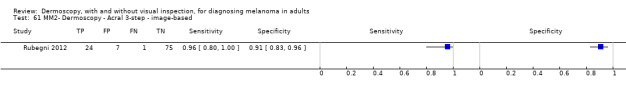
Rubegni 2012 {published data only}
-
- Rubegni P, Cevenini G, Nami N, Argenziano G, Saida T, Burroni M, et al. Dermoscopy and digital dermoscopy analysis of palmoplantar 'equivocal' pigmented skin lesions in Caucasians. Dermatology 2012;225(3):248‐55. [ER4:15466088; PUBMED: 23182753] - PubMed
Rubegni 2016 {published data only}
-
- Rubegni P, Tognetti L, Argenziano G, Nami N, Brancaccio G, Cinotti E, et al. A risk scoring system for the differentiation between melanoma with regression and regressing nevi. Journal of Dermatological Science 2016;83(2):138‐44. [ER4:25012293; PUBMED: 27157925] - PubMed
Sboner 2004 {published data only}
-
- Sboner A, Bauer P, Zumiani G, Eccher C, Blanzieri E, Forti S, et al. Clinical validation of an automated system for supporting the early diagnosis of melanoma. Skin Research & Technology 2004;10(3):184‐92. [ER4:15466104; PUBMED: 15225269] - PubMed
Seidenari 1998 {published data only}
-
- Seidenari S, Pellacani G, Pepe P. Digital videomicroscopy improves diagnostic accuracy for melanoma. Journal of the American Academy of Dermatology 1998;39(2 Pt 1):175‐81. [ER4:15466116; PUBMED: 9704824] - PubMed
Seidenari 2005 {published data only}
-
- Seidenari S, Pellacani G, Martella A. Acquired melanocytic lesions and the decision to excise: role of color variegation and distribution as assessed by dermoscopy. Dermatologic Surgery 2005;31(2):184‐9. [ER4:15466115; PUBMED: 15762212] - PubMed
Seidenari 2007 {published data only}
-
- Seidenari S, Grana C, Pellacani G. Colour clusters for computer diagnosis of melanocytic lesions. Dermatology 2007;214(2):137‐43. [ER4:15466111; PUBMED: 17341863] - PubMed
Skvara 2005 {published data only}
-
- Skvara H, Teban L, Fiebiger M, Binder M, Kittler H. Limitations of dermoscopy in the recognition of melanoma. Archives of Dermatology 2005;141(2):155‐60. [ER4:20569495; PUBMED: 15724011] - PubMed
Soyer 1995 {published data only}
-
- Soyer HP, Smolle J, Leitinger G, Rieger E, Kerl H. Diagnostic reliability of dermoscopic criteria for detecting malignant melanoma. Dermatology 1995;190(1):25‐30. [ER4:18375054; PUBMED: 7894091] - PubMed
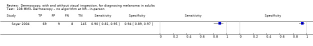
Soyer 2004 {published data only}
-
- Soyer HP, Argenziano G, Zalaudek I, Corona R, Sera F, Talamini R, et al. Three‐point checklist of dermoscopy. A new screening method for early detection of melanoma. Dermatology 2004;208(1):27‐31. [ER4:15466124; PUBMED: 14730233] - PubMed
Stanganelli 1998a {published data only}
-
- Stanganelli I, Serafini M, Cainelli T, Cristofolini M, Baldassari L, Staffa M, et al. Accuracy of epiluminescence microscopy among practical dermatologists: a study from the Emilia‐Romagna region of Italy. Tumori 1998;84(6):701‐5. [ER4:18375055; PUBMED: 10080681] - PubMed
Stanganelli 1999 {published data only}
-
- Stanganelli I, Seidenari S, Serafini M, Pellacani G, Bucchi L. Diagnosis of pigmented skin lesions by epiluminescence microscopy: determinants of accuracy improvement in a nationwide training programme for practical dermatologists. Public Health 1999;113(5):237‐42. [ER4:15466128; PUBMED: 10557118] - PubMed
Stanganelli 2000 {published data only}
-
- Stanganelli I, Serafini M, Bucch L. A cancer‐registry‐assisted evaluation of the accuracy of digital epiluminescence microscopy associated with clinical examination of pigmented skin lesions. Dermatology 2000;200(1):11‐6. [ER4:15466129; PUBMED: 10681607] - PubMed
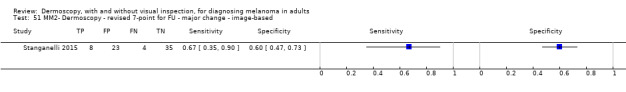
Stanganelli 2005 {published data only}
-
- Stanganelli I, Brucale A, Calori L, Gori R, Lovato A, Magi S, et al. Computer‐aided diagnosis of melanocytic lesions. Anticancer Research 2005;25(6C):4577‐82. [ER4:15466126; PUBMED: 16334145] - PubMed
Stanganelli 2015 {published data only}
-
- Stanganelli I, Longo C, Mazzoni L, Magi S, Medri M, Lanzanova G, et al. Integration of reflectance confocal microscopy in sequential dermoscopy follow‐up improves melanoma detection accuracy. British Journal of Dermatology 2015;172(2):365‐71. [ER4:20569496; PUBMED: 25154446] - PubMed
Stolz 1994a {published data only}
-
- Stolz W, Riemann A, Cognetta AB, Pillet L, Abmayer W, Holzel D, et al. ABCD rule of dermatoscopy: a new practical method for early recognition of malignant melanoma. European Journal of Dermatology 1994;4(7):521‐7. [EMBASE: 24349113; ER4:18375098]
Tan 2009 {published data only}
-
- Tan E, Levell NJ. Regular clinical dermatoscope use with training improves melanoma diagnosis by dermatologists. Clinical & Experimental Dermatology 2009;34(8):e876‐8. [ER4:17941000; PUBMED: 20055853] - PubMed
Tenenhaus 2010 {published data only}
-
- Tenenhaus A, Nkengne A, Horn JF, Serruys C, Giron A, Fertil B. Detection of melanoma from dermoscopic images of naevi acquired under uncontrolled conditions. Skin Research & Technology 2010;16(1):85‐97. [ER4:17941001; PUBMED: 20384887] - PubMed
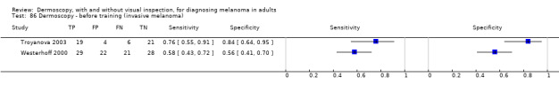
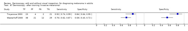
Troyanova 2003 {published data only}
-
- Troyanova P. A beneficial effect of a short‐term formal training course in epiluminescence microscopy on the diagnostic performance of dermatologists about cutaneous malignant melanoma. Skin Research & Technology 2003;9(3):269‐73. [ER4:17941004; PUBMED: 12877690] - PubMed
Unlu 2014 {published data only}
-
- Unlu E, Akay BN, Erdem C. Comparison of dermatoscopic diagnostic algorithms based on calculation: the ABCD rule of dermatoscopy, the seven‐point checklist, the three‐point checklist and the CASH algorithm in dermatoscopic evaluation of melanocytic lesions. Journal of Dermatology 2014;41(7):598‐603. [ER4:15466145; PUBMED: 24807635] - PubMed
Viglizzo 2004 {published data only}
-
- Viglizzo G, Rongioletti F. Clinical, dermoscopic and pathologic correlation of pigmentary lesions observed in a dermoscopy service in the year 2003 [Correlazione clinico‐dermoscopico‐patologica delle lesioni cutanee pigmentate osservate in un servizio di dermoscopia nell'anno 2003]. Giornale Italiano di Dermatologia e Venereologia 2004;139:339‐44. [EMBASE: 39456561; ER4:18375099]
Wells 2012 {published data only}
-
- Wells R, Gutkowicz‐Krusin D, Veledar E, Toledano A, Chen SC. Comparison of diagnostic and management sensitivity to melanoma between dermatologists and MelaFind: a pilot study. Archives of Dermatology 2012;148(9):1083‐4. [ER4:15466163; PUBMED: 22986873] - PubMed
Westerhoff 2000 {published data only}
-
- Westerhoff K, McCarthy WH, Menzies SW. Increase in the sensitivity for melanoma diagnosis by primary care physicians using skin surface microscopy. British Journal of Dermatology 2000;143(5):1016‐20. [ER4:15466164; PUBMED: 11069512] - PubMed
Winkelmann 2016 {published data only}
-
- Winkelmann RR, Farberg AS, Tucker N, White R, Rigel DS. Enhancement of international dermatologists' pigmented skin lesion biopsy decisions following dermoscopy with subsequent integration of multispectral digital skin lesion analysis. Journal of Clinical and Aesthetic Dermatology 2016;9(7):53‐5. [ER4:25701735; PUBMED: 27672411] - PMC - PubMed
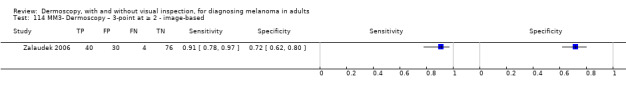
Zalaudek 2006 {published data only}
-
- Zalaudek I, Argenziano G, Soyer HP, Corona R, Sera F, Blum A, et al. Three‐point checklist of dermoscopy: an open internet study. British Journal of Dermatology 2006;154(3):431‐7. [ER4:15466171; PUBMED: 16445771] - PubMed
References to studies excluded from this review
Ahnlide 2013 {published data only}
-
- Ahnlide I, Bjellerup M. Accuracy of clinical skin tumour diagnosis in a dermatological setting. Acta Dermato‐Venereologica 2013;93(3):305‐8. - PubMed
Akasu 1996 {published data only}
-
- Akasu R, Sugiyama H, Araki M, Ohtake N, Furue M, Tamaki K. Dermatoscopic and videomicroscopic features of melanocytic plantar nevi. American Journal of Dermatopathology 1996;18(1):10‐8. - PubMed
Alendar 2009 {published data only}
Al Jalbout 2013 {published data only}
-
- Al Jalbout S, Moscarella E, Longo C, Argenziano G, Piana S, Zalaudek I. Dermoscopy should always be performed... even in clear‐cut cases!. Journal of the American Academy of Dermatology 2013;69(4):e159‐60. - PubMed
Altamura 2006 {published data only}
-
- Altamura D, Altobelli E, Micantonio T, Piccolo D, Fargnoli MC, Peris K. Dermoscopic patterns of acral melanocytic nevi and melanomas in a white population in central Italy. Archives of Dermatology 2006;142(9):1123‐8. - PubMed
Altamura 2010 {published data only}
-
- Altamura D, Menzies SW, Argenziano G, Zalaudek I, Soyer HP, Sera F, et al. Dermatoscopy of basal cell carcinoma: morphologic variability of global and local features and accuracy of diagnosis. Journal of the American Academy of Dermatology 2010;62(1):67‐75. - PubMed
Amirnia 2016 {published data only}
-
- Amirnia M, Ranjkesh MR, Azimpouran M, Karkon‐Shayan F, Alikhah H, Jafari‐Asl M, et al. Comparative study of dermatoscopic and histopathologic results in facial basal cell carcinoma and melanocytic nevi. Asian Pacific Journal of Cancer Prevention 2016;17(1):425‐9. - PubMed
Antonio 2013 {published data only}
-
- Antonio JR, Soubhia RM, D'Avila SC, Caldas AC, Tridico LA, Alves FT. Correlation between dermoscopic and histopathological diagnoses of atypical nevi in a dermatology outpatient clinic of the Medical School of Sao Jose do Rio Preto, SP, Brazil. Anais Brasileiros de Dermatologia 2013;88(2):199‐203. - PMC - PubMed
Antoszewski 2015 {published data only}
-
- Antoszewski B, Fijalkowska M, Stabryla P, Kasielska‐Trojan A. Dermatoscopy as a helpful tool in plastic surgeon's practice ‐ a preliminary study. Polski Przeglad Chirurgiczny 2015;87(12):609‐13. - PubMed
Aoyagi 2010 {published data only}
-
- Aoyagi S, Hata H, Izumi K, Iitani MM, Shimizu H. Diagnostic pitfalls of using dermoscopic features to differentiate between malignant melanoma and pigmented seborrhoeic keratosis. Acta Dermato‐Venereologica 2010;90(4):440‐1. - PubMed
Argenziano 1997 {published data only}
-
- Argenziano G, Fabbrocini G, Carli P, Giorgi V, Delfino M. Epiluminescence microscopy: criteria of cutaneous melanoma progression. Journal of the American Academy of Dermatology 1997;37(1):68‐74. - PubMed
Argenziano 1999 {published data only}
-
- Argenziano G, Fabbrocini G, Carli P, Giorgi V, Delfino M. Clinical and dermatoscopic criteria for the preoperative evaluation of cutaneous melanoma thickness. Journal of the American Academy of Dermatology 1999;40(1):61‐8. - PubMed
Argenziano 2002 {published data only}
-
- Argenziano G, Soyer HP, Chimenti S, Argenziano G, Ruocco V. Impact of dermoscopy on the clinical management of pigmented skin lesions. Clinics in Dermatology 2002;20(3):200‐2. - PubMed
Argenziano 2003 {published data only}
-
- Argenziano G, Soyer HP, Chimenti S, Talamini R, Corona R, Sera F, et al. Dermoscopy of pigmented skin lesions: results of a consensus meeting via the Internet. Journal of the American Academy of Dermatology 2003;48(5):679‐93. - PubMed
Argenziano 2004a {published data only}
-
- Argenziano G, Zalaudek I, Corona R, Sera F, Cicale L, Petrillo G, et al. Vascular structures in skin tumors: a dermoscopy study. Archives of Dermatology 2004;140(12):1485‐9. - PubMed
Argenziano 2004b {published data only}
-
- Argenziano G, Zalaudek I, Soyer HP. Which is the most reliable method for teaching dermoscopy for melanoma diagnosis to residents in dermatology?. British Journal of Dermatology 2004;151(2):512‐3. - PubMed
Argenziano 2008 {published data only}
-
- Argenziano G, Mordente I, Ferrara G, Sgambato A, Annese P, Zalaudek I. Dermoscopic monitoring of melanocytic skin lesions: clinical outcome and patient compliance vary according to follow‐up protocols. British Journal of Dermatology 2008;159(2):331‐6. - PubMed
Argenziano 2010 {published data only}
-
- Argenziano G, Kittler H, Ferrara G, Rubegni P, Malvehy J, Puig S, et al. Slow‐growing melanoma: a dermoscopy follow‐up study. British Journal of Dermatology 2010;162(2):267‐73. - PubMed
Argenziano 2011a {published data only}
-
- Argenziano G, Catricala C, Ardigo M, Buccini P, Simone P, Eibenschutz L, et al. Dermoscopy of patients with multiple nevi: improved management recommendations using a comparative diagnostic approach. Archives of Dermatology 2011;147(1):46‐9. - PubMed
Argenziano 2011b {published data only}
-
- Argenziano G, Longo C, Cameron A, Cavicchini S, Gourhant J Y, Lallas A, et al. Blue‐black rule: a simple dermoscopic clue to recognize pigmented nodular melanoma. British Journal of Dermatology 2011;165(6):1251‐5. - PubMed
Argenziano 2012 {published data only}
-
- Argenziano G, Zalaudek I, Hofmann‐Wellenhof R, Bakos RM, Bergman W, Blum A, et al. Total body skin examination for skin cancer screening in patients with focused symptoms. Journal of the American Academy of Dermatology 2012;66(2):212‐9. - PubMed
Armstrong 2011 {published data only}
-
- Armstrong A. Dermoscopy: an evidence‐based approach for the early detection of melanoma. UNF Graduate Theses and Dissertations. digitalcommons.unf.edu/etd/302 (accessed prior to 16 October 2018).
Ascierto 1998 {published data only}
-
- Ascierto PA, Satriano RA, Palmieri G, Parasole R, Bosco L, Castello G. Epiluminescence microscopy as a useful approach in the early diagnosis of cutaneous malignant melanoma. Melanoma Research 1998;8(6):529‐37. - PubMed
Ascierto 2000 {published data only}
-
- Ascierto PA, Palmieri G, Celentano E, Parasole R, Caraco C, Daponte A, et al. Sensitivity and specificity of epiluminescence microscopy: evaluation on a sample of 2731 excised cutaneous pigmented lesions. The Melanoma Cooperative Study. British Journal of Dermatology 2000;142(5):893‐8. - PubMed
Ascierto 2003 {published data only}
-
- Ascierto PA, Palmieri G, Botti G, Satriano RA, Stanganelli I, Bono R, et al. Early diagnosis of malignant melanoma: proposal of a working formulation for the management of cutaneous pigmented lesions from the Melanoma Cooperative Group. International Journal of Oncology 2003;22(6):1209‐15. - PubMed
Bafounta 2001 {published data only}
-
- Bafounta ML, Beauchet A, Aegerter P, Saiag P. Is dermoscopy (epiluminescence microscopy) useful for the diagnosis of melanoma? Results of a meta‐analysis using techniques adapted to the evaluation of diagnostic tests. Archives of Dermatology 2001;137(10):1343‐50. - PubMed
Bajaj 2016 {published data only}
Bauer 2005 {published data only}
-
- Bauer J, Blum A, Strohhacker U, Garbe C. Surveillance of patients at high risk for cutaneous malignant melanoma using digital dermoscopy. British Journal of Dermatology 2005;152(1):87‐92. - PubMed
Bauer 2006 {published data only}
-
- Bauer J, Leinweber B, Metzler G, Blum A, Hofmann‐Wellenhof R, Leitz N, et al. Correlation with digital dermoscopic images can help dermatopathologists to diagnose equivocal skin tumours. British Journal of Dermatology 2006;155(3):546‐51. - PubMed
Benati 2015 {published data only}
-
- Benati E, Argenziano G, Kyrgidis A, Moscarella E, Ciardo S, Bassoli S, et al. Melanoma and naevi with a globular pattern: confocal microscopy as an aid for diagnostic differentiation. British Journal of Dermatology 2015;173(5):1232‐8. - PubMed
Benelli 2000b {published data only}
-
- Benelli C, Roscetti E, Dal Pozzo V. Reproducibility of a dermoscopic method (7FFM) for the diagnosis of malignant melanoma. European Journal of Dermatology 2000;10(2):110‐4. - PubMed
Benvenuto‐Andrade 2006 {published data only}
-
- Benvenuto‐Andrade C, Dusza SW, Hay JL, Agero AL, Halpern AC, Kopf AW, et al. Level of confidence in diagnosis: clinical examination versus dermoscopy examination. Dermatologic Surgery 2006;32(5):738‐44. - PubMed
Benvenuto‐Andrade 2007 {published data only}
-
- Benvenuto‐Andrade C, Dusza SW, Agero AL, Scope A, Rajadhyaksha M, Halpern AC, et al. Differences between polarized light dermoscopy and immersion contact dermoscopy for the evaluation of skin lesions. Archives of Dermatology 2007;143(3):329‐38. - PubMed
Binder 1997 {published data only}
-
- Binder M, Puespoeck‐Schwarz M, Steiner A, Kittler H, Muellner M, Wolff K, et al. Epiluminescence microscopy of small pigmented skin lesions: short‐term formal training improves the diagnostic performance of dermatologists. Journal of the American Academy of Dermatology 1997;36(2 Pt 1):197‐202. - PubMed
Blum 2003c {published data only}
-
- Blum A. Amelanotic/hypomelanotic melanoma‐‐is dermatoscopy useful for diagnosis?. Journal der Deutschen Dermatologischen Gesellschaft 2003;1(8):666‐7. - PubMed
Blum 2004c {published data only}
-
- Blum A. Pattern analysis, not simplified algorithms, is the most reliable method for teaching dermoscopy for melanoma diagnosis to residents in dermatology. British Journal of Dermatology 2004;151(2):511‐2. - PubMed
Blum 2004d {published data only}
-
- Blum A, Clemens J, Argenziano G. Three‐colour test in dermoscopy: a re‐evaluation. British Journal of Dermatology 2004;150(5):1040. - PubMed
Blum 2004e {published data only}
-
- Blum A, Hofmann‐Wellenhof R. Simplified dermoscopic diagnosis of acral melanocytic lesions: mountains and valleys. Australasian Journal of Dermatology 2004;45(4):235‐6. - PubMed
Blum 2006 {published data only}
-
- Blum A, Clemens J, Argenziano G. Modified dermoscopic algorithm for the differentiation between melanocytic and nonmelanocytic skin tumors. Journal of Cutaneous Medicine & Surgery 2006;10(2):73‐8. - PubMed
Blum 2011 {published data only}
-
- Blum A, Simionescu O, Argenziano G, Braun R, Cabo H, Eichhorn A, et al. Dermoscopy of pigmented lesions of the mucosa and the mucocutaneous junction: results of a multicenter study by the International Dermoscopy Society (IDS). Archives of Dermatology 2011;147(10):1181‐7. - PubMed
Blum 2014 {published data only}
-
- Blum A, Ellwanger U, Luedtke H. Features Amplifying Dermoscopy (FAD) for better evaluation in difficult pigmented and non‐pigmented melanocytic skin tumors. Journal der Deutschen Dermatologischen Gesellschaft 2014;12(1):77‐9. - PubMed
Boespflug 2015 {published data only}
-
- Boespflug A, Guerra J, Dalle S, Thomas L. Enhancement of customary dermoscopy education with spaced education e‐learning: a prospective controlled trial. JAMA Dermatology 2015;151(8):847‐53. - PubMed
Bono 2001 {published data only}
-
- Bono A, Maurichi A, Moglia D, Camerini T, Tragni G, Lualdi M, et al. Clinical and dermatoscopic diagnosis of early amelanotic melanoma. Melanoma Research 2001;11(5):491‐4. - PubMed
Borsari 2010 {published data only}
-
- Borsari S, Longo C, Ferrari C, Benati E, Bassoli S, Schianchi S, et al. Dermoscopic island: a new descriptor for thin melanoma. Archives of Dermatology 2010;146(11):1257‐62. - PubMed
Bowns 2006 {published data only}
-
- Bowns IR, Collins K, Walters SJ, McDonagh AJG. Telemedicine in dermatology: a randomised controlled trial. Health Technology Assessment (Winchester, England) 2006;10(43):iii‐iv, ix‐xi, 1‐39. - PubMed
Braun 2000 {published data only}
-
- Braun RP, Krischer J, Saurat JH. The "wobble sign" in epiluminescence microscopy as a novel clue to the differential diagnosis of pigmented skin lesions. Archives of Dermatology 2000;136(7):940‐2. - PubMed
Braun 2007 {published data only}
-
- Braun RP, Gaide O, Oliviero M, Kopf AW, French LE, Saurat JH, et al. The significance of multiple blue‐grey dots (granularity) for the dermoscopic diagnosis of melanoma. British Journal of Dermatology 2007;157(5):907‐13. - PubMed
Braun‐Falco 1990 {published data only}
-
- Braun‐Falco O, Stolz W, Bilek P, Merkle T, Landthaler M. The dermatoscope. A simplification of epiluminescent microscopy of pigmented skin changes. Hautarzt 1990;41(3):131‐6. - PubMed
Brown 2000 {published data only}
-
- Brown N. Exploration of diagnostic techniques for malignant melanoma: an integrative review. Clinical Excellence for Nurse Practitioners 2000;4(5):263‐71. - PubMed
Buhl 2012 {published data only}
-
- Buhl T, Hansen‐Hagge C, Korpas B, Kaune KM, Haas E, Rosenberger A, et al. Integrating static and dynamic features of melanoma: the DynaMel algorithm. Journal of the American Academy of Dermatology 2012;66(1):27‐36. - PubMed
Bystryn 2003 {published data only}
-
- Bystryn JC. Dermoscopic and histopathologic diagnosis of equivocal melanocytic skin lesions: an interdisciplinary study on 107 cases. Cancer 2003;97(7):1817; author reply 1817‐8. - PubMed
Cabrijan 2008 {published data only}
-
- Cabrijan L, Lipozencic J, Batinac T, Lenkovic M, Gruber F, Stanic ZZ. Correlation between clinical‐dermatoscopic and histopathologic diagnosis of skin tumors in our patients. Collegium Antropologicum 2008;32 Suppl 2:195‐7. - PubMed
Canpolat 2011 {published data only}
-
- Canpolat F, Akış HK, Akay BN, Erdem C. Dermoscopic Features of Acral Melanocytic Nevi [Akral Melanositik Nevüslerin Dermoskopik Özellikleri]. Archives of the Turkish Dermatology & Venerology / Turkderm 2011;45(4):193‐7.
Cardenas 2009 {published data only}
-
- Cardenas E, Sosa A, Bezaury P, Madrid JV, Reyes E, Topete RO. Usefulness of high resolution ultrasound of 17 Mhz in palpable skin lesions. An analysis of 27 patients [Utilidad del ultrasonido de alta resolucion de 17 MHz en lesiones cutaneas palpables. Analisis de 27 pacientes]. Dermatologia Revista Mexicana 2009;53(3):119‐24.
Carli 1998 {published data only}
-
- Carli P, Giorgi V, Naldi L, Dosi G. Reliability and inter‐observer agreement of dermoscopic diagnosis of melanoma and melanocytic naevi. Dermoscopy Panel. European Journal of Cancer Prevention 1998;7(5):397‐402. - PubMed
Carli 2000 {published data only}
-
- Carli P, Giorgi V, Massi D, Giannotti B. The role of pattern analysis and the ABCD rule of dermoscopy in the detection of histological atypia in melanocytic naevi. British Journal of Dermatology 2000;143(2):290‐7. - PubMed
Carli 2003c {published data only}
-
- Carli P, Mannone F, Giorgi V, Nardini P, Chiarugi A, Giannotti B. The problem of false‐positive diagnosis in melanoma screening: the impact of dermoscopy. Melanoma Research 2003;13(2):179‐82. - PubMed
Carli 2004a {published data only}
-
- Carli P, Giorgi V, Chiarugi A, Nardini P, Weinstock MA, Crocetti E, et al. Addition of dermoscopy to conventional naked‐eye examination in melanoma screening: a randomized study. Journal of the American Academy of Dermatology 2004;50(5):683‐9. - PubMed
Carli 2004b {published data only}
-
- Carli P, Giorgi V, Crocetti E, Mannone F, Massi D, Chiarugi A, et al. Improvement of malignant/benign ratio in excised melanocytic lesions in the 'dermoscopy era': a retrospective study 1997‐2001. British Journal of Dermatology 2004;150(4):687‐92. - PubMed
Carli 2005 {published data only}
-
- Carli P, Chiarugi A, Giorgi V. Examination of lesions (including dermoscopy) without contact with the patient is associated with improper management in about 30% of equivocal melanomas. Dermatologic Surgery 2005;31(2):169‐72. - PubMed
Carlos‐Ortega 2007 {published data only}
-
- Carlos‐Ortega B, Sanchez‐Alva ME, Ysita‐Morales A, Angeles‐Garay U. Correlation among simple observation and dermoscopy in the study of pigmented lesions of the skin. Revista Medica del Instituto Mexicano del Seguro Social 2007;45(6):541‐8. - PubMed
Carroll 1998 {published data only}
-
- Carroll DM, Billingsley EM, Helm KF. Diagnosing basal cell carcinoma by dermatoscopy. Journal of Cutaneous Medicine & Surgery 1998;3(2):62‐7. - PubMed
Chen 2013 {published data only}
-
- Chen LL, Liebman TN, Soriano RP, Dusza SW, Halpern AC, Marghoob AA. One‐year follow‐up of dermoscopy education on the ability of medical students to detect skin cancer. Dermatology 2013;226(3):267‐73. - PubMed
Ciudad‐Blanco 2014 {published data only}
-
- Ciudad‐Blanco C, Aviles‐Izquierdo JA, Lazaro‐Ochaita P, Suarez‐Fernandez R. Dermoscopic findings for the early detection of melanoma: an analysis of 200 cases. Actas Dermo‐Sifiliograficas 2014;105(7):683‐93. - PubMed
de Giorgi 2006 {published data only}
-
- Giorgi V, Trez E, Salvini C, Duquia R, Villa D, Sestini S, et al. Dermoscopy in black people. British Journal of Dermatology 2006;155(4):695‐9. - PubMed
De Giorgi 2011 {published data only}
-
- Giorgi V, Grazzini M, Rossari S, Gori A, Alfaioli B, Papi F, et al. Adding dermatoscopy to naked eye examination of equivocal melanocytic skin lesions: effect on intention to excise by general dermatologists. Clinical & Experimental Dermatology 2011;36:255‐9. [ER4:15465901] - PubMed
Delfino 1997 {published data only}
-
- Delfino M, Fabbrocini G, Argenziano G, Magliocchetti N, Nofroni I. A statistical analysis of the characteristics of pigmented skin lesions using epiluminescence microscopy. Journal of the European Academy of Dermatology and Venereology 1997;9(3):243‐8.
de Troya‐Martin 2008 {published data only}
-
- Troya‐Martin M, Blazquez‐Sanchez N, Fernandez‐Canedo I, Frieyro‐Elicegui M, Funez‐Liebana R, Rivas‐Ruiz F. Dermoscopic study of cutaneous malignant melanoma: descriptive analysis of 45 cases. Actas Dermo‐Sifiliograficas 2008;99(1):44‐53. - PubMed
Di Chiacchio 2010 {published data only}
-
- Chiacchio N, Hirata SH, Enokihara MY, Michalany NS, Fabbrocini G, Tosti A. Dermatologists' accuracy in early diagnosis of melanoma of the nail matrix. Archives of Dermatology 2010;146(4):382‐7. - PubMed
Di Stefani 2007 {published data only}
-
- Stefani A, Zalaudek I, Argenziano G, Chimenti S, Soyer HP. Feasibility of a two‐step teledermatologic approach for the management of patients with multiple pigmented skin lesions. Dermatologic Surgery 2007;33(6):686‐92. - PubMed
Dummer 1995 {published data only}
-
- Dummer W, Blaheta HJ, Bastian BC, Schenk T, Brocker EV, Remy W. Preoperative characterization of pigmented skin lesions by epiluminescence microscopy and high‐frequency ultrasound. Archives of Dermatology 1995;131(3):279‐85. - PubMed
Elwan 2016 {published data only}
-
- Elwan NM, Eltatawy RA, Elfar NN, Elsakka OM. Dermoscopic features of acral pigmented lesions in Egyptian patients: a descriptive study. International Journal of Dermatology 2016;55(2):187‐92. - PubMed
Fabbrocini 2008 {published data only}
-
- Fabbrocini G, Balato A, Rescigno O, Mariano M, Scalvenzi M, Brunetti B. Telediagnosis and face‐to‐face diagnosis reliability for melanocytic and non‐melanocytic 'pink' lesions. Journal of the European Academy of Dermatology & Venereology 2008;22(2):229‐34. - PubMed
Ferrara 2002 {published data only}
-
- Ferrara G, Argenziano G, Soyer HP, Corona R, Sera F, Brunetti B, et al. Dermoscopic and histopathologic diagnosis of equivocal melanocytic skin lesions: an interdisciplinary study on 107 cases. Cancer 2002;95(5):1094‐100. - PubMed
Fidalgo 2003 {published data only}
-
- Fidalgo A, Caldas Lopes L, Macedo Ferreira A. Digital dermatoscopy: one‐year experience with the DANAOS system. Skin Cancer 2003;18(4):211‐8.
Fruhauf 2012 {published data only}
-
- Fruhauf J, Leinweber B, Fink‐Puches R, Ahlgrimm‐Siess V, Richtig E, Wolf I H, et al. Patient acceptance and diagnostic utility of automated digital image analysis of pigmented skin lesions. Journal of the European Academy of Dermatology & Venereology 2012;26(3):368‐72. - PubMed
Fueyo‐Casado 2009 {published data only}
-
- Fueyo‐Casado A, Vazquez‐Lopez F, Sanchez‐Martin J, Garcia‐Garcia B, Perez‐Oliva N. Evaluation of a program for the automatic dermoscopic diagnosis of melanoma in a general dermatology setting. Dermatologic Surgery 2009;35(2):257‐9; discussion 260‐2. - PubMed
Giacomel 2005 {published data only}
-
- Giacomel J, Zalaudek I. Dermoscopy of superficial basal cell carcinoma. Dermatologic Surgery 2005;31(12):1710‐3. - PubMed
Giacomel 2014 {published data only}
-
- Giacomel J, Lallas A, Zalaudek I, Argenziano G. Dermoscopic "signature" pattern of pigmented and nonpigmented lentigo maligna. Journal of the American Academy of Dermatology 2014;70(2):e33‐5. - PubMed
Giannotti 2004 {published data only}
-
- Giannotti B, Carli P. Improvement of early diagnosis of melanoma in a Mediterranean population: the experience of the Florence melanoma clinic [Novita in tema di diagnosi precoce del melanoma cutaneo: l'esperienza del gruppo Fiorentino]. Giornale Italiano di Dermatologia e Venereologia 2004;139(2):89‐96.
Gill 2015 {published data only}
-
- Gill L, Wang S, Mancebo SE, Lim HW, Kohen LL. Dermoscopic features of acral melanocytic nevi in patients with skin types V and VI: a cross‐sectional study. Journal of the American Academy of Dermatology 2015;73(6):1059‐61. - PubMed
Gilmore 2009 {published data only}
Grichnik 2003 {published data only}
-
- Grichnik JM. Dermoscopy of melanocytic neoplasms: subpatterns of dysplastic/atypical nevi. Archives of Dermatology 2003;139(12):1696. - PubMed
Grichnik 2004 {published data only}
-
- Grichnik JM. Dermoscopy of melanocytic neoplasms: familial patterns. Archives of Dermatology 2004;140(5):642. - PubMed
Guillod 1996 {published data only}
-
- Guillod JF, Schmid Ph, Fischer S, Salomon D, Saurat JH. Detection and classification of pigmented skin lesions by dermatoscopic digital image processing. Dermatology 1996;193(2):169.
Gunduz 2003 {published data only}
-
- Gunduz K, Koltan S, Sahin MT, E Filiz E. Analysis of melanocytic naevi by dermoscopy during pregnancy. Journal of the European Academy of Dermatology & Venereology 2003;17(3):349‐51. - PubMed
Hacioglu 2013 {published data only}
-
- Hacioglu S, Saricaoglu H, Baskan EB, Uner SI, Aydogan K, Tunali S. The value of spectrophotometric intracutaneous analysis in the noninvasive diagnosis of nonmelanoma skin cancers. Clinical & Experimental Dermatology 2013;38(5):464‐9. - PubMed
Haenssle 2006 {published data only}
-
- Haenssle HA, Krueger U, Vente C, Thoms KM, Bertsch HP, Zutt M, et al. Results from an observational trial: digital epiluminescence microscopy follow‐up of atypical nevi increases the sensitivity and the chance of success of conventional dermoscopy in detecting melanoma. Journal of Investigative Dermatology 2006;126(5):980‐5. - PubMed
Haenssle 2010 {published data only}
-
- Haenssle HA, Korpas B, Hansen‐Hagge C, Buhl T, Kaune KM, Johnsen S, et al. Selection of patients for long‐term surveillance with digital dermoscopy by assessment of melanoma risk factors. Archives of Dermatology 2010;146(3):257‐64. - PubMed
Haspeslagh 2016 {published data only}
-
- Haspeslagh M, Vossaert K, Lanssens S, Noe M, Hoorens I, Chevolet I, et al. Comparison of ex vivo and in vivo dermoscopy in dermatopathologic evaluation of skin tumors. JAMA Dermatology 2016;152(3):312‐7. - PubMed
Henning 2007 {published data only}
-
- Henning JS, Dusza SW, Wang SQ, Marghoob AA, Rabinovitz HS, Polsky D, et al. The CASH (color, architecture, symmetry, and homogeneity) algorithm for dermoscopy. Journal of the American Academy of Dermatology 2007;56(1):45‐52. - PubMed
Henning 2008 {published data only}
-
- Henning JS, Stein JA, Yeung J, Dusza SW, Marghoob AA, Rabinovitz HS, et al. CASH algorithm for dermoscopy revisited. Archives of Dermatology 2008;144(4):554‐5. [PUBMED: 18427058] - PubMed
Herschorn 2012 {published data only}
Hirata 2011 {published data only}
-
- Hirata SH, Yamada S, Enokihara MY, Chiacchio N, Almeida FA, Enokihara MM, et al. Patterns of nail matrix and bed of longitudinal melanonychia by intraoperative dermatoscopy. Journal of the American Academy of Dermatology 2011;65(2):297‐303. - PubMed
Hoffmann 2003 {published data only}
-
- Hoffmann K, Gambichler T, Rick A, Kreutz M, Anschuetz M, Grunendick T, et al. Diagnostic and neural analysis of skin cancer (DANAOS). A multicentre study for collection and computer‐aided analysis of data from pigmented skin lesions using digital dermoscopy. British Journal of Dermatology 2003;149(4):801‐9. - PubMed
Hoorens 2016 {published data only}
-
- Hoorens I, Vossaert K, Pil L, Boone B, Schepper S, Ongenae K, et al. Total‐body examination vs lesion‐directed skin cancer screening. JAMA Dermatology 2016;152(1):27‐34. - PubMed
Ishioka 2009 {published data only}
-
- Ishioka P, Tenorio JM, Lopes PRl, Yamada S, Michalany NS, Amaral MB, et al. A comparative study of teledermatoscopy and face‐to‐face examination of pigmented skin lesions. Journal of Telemedicine & Telecare 2009;15(5):221‐5. - PubMed
Iyatomi 2006 {published data only}
-
- Iyatomi H, Oka H, Saito M, Miyake A, Kimoto M, Yamagami J, et al. Quantitative assessment of tumour extraction from dermoscopy images and evaluation of computer‐based extraction methods for an automatic melanoma diagnostic system. Melanoma Research 2006;16(2):183‐90. - PubMed
Iyatomi 2008 {published data only}
-
- Iyatomi H, Oka H, Celebi ME, Ogawa K, Argenziano G, Soyer HP, et al. Computer‐based classification of dermoscopy images of melanocytic lesions on acral volar skin. Journal of Investigative Dermatology 2008;128(8):2049‐54. - PubMed
Johr 2002 {published data only}
-
- Johr RH. Dermoscopy: alternative melanocytic algorithms‐the ABCD rule of dermatoscopy, Menzies scoring method, and 7‐point checklist. Clinics in Dermatology 2002;20(3):240‐7. - PubMed
Kawabata 1998 {published data only}
-
- Kawabata Y, Tamaki K. Distinctive dermatoscopic features of acral lentiginous melanoma in situ from plantar melanocytic nevi and their histopathologic correlation. Journal of Cutaneous Medicine & Surgery 1998;2(4):199‐204. - PubMed
Kawabata 2001 {published data only}
-
- Kawabata Y, Ohara K, Hino H, Tamaki K. Two kinds of Hutchinson's sign, benign and malignant. Journal of the American Academy of Dermatology 2001;44(2, Part 1):305‐7. - PubMed
Kefel 2012 {published data only}
Kenet 1994 {published data only}
-
- Kenet RO, Fitzpatrick TB. Reducing mortality and morbidity of cutaneous melanoma: a six year plan. B). Identifying high and low risk pigmented lesions using epiluminescence microscopy. Journal of Dermatology 1994;21(11):881‐4. - PubMed
Kittler 2002 {published data only}
-
- Kittler H, Pehamberger H, Wolff K, Binder M. Diagnostic accuracy of dermoscopy. Lancet Oncology 2002;3(3):159‐65. - PubMed
Kittler 2006 {published data only}
-
- Kittler H. Value of follow‐up of pigmented skin lesions by digital dermatoscopy. Journal of Investigative Dermatology 2006;126:S20.
Koga 2011 {published data only}
-
- Koga H, Saida T. Revised 3‐step dermoscopic algorithm for the management of acral melanocytic lesions. Archives of Dermatology 2011;147(6):741‐3. - PubMed
Korotkov 2012 {published data only}
-
- Korotkov K, Garcia R. Computerized analysis of pigmented skin lesions: a review. Artificial Intelligence in Medicine 2012;56(2):69‐90. - PubMed
Lallas 2015 {published data only}
-
- Lallas A, Kyrgidis A, Koga H, Moscarella E, Tschandl P, Apalla Z, et al. The BRAAFF checklist: a new dermoscopic algorithm for diagnosing acral melanoma. British Journal of Dermatology 2015;173(4):1041‐9. - PubMed
Liebman 2011 {published data only}
-
- Liebman TN, Scope A, Rabinovitz H, Braun RP, Marghoob AA. Rosettes may be observed in a range of conditions. Archives of Dermatology 2011;147(12):1468. - PubMed
Liebman 2012 {published data only}
-
- Liebman TN, Rabinovitz HS, Balagula Y, Jaimes‐Lopez N, Marghoob AA. White shiny structures in melanoma and BCC. Archives of Dermatology 2012;148(1):146. - PubMed
Lipoff 2008 {published data only}
-
- Lipoff JB, Scope A, Dusza SW, Marghoob AA, Oliveria SA, Halpern AC. Complex dermoscopic pattern: a potential risk marker for melanoma. British Journal of Dermatology 2008;158(4):821‐4. - PubMed
Liu 2012 {published data only}
-
- Liu Z, Sun J, Smith L, Smith M, Warr R. Distribution quantification on dermoscopy images for computer‐assisted diagnosis of cutaneous melanomas. Medical & Biological Engineering & Computing 2012;50(5):503‐13. - PubMed
Lorentzen 1999b {published data only}
-
- Lorentzen H, Weismann K, Secher L, Petersen CS, Larsen FG. The dermatoscopic ABCD rule does not improve diagnostic accuracy of malignant melanoma. Acta Dermato‐Venereologica 1999;79(6):469‐72. - PubMed
Luttrell 2012 {published data only}
-
- Luttrell MJ, McClenahan P, Hofmann‐Wellenhof R, Fink‐Puches R, Soyer HP. Laypersons' sensitivity for melanoma identification is higher with dermoscopy images than clinical photographs. British Journal of Dermatology 2012;167(5):1037‐41. - PubMed
MacKie 1971 {published data only}
-
- MacKie RM. An aid to the preoperative assessment of pigmented lesions of the skin. British Journal of Dermatology 1971;85(3):232‐8. - PubMed
MacKie 2002 {published data only}
-
- Mackie RM, Fleming C, McMahon AD, Jarrett P. The use of the dermatoscope to identify early melanoma using the three‐colour test. British Journal of Dermatology 2002;146(3):481‐4. - PubMed
Markowitz 2015 {published data only}
-
- Markowitz O, Schwartz M, Feldman E, Bienenfeld A, Bieber AK, Ellis J, et al. Evaluation of optical coherence tomography as a means of identifying earlier stage basal cell carcinomas while reducing the use of diagnostic biopsy. The Journal of Clinical & Aesthetic Dermatology 2015;8(10):14‐20. - PMC - PubMed
Massi 2001 {published data only}
-
- Massi D, Giorgi V, Carli P, Santucci M. Diagnostic significance of the blue hue in dermoscopy of melanocytic lesions: a dermoscopic‐pathologic study. American Journal of Dermatopathology 2001;23(5):463‐9. - PubMed
Mayer 1997 {published data only}
-
- Mayer J. Systematic review of the diagnostic accuracy of dermatoscopy in detecting malignant melanoma. Medical Journal of Australia 1997;167(4):206‐10. - PubMed
Menzies 1996a {published data only}
-
- Menzies SW, Ingvar C, McCarthy WH. A sensitivity and specificity analysis of the surface microscopy features of invasive melanoma. Melanoma Research 1996;6(1):55‐62. - PubMed
Menzies 1999 {published data only}
-
- Menzies SW. Automated epiluminescence microscopy: human vs machine in the diagnosis of melanoma. Archives of Dermatology 1999;135(12):1538‐40. - PubMed
Menzies 2000 {published data only}
-
- Menzies SW, Westerhoff K, Rabinovitz H, Kopf AW, McCarthy WH, Katz B. Surface microscopy of pigmented basal cell carcinoma. Archives of Dermatology 2000;136(8):1012‐6. - PubMed
Menzies 2001 {published data only}
-
- Menzies SW, Gutenev A, Avramidis M, Batrac A, McCarthy WH. Short‐term digital surface microscopic monitoring of atypical or changing melanocytic lesions. Archives of Dermatology 2001;137(12):1583‐9. - PubMed
Mun 2016 {published data only}
Nathansohn 2007 {published data only}
-
- Nathansohn N, Orenstein A, Trau H, Liran A, Schachter J. Pigmented lesions clinic for early detection of melanoma: preliminary results. Israel Medical Association Journal 2007;9(10):708‐12. - PubMed
Navarrete‐Dechent 2016 {published data only}
Pan 2008 {published data only}
-
- Pan Y, Chamberlain AJ, Bailey M, Chong AH, Haskett M, Kelly JW. Dermatoscopy aids in the diagnosis of the solitary red scaly patch or plaque‐features distinguishing superficial basal cell carcinoma, intraepidermal carcinoma, and psoriasis. Journal of the American Academy of Dermatology 2008;59(2):268‐74. - PubMed
Panasiti 2009 {published data only}
-
- Panasiti V, Devirgiliis V, Curzio M, Roberti V, Gobbi S, Masciangelo R, et al. The reticular point of view in dermatoscopy. Journal of the American Academy of Dermatology 2009;61(4):605‐10. - PubMed
Pazzini 1996 {published data only}
-
- Pazzini C, Pozzi M, Betti R, Vergani R, Crosti C. Improvement of diagnostic accuracy in the clinical diagnosis of pigmented skin lesions by epiluminescence microscopy. Skin Cancer 1996;11(2):159‐61.
Pehamberger 1987 {published data only}
-
- Pehamberger H, Steiner A, Wolff K. In vivo epiluminescence microscopy of pigmented skin lesions. I. Pattern analysis of pigmented skin lesions. Journal of the American Academy of Dermatology 1987;17(4):571‐83. - PubMed
Pellacani 2002 {published data only}
-
- Pellacani G, Seidenari S. Comparison between morphological parameters in pigmented skin lesion images acquired by means of epiluminescence surface microscopy and polarized‐light videomicroscopy. Clinics in Dermatology 2002;20(3):222‐7. - PubMed
Pellacani 2006 {published data only}
-
- Pellacani G, Grana C, Seidenari S. Algorithmic reproduction of asymmetry and border cut‐off parameters according to the ABCD rule for dermoscopy. Journal of the European Academy of Dermatology & Venereology 2006;20(10):1214‐9. - PubMed
Pellacani 2007 {published data only}
-
- Pellacani G, Bassoli S, Longo C, Cesinaro AM, Seidenari S. Diving into the blue: in vivo microscopic characterization of the dermoscopic blue hue. Journal of the American Academy of Dermatology 2007;57(1):96‐104. - PubMed
Pellacani 2009 {published data only}
-
- Pellacani G, Longo C, Ferrara G, Cesinaro AM, Bassoli S, Guitera P, et al. Spitz nevi: in vivo confocal microscopic features, dermatoscopic aspects, histopathologic correlates, and diagnostic significance. Journal of the American Academy of Dermatology 2009;60(2):236‐47. - PubMed
Peris 2002a {published data only}
-
- Peris K, Altobelli E, Ferrari A, Fargnoli MC, Piccolo D, Esposito M, et al. Interobserver agreement on dermoscopic features of pigmented basal cell carcinoma. Dermatologic Surgery 2002;28(7):643‐5. - PubMed
Peris 2002b {published data only}
-
- Peris K, Ferrari A, Argenziano G, Soyer HP, Chimenti S. Dermoscopic classification of Spitz/Reed nevi. Clinics in Dermatology 2002;20(3):259‐62. - PubMed
Phan 2010 {published data only}
-
- Phan A, Dalle S, Touzet S, Ronger‐Savle S, Balme B, Thomas L. Dermoscopic features of acral lentiginous melanoma in a large series of 110 cases in a white population. British Journal of Dermatology 2010;162(4):765‐71. - PubMed
Piccolo 2002b {published data only}
-
- Piccolo D, Peris K, Chimenti S, Argenziano G, Soyer HP. Jumping into the future using teledermoscopy. Skinmed 2002;1(1):20‐4. - PubMed
Piccolo 2004 {published data only}
-
- Piccolo D, Soyer HP, Chimenti S, Argenziano G, Bartenjev I, Hofmann‐Wellenhof R, et al. Diagnosis and categorization of acral melanocytic lesions using teledermoscopy. Journal of Telemedicine & Telecare 2004;10(6):346‐50. - PubMed
Piccolo 2006 {published data only}
-
- Piccolo D, Fargnoli MC, Ferrara G, Lozzi GP, Altamura D, Ventura T, et al. Hypoepiluminescence microscopy of pigmented skin lesions: new approach to improve recognition of dermoscopic structures. Dermatologic Surgery 2006;32(11):1391‐7. - PubMed
Pizzichetta 2001a {published data only}
-
- Pizzichetta MA, Argenziano G, Talamini R, Piccolo D, Gatti A, Trevisan G, et al. Dermoscopic criteria for melanoma in situ are similar to those for early invasive melanoma. Cancer 2001;91(5):992‐7. - PubMed
Pizzichetta 2001b {published data only}
-
- Pizzichetta MA, Talamini R, Piccolo D, Argenziano G, Pagnanelli G, Burgdorf T, et al. The ABCD rule of dermatoscopy does not apply to small melanocytic skin lesions. Archives of Dermatology 2001;137(10):1376‐8. - PubMed
Pizzichetta 2007 {published data only}
-
- Pizzichetta MA, Stanganelli I, Bono R, Soyer HP, Magi S, Canzonieri V, et al. Dermoscopic features of difficult melanoma. Dermatologic Surgery 2007;33(1):91‐9. - PubMed
Pizzichetta 2010 {published data only}
-
- Pizzichetta MA, Canzonieri V, Massarut S, Baresic T, Borsatti E, Menzies SW. Pitfalls in the dermoscopic diagnosis of amelanotic melanoma. Journal of the American Academy of Dermatology 2010;62(5):893‐4. - PubMed
Pizzichetta 2013 {published data only}
-
- Pizzichetta MA, Talamini R, Marghoob AA, Soyer HP, Argenziano G, Bono R, et al. Negative pigment network: an additional dermoscopic feature for the diagnosis of melanoma. Journal of the American Academy of Dermatology 2013;68(4):552‐9. - PubMed
Pralong 2012 {published data only}
-
- Pralong P, Bathelier E, Dalle S, Poulalhon N, Debarbieux S, Thomas L. Dermoscopy of lentigo maligna melanoma: report of 125 cases. British Journal of Dermatology 2012;167(2):280‐7. - PubMed
Provost 1998 {published data only}
-
- Provost N, Kopf AW, Rabinovitz HS, Stolz W, DeDavid M, Wasti Q, et al. Comparison of conventional photographs and telephonically transmitted compressed digitized images of melanomas and dysplastic nevi. Dermatology 1998;196(3):299‐304. - PubMed
Rader 2014 {published data only}
Rajpara 2009 {published data only}
-
- Rajpara SM, Botello AP, Townend J, Ormerod AD. Systematic review of dermoscopy and digital dermoscopy/artificial intelligence for the diagnosis of melanoma. British Journal of Dermatology 2009;161(3):591‐604. - PubMed
Reggiani 2015 {published data only}
-
- Reggiani C, Manfredini M, Mandel VD, Farnetani F, Ciardo S, Bassoli S, et al. Update on non‐invasive imaging techniques in early diagnosis of non‐melanoma skin cancer. Giornale Italiano di Dermatologia e Venereologia 2015;150(4):393‐405. - PubMed
Rigel 1997 {published data only}
-
- Rigel DS. Epiluminescence microscopy in clinical diagnosis of pigmented skin lesions?. Lancet 1997;349(9065):1566‐7. - PubMed
Ronger 2002 {published data only}
-
- Ronger S, Touzet S, Ligeron C, Balme B, Viallard AM, Barrut D, et al. Dermoscopic examination of nail pigmentation. Archives of Dermatology 2002;138(10):1327‐33. - PubMed
Rosendahl 2012a {published data only}
-
- Rosendahl C, Cameron A, Argenziano G, Zalaudek I, Tschandl P, Kittler H. Dermoscopy of squamous cell carcinoma and keratoacanthoma. Archives of Dermatology 2012;148(12):1386‐92. - PubMed
Rosendahl 2012b {published data only}
-
- Rosendahl C, Cameron A, McColl I, Wilkinson D. Dermatoscopy in routine practice: 'Chaos and Clues'. Australian Family Physician 2012;41(7):482‐7. - PubMed
Rossi 2000 {published data only}
-
- Rossi CR, Vecchiato A, Bezze G, Mastrangelo G, Montesco MC, Mocellin S, et al. Early detection of melanoma: an educational campaign in Padova, Italy. Melanoma Research 2000;10(2):181‐7. - PubMed
Rubegni 2002 {published data only}
-
- Rubegni P, Burroni M, Dell'eva G, Andreassi L. Digital dermoscopy analysis for automated diagnosis of pigmented skin lesions. Clinics in Dermatology 2002;20(3):309‐12. - PubMed
Rubegni 2005a {published data only}
-
- Rubegni P, Burroni M, Andreassi A, Fimiani M. The role of dermoscopy and digital dermoscopy analysis in the diagnosis of pigmented skin lesions. Archives of Dermatology 2005;141(11):1444‐6. - PubMed
Rubegni 2005b {published data only}
-
- Rubegni P, Burroni M, Sbano P, Andreassi L. Digital dermoscopy analysis and internet‐based program for discrimination of pigmented skin lesion dermoscopic images. British Journal of Dermatology 2005;152(2):395‐6. - PubMed
Rubegni 2010 {published data only}
-
- Rubegni P, Cevenini G, Burroni M, Bono R, Sbano P, Biagioli M, et al. Objective follow‐up of atypical melanocytic skin lesions: a retrospective study. Archives of Dermatological Research 2010;302(7):551‐60. - PubMed
Sahin 2004 {published data only}
-
- Sahin MT, Ozturkcan S, Ermertcan AT, Gunes AT. A comparison of dermoscopic features among lentigo senilis/initial seborrheic keratosis, seborrheic keratosis, lentigo maligna and lentigo maligna melanoma on the face. Journal of Dermatology 2004;31(11):884‐9. - PubMed
Saida 2002 {published data only}
-
- Saida T, Oguchi S, Miyazaki A. Dermoscopy for acral pigmented skin lesions. Clinics in Dermatology 2002;20(3):279‐85. - PubMed
Saida 2004 {published data only}
-
- Saida T, Miyazaki A, Oguchi S, Ishihara Y, Yamazaki Y, Murase S, et al. Significance of dermoscopic patterns in detecting malignant melanoma on acral volar skin: results of a multicenter study in Japan. Archives of Dermatology 2004;140(10):1233‐8. - PubMed
Sakakibara 2010 {published data only}
-
- Sakakibara A, Kamijima M, Shibata S, Yasue S, Kono M, Tomita Y. Dermoscopic evaluation of vascular structures of various skin tumors in Japanese patients. Journal of Dermatology 2010;37(4):316‐22. - PubMed
Salerni 2011 {published data only}
-
- Salerni G, Lovatto L, Carrera C, Palou J, Alos L, Puig‐Butille JA, et al. Correlation among dermoscopy, confocal reflectance microscopy, and histologic features of melanoma and basal cell carcinoma collision tumor. Dermatologic Surgery 2011;37(2):275‐9. - PubMed
Salerni 2012 {published data only}
-
- Salerni G, Carrera C, Lovatto L, Puig‐Butille JA, Badenas C, Plana E, et al. Benefits of total body photography and digital dermatoscopy ("two‐step method of digital follow‐up") in the early diagnosis of melanoma in patients at high risk for melanoma. Journal of the American Academy of Dermatology 2012;67(1):e17‐27. - PMC - PubMed
Salerni 2013 {published data only}
-
- Salerni G, Teran T, Puig S, Malvehy J, Zalaudek I, Argenziano G, et al. Meta‐analysis of digital dermoscopy follow‐up of melanocytic skin lesions: a study on behalf of the International Dermoscopy Society. Journal of the European Academy of Dermatology & Venereology 2013;27(7):805‐14. - PubMed
Salvio 2011 {published data only}
-
- Salvio AG, Assumpcao JA, Segalla JGM, Panfilo BL, Nicolini HR, Didone R. One year experience of a model for melanoma continuous prevention in the city of Jau (Sao Paulo), Brazil. Anais Brasileiros de Dermatologia 2011;86(4):669‐74. - PubMed
Sanchez‐Martin 2012 {published data only}
-
- Sanchez‐Martin J, Vazquez‐Lopez F, Perez‐Oliva N, Argenziano G. Dermoscopy of small basal cell carcinoma: study of 100 lesions 5 mm or less in diameter. Dermatologic Surgery 2012;38(6):947‐50. - PubMed
Savk 2004 {published data only}
-
- Savk E, Sahinkaras E, Okyay P, Karaman G, Erkek M, Sendur N. Interobserver agreement in the use of the ABCD rule for dermoscopy. Journal of Dermatology 2004;31(12):1041‐3. - PubMed
Sawada 2013 {published data only}
Sboner 2003 {published data only}
-
- Sboner A, Eccher C, Blanzieri E, Bauer P, Cristofolini M, Zumiani G, et al. A multiple classifier system for early melanoma diagnosis. Artificial Intelligence in Medicine 2003;27(1):29‐44. - PubMed
Schulz 2001 {published data only}
-
- Schulz H. Epiluminescent microscopy aspects of initial cutaneous melanoma metastases. Hautarzt 2001;52(1):21‐5. - PubMed
Scope 2015 {published data only}
-
- Scope A, Braun RP. The recognition process in dermoscopy: analytic approach vs heuristic approach. JAMA Dermatology 2015;151(7):704‐6. - PubMed
Segura 2009 {published data only}
-
- Segura S, Puig S, Carrera C, Palou J, Malvehy J. Development of a two‐step method for the diagnosis of melanoma by reflectance confocal microscopy. Journal of the American Academy of Dermatology 2009;61(2):216‐29. - PubMed
Seidenari 2004 {published data only}
-
- Seidenari S, Pellacani G, Righi E, Nardo A. Is JPEG compression of videomicroscopic images compatible with telediagnosis? Comparison between diagnostic performance and pattern recognition on uncompressed TIFF images and JPEG compressed ones. Telemedicine Journal & E‐Health 2004;10(3):294‐303. - PubMed
Seidenari 2006a {published data only}
-
- Seidenari S, Longo C, Giusti F, Pellacani G. Clinical selection of melanocytic lesions for dermoscopy decreases the identification of suspicious lesions in comparison with dermoscopy without clinical preselection. British Journal of Dermatology 2006;154(5):873‐9. - PubMed
Seidenari 2006b {published data only}
-
- Seidenari S, Pellacani G, Grana C. Asymmetry in dermoscopic melanocytic lesion images: a computer description based on colour distribution. Acta Dermato‐Venereologica 2006;86(2):123‐8. - PubMed
Seidenari 2012 {published data only}
-
- Seidenari S, Ferrari C, Borsari S, Bassoli S, Cesinaro AM, Giusti F, et al. The dermoscopic variability of pigment network in melanoma in situ. Melanoma Research 2012;22(2):151‐7. - PubMed
Seidenari 2013 {published data only}
Serrao 2006 {published data only}
-
- Serrao VV, Baptista J, Paris F, Lopes LC, Fidalgo A, Ferreira A. Digital dermoscopy. Review of 652 lesions analysed by the DANAOS system. Skin Cancer 2006;21(4):185‐98.
Sgouros 2014 {published data only}
-
- Sgouros D, Lallas A, Julian Y, Rigopoulos D, Zalaudek I, Longo C, et al. Assessment of SIAscopy in the triage of suspicious skin tumours. Skin Research & Technology 2014;20(4):440‐4. - PubMed
Shakya 2012 {published data only}
-
- Shakya NM, LeAnder RW, Hinton KA, Stricklin SM, Rader RK, Hagerty J, et al. Discrimination of squamous cell carcinoma in situ from seborrheic keratosis by color analysis techniques requires information from scale, scale‐crust and surrounding areas in dermoscopy images. Computers in Biology & Medicine 2012;42(12):1165‐9. - PubMed
Shitara 2014 {published data only}
-
- Shitara D, Ishioka P, Alonso‐Pinedo Y, Palacios‐Bejarano L, Carrera C, Malvehy J, et al. Shiny white streaks: a sign of malignancy at dermoscopy of pigmented skin lesions. Acta Dermato‐Venereologica 2014;94(2):132‐7. - PubMed
Shitara 2015 {published data only}
-
- Shitara D, Nascimento M, Ishioka P, Carrera C, Alos L, Malvehy J, et al. Dermoscopy of naevus‐associated melanomas. Acta Dermato‐Venereologica 2015;95(6):671‐5. - PubMed
Sondak 2015 {published data only}
-
- Sondak VK, Glass LF, Geller AC. Risk‐stratified screening for detection of melanoma. JAMA 2015;313(6):616‐7. - PubMed
Soyer 1987 {published data only}
-
- Soyer HP, Smolle J, Kerl H, Stettner H. Early diagnosis of malignant melanoma by surface microscopy. Lancet 1987;2(8562):803. - PubMed
Soyer 2001 {published data only}
-
- Soyer HP, Argenziano G, Talamini R, Chimenti S. Is dermoscopy useful for the diagnosis of melanoma?. Archives of Dermatology 2001;137(10):1361‐3. - PubMed
Stanganelli 1998b {published data only}
-
- Stanganelli I, Bucchi L. Epiluminescence microscopy versus clinical evaluation of pigmented skin lesions: effects of operator's training on reproducibility and accuracy. Dermatology and Venereology Society of the Canton of Ticino. Dermatology 1998;196(2):199‐203. - PubMed
Steiner 1987a {published data only}
-
- Steiner A, Pehamberger H, Wolff K. In vivo epiluminescence microscopy of pigmented skin lesions. II. Diagnosis of small pigmented skin lesions and early detection of malignant melanoma. Journal of the American Academy of Dermatology 1987;17(4):584‐91. [ER4:17940992; PUBMED: 3668003] - PubMed
Steiner 1987b {published data only}
-
- Steiner A, Pehamberger H, Wolff K. Improvement of the diagnostic accuracy in pigmented skin lesions by epiluminescent light microscopy. Anticancer research 1987;7(3 Pt B):433‐4. - PubMed
Steiner 1993 {published data only}
-
- Steiner A, Binder M, Schemper M, Wolff K, Pehamberger H. Statistical evaluation of epiluminescence microscopy criteria for melanocytic pigmented skin lesions. Journal of the American Academy of Dermatology 1993;29(4):581‐8. - PubMed
Stephens 2013 {published data only}
-
- Stephens A, Fraga‐Braghiroli N, Oliviero M, Rabinovitz H, Scope A. Spoke wheel‐like structures in superficial basal cell carcinoma: a correlation between dermoscopy, histopathology, and reflective confocal microscopy. Journal of the American Academy of Dermatology 2013;69(5):e219‐21. - PubMed
Stoecker 2009a {published data only}
Stoecker 2009b {published data only}
Stoecker 2011 {published data only}
Stolz 2002 {published data only}
-
- Stolz W, Schiffner R, Burgdorf WHC. Dermatoscopy for facial pigmented skin lesions. Clinics in Dermatology 2002;20(3):276‐8. - PubMed
Stratigos 2007 {published data only}
-
- Stratigos A, Nikolaou V, Kedicoglou S, Antoniou C, Stefanaki I, Haidemenos G, et al. Melanoma/skin cancer screening in a Mediterranean country: results of the Euromelanoma Screening Day Campaign in Greece. Journal of the European Academy of Dermatology & Venereology 2007;21(1):56‐62. - PubMed
Stricklin 2011 {published data only}
Strumia 2003 {published data only}
-
- Strumia R, Montanari A. Low positive predictive value of ABCD‐E rule for dermatoscopy of small melanocytic naevi. Melanoma Research 2003;13:631‐2. - PubMed
Tasli 2012 {published data only}
-
- Tasli L, Kacar N, Argenziano G. A scientometric analysis of dermoscopy literature over the past 25 years. Journal of the European Academy of Dermatology & Venereology 2012;26(9):1142‐8. - PubMed
Teban 2003 {published data only}
-
- Teban L, Pehamberger H, Wolff K, Binder M, Kittler H. Clinical value of a dermatoscopic classification of Clark nevi. Journal der Deutschen Dermatologischen Gesellschaft 2003;1(4):292‐6. - PubMed
Terstappen 2007 {published data only}
-
- Terstappen K, Larko O, Wennberg AM. Pigmented basal cell carcinoma‐‐comparing the diagnostic methods of SIAscopy and dermoscopy. Acta Dermato‐venereologica 2007;87(3):238‐42. - PubMed
Terushkin 2010a {published data only}
-
- Terushkin V, Braga JC, Dusza SW, Scope A, Busam K, Marghoob AA, et al. Agreement on the clinical diagnosis and management of cutaneous squamous neoplasms. Dermatologic Surgery 2010;36(10):1514‐20. - PubMed
Terushkin 2010b {published data only}
-
- Terushkin V, Warycha M, Levy M, Kopf AW, Cohen DE, Polsky D. Analysis of the benign to malignant ratio of lesions biopsied by a general dermatologist before and after the adoption of dermoscopy. Archives of Dermatology 2010;146(3):343‐4. - PubMed
Tromme 2012 {published data only}
-
- Tromme I, Sacre L, Hammouch F, Legrand C, Marot L, Vereecken P, et al. Availability of digital dermoscopy in daily practice dramatically reduces the number of excised melanocytic lesions: results from an observational study. British Journal of Dermatology 2012;167(4):778‐86. - PubMed
Tschandl 2012 {published data only}
Tschandl 2015a {published data only}
-
- Tschandl P, Kittler H, Schmid K, Zalaudek I, Argenziano G. Teaching dermatoscopy of pigmented skin tumours to novices: comparison of analytic vs. heuristic approach. Journal of the European Academy of Dermatology & Venereology 2015;29(6):1198‐204. - PubMed
Tschandl 2015b {published data only}
-
- Tschandl P, Rosendahl C, Kittler H. Dermatoscopy of flat pigmented facial lesions. Journal of the European Academy of Dermatology & Venereology 2015;29(1):120‐7. - PubMed
Ulrich 2015 {published data only}
-
- Ulrich M, Braunmuehl T, Kurzen H, Dirschka T, Kellner C, Sattler E, et al. The sensitivity and specificity of optical coherence tomography for the assisted diagnosis of nonpigmented basal cell carcinoma: an observational study. British Journal of Dermatology 2015;173(2):428‐35. - PubMed
Van der Leest 2011 {published data only}
-
- Leest RJT, Vries E, Bulliard JL, Paoli J, Peris K, Stratigos AJ, et al. The Euromelanoma skin cancer prevention campaign in Europe: characteristics and results of 2009 and 2010. Journal of the European Academy of Dermatology & Venereology 2011;25(12):1455‐65. - PubMed
Van der Rhee 2010 {published data only}
-
- Rhee JI, Bergman W, Kukutsch NA. The impact of dermoscopy on the management of pigmented lesions in everyday clinical practice of general dermatologists: a prospective study. British Journal of Dermatology 2010;162(3):563‐7. - PubMed
Van der Rhee 2011 {published data only}
-
- Rhee JI, Bergman W, Kukutsch NA. Impact of dermoscopy on the management of high‐risk patients from melanoma families: a prospective study. Acta Dermato‐Venereologica 2011;91(4):428‐31. - PubMed
Vasili 2010 {published data only}
-
- Vasili E, Shkodrani E, Harja D, Labinoti L, Zoto A. Retrospective study of 70 patients with NMSC. Melanoma Research 2010;20:e63.
Verduzco‐Martinez 2013 {published data only}
-
- Verduzco‐Martinez AP, Quinones‐Venegas R, Guevara‐Gutierrez E, Tlacuilo‐Parra A. Correlation of dermoscopic findings with histopathologic variants of basal cell carcinoma. International Journal of Dermatology 2013;52(6):718‐21. - PubMed
Vestergaard 2008 {published data only}
-
- Vestergaard ME, Macaskill P, Holt PE, Menzies SW. Dermoscopy compared with naked eye examination for the diagnosis of primary melanoma: a meta‐analysis of studies performed in a clinical setting. British Journal of Dermatology 2008;159(3):669‐76. - PubMed
Wang 2008 {published data only}
-
- Wang SQ, Dusza SW, Scope A, Braun RP, Kopf AW, Marghoob AA. Differences in dermoscopic images from nonpolarized dermoscope and polarized dermoscope influence the diagnostic accuracy and confidence level: a pilot study. Dermatologic Surgery 2008;34(10):1389‐95. - PubMed
Warshaw 2009a {published data only}
-
- Warshaw EM, Lederle FA, Grill JP, Gravely AA, Bangerter AK, Fortier LA, et al. Accuracy of teledermatology for pigmented neoplasms. Journal of the American Academy of Dermatology 2009;61(5):753‐65. - PubMed
Warshaw 2009b {published data only}
-
- Warshaw EM, Lederle FA, Grill JP, Gravely AA, Bangerter AK, Fortier LA, et al. Accuracy of teledermatology for nonpigmented neoplasms. Journal of the American Academy of Dermatology 2009;60(4):579‐88. - PubMed
Warshaw 2010 {published data only}
-
- Warshaw EM, Gravely AA, Bohjanen KA, Chen K, Lee PK, Rabinovitz HS, et al. Interobserver accuracy of store and forward teledermatology for skin neoplasms. Journal of the American Academy of Dermatology 2010;62(3):513‐6. - PubMed
Weismann 2002 {published data only}
-
- Weismann K, Lorentzen HF, Larsen FG. Diagnostic pearl: bright field globe magnifier diascopy for large pigmented skin lesions: a practical approach to epiluminescence microscopy. Journal of the American Academy of Dermatology 2002;47(2):304‐6. - PubMed
Wilkes 2010 {published data only}
-
- Wilkes D. The use of dermoscopy in medical photography for the early detection of skin cancer. Journal of Visual Communication in Medicine 2010;33(4):169‐73. - PubMed
Winkelmann 2015a {published data only}
-
- Winkelmann RR, Hauschild A, Tucker N, White R, Rigel DS. The impact of multispectral digital skin lesion analysis on German dermatologist decisions to biopsy atypical pigmented lesions with clinical characteristics of melanoma. The Journal of Clinical & Aesthetic Dermatology 2015;8(10):27‐9. - PMC - PubMed
Winkelmann 2015b {published data only}
Witkowski 2016 {published data only}
-
- Witkowski AM, Ludzik J, DeCarvalho N, Ciardo S, Longo C, DiNardo A, et al. Non‐invasive diagnosis of pink basal cell carcinoma: how much can we rely on dermoscopy and reflectance confocal microscopy?. Skin Research & Technology 2016;22(2):230‐7. - PubMed
Yadav 1993 {published data only}
-
- Yadav S, Vossaert KA, Kopf AW, Silverman M, Grin‐Jorgensen C. Histopathologic correlates of structures seen on dermoscopy (epiluminescence microscopy). American Journal of Dermatopathology 1993;15(4):297‐305. - PubMed
Yamaura 2005 {published data only}
-
- Yamaura M, Takata M, Miyazaki A, Saida T. Specific dermoscopy patterns and amplifications of the cyclin D1 gene to define histopathologically unrecognizable early lesions of acral melanoma in situ. Archives of Dermatology 2005;141(11):1413‐8. - PubMed
Yelamos 2016 {published data only}
-
- Yelamos O, Nehal KS. Integrating clinical information, dermoscopy and reflectance confocal microscopy to improve the diagnostic accuracy and confidence of amelanotic and lightly pigmented melanomas. British Journal of Dermatology 2016;175(6):1147‐8. - PubMed
Yoo 2015 {published data only}
-
- Yoo J, Tucker N, White R, Rigel D. The impact of probability of melanoma information provided by a multispectral digital skin lesion analysis device (MSDSLA) on resident dermatologists' decisions to biopsy clinical atypical lesions. Journal of the American Academy of Dermatology 2015;1:AB177.
Youl 2007a {published data only}
-
- Youl PH, Raasch BA, Janda M, Aitken JF. The effect of an educational programme to improve the skills of general practitioners in diagnosing melanocytic/pigmented lesions. Clinical and Experimental Dermatology 2007;32(4):365‐70. - PubMed
Youl 2007b {published data only}
-
- Youl PH, Baade PD, Janda M, Mar CB, Whiteman DC, Aitken JF. Diagnosing skin cancer in primary care: how do mainstream general practitioners compare with primary care skin cancer clinic doctors?. Medical Journal of Australia 2007;187(4):215‐20. - PubMed
Zaballos 2013 {published data only}
-
- Zaballos P, Banuls J, Cabo H, Llambrich A, Salsench E, Puig S, et al. The usefulness of dermoscopy for the recognition of basal cell carcinoma‐‐seborrhoeic keratosis compound tumours. Australasian Journal of Dermatology 2013;54(3):208‐12. - PubMed
Zalaudek 2010 {published data only}
Zell 2008 {published data only}
-
- Zell D, Kim N, Olivero M, Elgart G, Rabinovitz H. Early diagnosis of multiple primary amelanotic/hypomelanotic melanoma using dermoscopy. Dermatologic Surgery 2008;34(9):1254‐7. - PubMed
Zortea 2014 {published data only}
-
- Zortea M, Schopf TR, Thon K, Geilhufe M, Hindberg K, Kirchesch H, et al. Performance of a dermoscopy‐based computer vision system for the diagnosis of pigmented skin lesions compared with visual evaluation by experienced dermatologists. Artificial Intelligence in Medicine 2014;60(1):13‐26. - PubMed
Zou 2001 {published data only}
-
- Zou KH. Comparison of correlated receiver operating characteristic curves derived from repeated diagnostic test data. Academic Radiology 2001;8(3):225‐33. - PubMed
Additional references
ACIM 2017
-
- Australian Cancer Database. www.aihw.gov.au/acim‐books/. Canberra: Australian Institute of Health and Welfare, (accessed prior to 12 November 2018).
Ahmadi 2017
-
- Ahmadi K, Prickaerts E, Smeets JG, Joosten VH, Kelleners‐Smeets NW, Dinant GJ. Current approach of skin lesions suspected of malignancy in general practice in the Netherlands: a quantitative overview. Journal of the European Academy of Dermatology and Venereology 2017 Jul 27 [Epub ahead of print]. [DOI: 10.1111/jdv.14484; PUBMED: 28750138] - DOI - PubMed
Aldridge 2013
-
- Aldridge RB, Naysmith L, Ooi ET, Murray CS, Rees JL. The importance of a full clinical examination: assessment of index lesions referred to a skin cancer clinic without a total body skin examination would miss one in three melanomas. Acta Dermato‐Venereologica 2013;93(6):689‐92. [PUBMED: 23695107] - PMC - PubMed
Altamura 2008
-
- Altamura D, Avramidis M, Menzies SW. Assessment of the optimal interval for and sensitivity of short‐term sequential digital dermoscopy monitoring for the diagnosis of melanoma. Archives of Dermatology 2008;144(4):502‐6. [PUBMED: 18427044] - PubMed
Argenziano 2001
-
- Argenziano G, Soyer HP. Dermoscopy of pigmented skin lesions‐‐a valuable tool for early diagnosis of melanoma. Lancet Oncology 2001;2(7):443‐9. [PUBMED: 11905739] - PubMed
Armstrong 2017
-
- Armstrong BK, Cust AE. Sun exposure and skin cancer, and the puzzle of cutaneous melanoma: a perspective on Fears et al. Mathematical models of age and ultraviolet effects on the incidence of skin cancer among whites in the United States. American Journal of Epidemiology 1977; 105: 420‐427. Cancer Epidemiology 2017;48:147‐56. [PUBMED: 28478931] - PubMed
Arnold 2014
-
- Arnold M, Holterhues C, Hollestein LM, Coebergh JW, Nijsten T, Pukkala E, et al. Trends in incidence and predictions of cutaneous melanoma across Europe up to 2015. Journal of the European Academy of Dermatology & Venereology 2014;28(9):1170‐8. [PUBMED: 23962170] - PubMed
BAD 2013
-
- British Association of Dermatology. Quality standards for Teledermatology using ‘store and forward’ images. www.bad.org.uk/shared/get‐file.ashx?itemtype=document&id=794. London: British Association of Dermatology, (accessed prior to 29 May 2018).
Bahmer 1990
-
- Bahmer FA, Fritsch P, Kreusch I, Pehamberger H, Rohrer C, Schindera I, et al. Terminology in surface microscopy. J Am Acad Dermatol 1990;23:1159‐62. - PubMed
Balch 2001
-
- Balch CM, Soong SJ, Gershenwald JE, Thompson JF, Reintgen DS, Cascinelli N, et al. Prognostic factors analysis of 17,600 melanoma patients: validation of the American Joint Committee on Cancer melanoma staging system. Journal of Clinical Oncology 2001;19(16):3622‐34. [PUBMED: 11504744] - PubMed
Balch 2009
Beer 2011
Belbasis 2016
-
- Belbasis L, Stefanaki I, Stratigos AJ, Evangelou E. Non‐genetic risk factors for cutaneous melanoma and keratinocyte skin cancers: an umbrella review of meta‐analyses. Journal of Dermatological Science 2016;84(3):330‐9. [PUBMED: 27663092] - PubMed
Boniol 2012
Boring 1994
-
- Boring CC, Squires TS, Tong T, Montgomery S. Cancer statistics, 1994. CA: a Cancer Journal for Clinicians 1994;44(1):7‐26. [PUBMED: 8281473] - PubMed
Bossuyt 2015
Braun 2005
-
- Braun RP, Rabinovitz HS, Oliviero M, Kopf AW, Saurat JH. Dermoscopy of pigmented skin lesions. Journal of the American Academy of Dermatology 2005;52(1):109‐21. [PUBMED: 15627088] - PubMed
Cancer Research UK 2017a
-
- Cancer Research UK. Skin cancer statistics. www.cancerresearchuk.org/health‐professional/cancer‐statistics/statistic... (accessed prior to 19 July 2017).
Cancer Research UK 2017b
-
- Cancer Research UK. Skin cancer incidence statistics. www.cancerresearchuk.org/health‐professional/cancer‐statistics/statistic... (accessed prior to 19 July 2017).
Chao 2013
-
- Chao D, London Cancer North and East. London Cancer, Guidelines for Cutaneous Malignant Melanoma Management August 2014. www.londoncancer.org/media/76373/london‐cancer‐melanoma‐guidelines‐2013‐.... London: London Cancer North and East Alliance, (accessed 25 February 2015).
Chappuis 2016
-
- Chappuis P, Duru G, Marchal O, Girier P, Dalle S, Thomas L. Dermoscopy, a useful tool for general practitioners in melanoma screening: a nationwide survey. British Journal of Dermatology 2016;175(4):744‐50. [PUBMED: 26914613] - PubMed
Cho 2014
Chu 2006
-
- Chu H, Cole SR. Bivariate meta‐analysis for sensitivity and specificity with sparse data: a generalized linear mixed model approach (comment). Journal of Clinical Epidemiology 2006;59(12):1331‐2. [PUBMED: 17098577] - PubMed
Chuchu 2018a
Chuchu 2018b
Clemente 1991
-
- Clemente C, Cochran AJ, Elder DE, Levene A, MacKie RM, Mihm MC, et al. Histopathologic diagnosis of dysplastic nevi: concordance among pathologists convened by the World Health Organization Melanoma Programme. Human Pathology 1991;22(4):313‐9. [PUBMED: 1741810] - PubMed
Corbo 2012
-
- Corbo MD, Wismer J. Agreement between dermatologists and primary care practitioners in the diagnosis of malignant melanoma: review of the literature. Journal of Cutaneous Medicine & Surgery 2012;16(5):306‐10. [PUBMED: 22971304] - PubMed
Dal Pozzo 1994
-
- Dal Pozzo V, Benelli C, Gianotti R, Restano L. Atlante di Dermoscopia. Guida alla diagnosi delle lesioni pigmentate. Milano: Raffaello Cortina Editore, 1994.
Deeks 2005
-
- Deeks JJ, Macaskill P, Irwig L. The performance of tests of publication bias and other sample size effects in systematic reviews of diagnostic test accuracy was assessed. Journal of Clinical Epidemiology 2005;58(9):882‐93. [PUBMED: 16085191] - PubMed
DePry 2011
-
- DePry JL, Reed KB, Cook‐Norris RH, Brewer JD. Iatrogenic immunosuppression and cutaneous malignancy. Clinics in Dermatology 2011;29(6):602‐13. [PUBMED: 22014982] - PubMed
Dinnes 2018a
Dinnes 2018b
Dinnes 2018c
Dinnes 2018d
Efron 1983
-
- Efron B. Estimating the error rate of a prediction rule: improvement on cross‐validation. Journal of the American Statistical Association 1983;78(382):316‐31. [DOI: 10.1080/01621459.1983.10477973] - DOI
Elstein 2002
Erdmann 2013
-
- Erdmann F, Lortet‐Tieulent J, Schuz J, Zeeb H, Greinert R, Breitbart EW, et al. International trends in the incidence of malignant melanoma 1953‐2008‐‐are recent generations at higher or lower risk?. International Journal of Cancer 2013;132(2):385‐400. [PUBMED: 22532371] - PubMed
EUCAN 2012
-
- EUCAN, International Agency for Research on Cancer. Malignant melanoma of skin: estimated incidence, mortality & prevalence for both sexes, 2012. eco.iarc.fr/eucan/Cancer.aspx?Cancer=20. International Agency for Research on Cancer, (accessed 29 July 2015).
Farina 2000
-
- Farina B, Bartoli C, Bono A, Colombo A, Lualdi M, Tragni G, et al. Multispectral imaging approach in the diagnosis of cutaneous melanoma: potentiality and limits. Physics in Medicine and Biology 2000;45(5):1243‐54. [PUBMED: 10843103] - PubMed
Ferlay 2015
-
- Ferlay J, Soerjomataram I, Dikshit R, Eser S, Mathers C, Rebelo M, et al. Cancer incidence and mortality worldwide: Sources, methods and major patterns in GLOBOCAN 2012. International Journal of Cancer 2015;136(5):E359‐86. [PUBMED: 25220842] - PubMed
Ferrante di Ruffano 2018a
Ferrante di Ruffano 2018b
-
- Ferrante di Ruffano L, Takwoingi Y, Dinnes J, Chuchu N, Bayliss SE, Davenport C, et al. Computer‐assisted diagnosis techniques (dermoscopy and spectroscopy‐based) for diagnosing skin cancer in adults. Cochrane Database of Systematic Reviews 2018, Issue 12. [DOI: 10.1002/14651858.CD013186] - DOI - PMC - PubMed
Friedman 1985
-
- Friedman RJ, Rigel DS. The clinical features of malignant melanoma. Dermatologic Clinics 1985;3(2):271‐83. [PUBMED: 3830490] - PubMed
Gandini 2005
-
- Gandini S, Sera F, Cattaruzza MS, Pasquini P, Abeni D, Boyle P, et al. Meta‐analysis of risk factors for cutaneous melanoma: I. Common and atypical naevi. European Journal of Cancer 2005;41(1):28‐44. [PUBMED: 15617989] - PubMed
Garbe 2016
-
- Garbe C, Peris K, Hauschild A, Saiag P, Middleton M, Bastholt L, et al. Diagnosis and treatment of melanoma. European consensus‐based interdisciplinary guideline ‐ Update 2016. European Journal of Cancer 2016;63:201‐17. [PUBMED: 27367293] - PubMed
Geller 2002
-
- Geller AC, Miller DR, Annas GD, Demierre MF, Gilchrest BA, Koh HK. Melanoma incidence and mortality among US whites, 1969‐1999. JAMA 2002;288(14):1719‐20. [PUBMED: 12365954] - PubMed
Gershenwald 2017
Grob 1998
-
- Grob JJ, Bonerandi JJ. The 'ugly duckling' sign: identification of the common characteristics of nevi in an individual as a basis for melanoma screening. Archives of Dermatology 1998;134(1):103‐4. [PUBMED: 9449921] - PubMed
Harrington 2017
Hofmann‐Wellenhof 2001
-
- Hofmann‐Wellenhof R, Blum A, Wolf IH, Piccolo D, Kerl H, Garbe C, et al. Dermoscopic classification of atypical melanocytic nevi (Clark nevi). Archives of Dermatology 2001;137(12):1575‐80. [PUBMED: 11735707] - PubMed
HPA and MelNet NZ 2014
-
- Health Promotion Agency and the Melanoma Network of New Zealand (MelNet). New Zealand Skin Cancer Primary Prevention and Early Detection Strategy 2014 to 2017. www.sunsmart.org.nz//sites/default/files/documents/NZ%20Skin%20Cancer%20.... Cancer Society of New Zealand, (accessed 29 May 2018).
Kasprzak 2015
Kenet 1993
-
- Kenet R, Kang S, Kenet BJ, Fitzpatrick TB, Sober AJ, Barnhill RL. Clinical diagnosis of pigmented lesions using digital epiluminescence microscopy. Archives of Dermatology 1993;129(2):157‐74. [PUBMED: 8434973] - PubMed
Kenet 2001
-
- Kenet RO, Kenet BJ. Risk stratification. A practical approach to using epiluminescence microscopy/dermoscopy in melanoma screening. Dermatologic Clinics 2001;19(2):327‐35. [PUBMED: 11556241] - PubMed
Kirkpatrick 1995
-
- Kirkpatrick JJ, Taggart I, Rigby HS, Townsend PL. A pigmented lesion clinic: analysis of the first year’s 1055 patients. British Journal of Plastic Surgery 1995;48(4):247‐51. [PUBMED: 7640860] - PubMed
Kittler 2007
-
- Kittler H. Dermatoscopy: introduction of a new algorithmic method based on pattern analysis for diagnosis of pigmented skin lesions. Dermatopathology: Practical and Conceptual 2007;13(1):3.
Kittler 2011
-
- Kittler H, Rosendahl C, Cameron A, Tschandl P. Dermatoscopy. An algorithmic method based on pattern analysis. Austria: Facultas.WUV, 2011. [ISBN‐10: 3708907175]
Korn 2008
-
- Korn EL, Liu PY, Lee SJ, Chapman JA, Niedzwiecki D, Suman VJ, et al. Meta‐analysis of phase II cooperative group trials in metastatic stage IV melanoma to determine progression‐free and overall survival benchmarks for future phase II trials. Journal of Clinical Oncology 2008;26(4):527‐34. [PUBMED: 18235113] - PubMed
Kreusch 1991
-
- Kreusch J, Rassner G. Standardized differentiation of melanocytic and non‐melanocytic pigmented lesions by means of epiluminescent microscopy. Der Hautarzt; Zeitschrift fur Dermatologie, Venerologie, und Verwandte Gebiete 1991;42(2):77‐83. [PUBMED: 1828065] - PubMed
Lachs 1992
-
- Lachs MS, Nachamkin I, Edelstein PH, Goldman J, Feinstein AR, Schwartz JS. Spectrum bias in the evaluation of diagnostic tests: lessons from the rapid dipstick test for urinary tract infection. Annals of Internal Medicine 1992;117(2):135‐40. [PUBMED: 1605428] - PubMed
Leeflang 2013
Leff 2008
-
- Leff B, Finucane TE. Gizmo idolatry. JAMA 2008;299(15):1830‐2. [PUBMED: 18413879] - PubMed
Lehmann 2011
Linos 2009
Loescher 2011
-
- Loescher LJ, Harris JM Jr, Curiel‐Lewandrowski C. A systematic review of advanced practice nurses' skin cancer assessment barriers, skin lesion recognition skills, and skin cancer training activities. Journal of the American Academy of Nurse Practitioners 2011;23(12):667‐73. [PUBMED: 22145657] - PubMed
MacKie 1985
-
- MacKie RM, English J, Aitchison TC, Fitzsimons CP, Wilson P. The number and distribution of benign pigmented moles (melanocytic naevi) in a healthy British population. British Journal of Dermatology 1985;113(2):167‐74. [PUBMED: 4027184] - PubMed
MacKie 1990
Maley 2014
-
- Maley A, Rhodes AR. Cutaneous melanoma: preoperative tumor diameter in a general dermatology outpatient setting. Dermatologic Surgery 2014;40(4):446‐54. [PUBMED: 24479783] - PubMed
Marsden 2010
-
- Marsden JR, Newton‐Bishop JA, Burrows L, Cook M, Corrie PG, Cox NH, et al. BAD Guidelines: revised UK guidelines for the management of cutaneous melanoma 2010. British Journal of Dermatology 2010;163(2):238‐56. [PUBMED: 20608932] - PubMed
Menzies 1996b
-
- Menzies SW, Crotty KA, Ingvar C, McCarthy WH. An Atlas of Surface Microscopy of Pigmented Skin Lesions. Sydney, Australia: McGraw‐Hill International Book Co., 1996.
Menzies 2003
-
- Menzies SW, Crotty KA, Ingvar C, McCarthy WH. An Atlas of Surface Microscopy of Pigmented Skin Lesions. 2nd Edition. Sydney, Australia: McGraw‐Hill Medical, 2003.
Mistry 2011
Moher 2009
Monheit 2011
-
- Monheit G, Cognetta AB, Ferris L, Rabinovitz H, Gross K, Martini M, et al. The performance of MelaFind: a prospective multicenter study. Archives of Dermatology 2011;147(2):188‐94. [PUBMED: 20956633] - PubMed
Moons 1997
-
- Moons KG, Es GA, Deckers JW, Habbema JD, Grobbee DE. Limitations of sensitivity, specificity, likelihood ratio, and Bayes' theorem in assessing diagnostic probabilities: a clinical example. Epidemiology 1997;8(1):12‐7. [PUBMED: 9116087] - PubMed
Ndegwa 2010
-
- Ndegwa S, Prichett‐Pejic W, McGill S, Murphy G, Severn M. Teledermatology services: rapid review of diagnostic, clinical management, and economic outcomes. www.cadth.ca/media/pdf/H0502_Teledermatology_Report_e.pdf. Ottawa: Canadian Agency for Drugs and Technologies in Health (CADTH), (accessed prior to 29 May 2018).
NICE 2015a
-
- National Institute for Health and Care Excellence. Melanoma: assessment and management. www.nice.org.uk/guidance/ng14. London: National Institute for Health and Care Excellence, (accessed prior to 19 July 2017).
NICE 2015b
-
- National Institute for Health and Clinical Excellence. Suspected cancer: recognition and referral. www.nice.org.uk/guidance/ng12. London: National Institute for Health and Clinical Excellence, (accessed prior to 28 March 2018).
Norman 1989
-
- Norman GR, Rosenthal D, Brooks LR, Allen SW, Muzzin LJ. The development of expertise in dermatology. Archives of Dermatology 1989;125(8):1063‐8. [PUBMED: 2757402] - PubMed
Norman 2009
-
- Norman G, Barraclough K, Dolovich L, Price D. Iterative diagnosis. BMJ 2009;339:b3490. [PUBMED: 19773326] - PubMed
Pasquali 2018
Pehamberger 1993
-
- Pehamberger H, Binder M, Steiner A, Wolff K. In vivo epiluminescence microscopy: improvement of early diagnosis of melanoma. Journal of Investigative Dermatology 1993;100(3):356s‐62s. [PUBMED: 8440924] - PubMed
Reitsma 2005
-
- Reitsma JB, Glas AS, Rutjes AW, Scholten RJ, Bossuyt PM, Zwinderman AH. Bivariate analysis of sensitivity and specificity produces informative summary measures in diagnostic reviews. Journal of Clinical Epidemiology 2005;58(10):982‐90. [PUBMED: 16168343] - PubMed
Review Manager 2014 [Computer program]
-
- Nordic Cochrane Centre, The Cochrane Collaboration. Review Manager 5 (RevMan 5). Version 5.3. Copenhagen: Nordic Cochrane Centre, The Cochrane Collaboration, 2014.
Reyes‐Ortiz 2006
Rozeman 2017
Rutjes 2005
-
- Rutjes AW, Reitsma JB, Vandenbroucke JP, Glas AS, Bossuyt PM. Case‐control and two‐gate designs in diagnostic accuracy studies. Clinical Chemistry 2005;51(8):1335‐41. [PUBMED: 15961549] - PubMed
Rutjes 2006
Rutter 2001
-
- Rutter CM, Gatsonis CA. A hierarchical regression approach to meta‐analysis of diagnostic test accuracy evaluations. Statistics in Medicine 2001;20(19):2865‐84. [PUBMED: 11568945] - PubMed
SAS 2012 [Computer program]
-
- SAS Institute Inc.. SAS 2012. Version 9.3. Cary, NC, USA: SAS Institute Inc., 2012.
Scope 2006
-
- Scope A, Burroni M, Agero AL, Benvenuto‐Andrade C, Dusza SW, Rubegni P, et al. Predominant dermoscopic patterns observed among nevi. Journal of Cutaneous Medicine and Surgery 2006;10(4):170‐4. [PUBMED: 17234115] - PubMed
Seidenari 2010
-
- Seidenari S, Ferrari C, Borsari S, et al. Reticular grey‐blue areas of regression as a dermoscopic marker of melanoma in situ. Br J Dermatol 2010;163:302‐309. - PubMed
Siegel 2015
-
- Siegel R, Miller K, Jemal A. Cancer statistics, 2015. CA: a Cancer Journal for Clinicians 2015;65(1):5‐29. [PUBMED: 25559415] - PubMed
SIGN 2017
-
- Scottish Intercollegiate Guidelines Network. Cutaneous Melanoma. www.sign.ac.uk/sign‐146‐melanoma.html. Scotland: SIGN, (accessed prior to 19 July 2017).
Sladden 2009
Slater 2014
-
- Slater D, Walsh M. Standards and datasets for reporting cancers: dataset for the histological reporting of primary cutaneous malignant melanoma and regional lymph nodes, May 2014. www.rcpath.org/Resources/RCPath/Migrated%20Resources/Documents/G/G125_Da.... London: Royal College of Pathologists, (accessed 29 July 2015).
Sober 1979
-
- Sober AJ, Fitzpatrick TB, Mihm MC, Wise TG, Pearson BJ, Clark WH, et al. Early recognition of cutaneous melanoma. JAMA 1979;242(25):2795‐9. [PUBMED: 501893] - PubMed
STATA 15 [Computer program]
-
- StataCorp. Stata. Version 15. College Station, TX, USA: StataCorp, 2017.
Steiner 1987
-
- Steiner A, Pehamberger H, Wolff K. In vivo epiluminescence microscopy of pigmented skin lesions. II diagnosis of small pigmented skin lesions and early detection of malignant melanoma. Journal of the American Academy of Dermatology 1987;17(4):534‐91. [PUBMED: 3668003] - PubMed
Stolz 1989
-
- Stolz W, Bilek P, Landthaler M, Merkle T, Braun‐Falco O. Skin surface microscopy. Lancet 1989;2(8667):864‐5. [PUBMED: 2571785] - PubMed
Stolz 1994b
-
- Stolz W, Braun‐Falco O, Bilek P. Color Atlas of Dermatoscopy. Cambridge: Blackwell Science Inc, 1994.
Swerdlow 1995
-
- Swerdlow AJ, English JS, Qiao Z. The risk of melanoma in patients with congenital nevi: a cohort study. Journal of the American Academy of Dermatology 1995;32(4):595‐9. [PUBMED: 7896948] - PubMed
Takwoingi 2010
-
- Takwoingi Y, Deeks J. MetaDAS: a SAS macro for meta‐analysis of diagnostic accuracy studies. User Guide Version 1.3. 2010. www.methods.cochrane.org/sites/methods.cochrane.org.sdt/files/public/upl... (accessed prior to 17 July 2017).
Takwoingi 2013
-
- Takwoingi Y, Leeflang MM, Deeks JJ. Empirical evidence of the importance of comparative studies of diagnostic test accuracy. Annals of Internal Medicine 2013;158(7):544‐54. [PUBMED: 23546566] - PubMed
Takwoingi 2015
Terushkin 2012
-
- Terushkin V, Dusza SW, Scope A, et al. Changes observed in slow growing melanomas during long‐term dermoscopic monitoring. BrJ Dermatol 2012;16:1213‐20. - PubMed
Tucker 1985
-
- Tucker MA, Boice JD Jr, Hoffman DA. Second cancer following cutaneous melanoma and cancers of the brain, thyroid, connective tissue, bone, and eye in Connecticut, 1935‐82. National Cancer Institute Monographs 1985;68:161‐89. [PUBMED: 4088297] - PubMed
Usher‐Smith 2016
Whiting 2011
-
- Whiting PF, Rutjes AW, Westwood ME, Mallett S, Deeks JJ, Reitsma JB, et al. QUADAS‐2: a revised tool for the quality assessment of diagnostic accuracy studies. Annals of Internal Medicine 2011;155(8):529‐36. [PUBMED: 22007046] - PubMed
Zalaudek 2004
-
- Zalaudeck I, Argenziano G, Ferrara G, et al. Clinically equivocal melanocytic skin lesions with features of regression: a dermoscopic–pathological study. Br J Dermatol 2004;150:64‐71. - PubMed
Zalaudek 2008
-
- Zalaudek I, Giacomel J, Cabo H, Stefani A, Ferrara G, Hofmann‐Wellenhof R, et al. Entodermoscopy: a new tool for diagnosing skin infections and infestations. Dermatology 2008;216(1):14‐23. [PUBMED: 18032894] - PubMed
References to other published versions of this review
Dinnes 2015a
-
- Dinnes J, Matin RN, Moreau JF, Patel L, Chan SA, Wong KY, et al. Tests to assist in the diagnosis of cutaneous melanoma in adults: a generic protocol. Cochrane Database of Systematic Reviews 2015, Issue 10. [DOI: 10.1002/14651858.CD011902] - DOI
Dinnes 2015b
-
- Dinnes J, Wong KY, Gulati A, Chuchu N, Leonardi‐Bee J, Bayliss SE, et al. Tests to assist in the diagnosis of keratinocyte skin cancers in adults: a generic protocol. Cochrane Database of Systematic Reviews 2015, Issue 10. [DOI: 10.1002/14651858.CD011901] - DOI
Publication types
MeSH terms
Grants and funding
LinkOut - more resources
Full Text Sources
Medical