

Past and future trends of Cryptosporidium in vitro research
- PMID: 30521793
- PMCID: PMC6333944
- DOI: 10.1016/j.exppara.2018.12.001
Past and future trends of Cryptosporidium in vitro research
Abstract
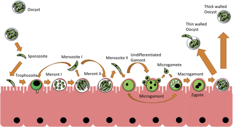
Cryptosporidium is a genus of single celled parasites capable of infecting a wide range of animals including humans. Cryptosporidium species are members of the phylum apicomplexa, which includes well-known genera such as Plasmodium and Toxoplasma. Cryptosporidium parasites cause a severe gastro-intestinal disease known as cryptosporidiosis. They are one of the most common causes of childhood diarrhoea worldwide, and infection can have prolonged detrimental effects on the development of children, but also can be life threatening to HIV/AIDS patients and transplant recipients. A variety of hosts can act as reservoirs, and Cryptosporidium can persist in the environment for prolonged times as oocysts. While there has been substantial interest in these parasites, there is very little progress in terms of treatment development and understanding the majority of the life cycle of this unusual organism. In this review, we will provide an overview on the existing knowledge of the biology of the parasite and the current progress in developing in vitro cultivation systems. We will then describe a synopsis of current and next generation approaches that could spearhead further research in combating the parasite.
Copyright © 2018. Published by Elsevier Inc.
Figures
References
-
- Abrahamsen M.S., Templeton T.J., Enomoto S., Abrahante J.E., Zhu G., Lancto C.A., Deng M., Liu C., Widmer G., Tzipori S., Buck G.A., Xu P., Bankier A.T., Dear P.H., Konfortov B.A., Spriggs H.F., Iyer L., Anantharaman V., Aravind L., Kapur V. Complete genome sequence of the apicomplexan, Cryptosporidium parvum. Science (New York, N.Y.) 2004;304:441–445. - PubMed
-
- Aldeyarbi H.M., Karanis P. Electron microscopic observation of the early stages of Cryptosporidium parvum asexual multiplication and development in in vitro axenic culture. Eur. J. Protistol. 2016;52:36–44. - PubMed
-
- Aldeyarbi H.M., Karanis P. The fine structure of sexual stage development and sporogony of Cryptosporidium parvum in cell-free culture. Parasitology. 2016;143:749–761. - PubMed
-
- Aldeyarbi H.M., Karanis P. The ultra-structural similarities between Cryptosporidium parvum and the gregarines. J. Eukaryot. Microbiol. 2016;63:79–85. - PubMed
Publication types
MeSH terms
Grants and funding
LinkOut - more resources
Full Text Sources
Medical
Miscellaneous
