

Cardiovascular development and survival require Mef2c function in the myocardial but not the endothelial lineage
- PMID: 30521808
- PMCID: PMC6370303
- DOI: 10.1016/j.ydbio.2018.12.002
Cardiovascular development and survival require Mef2c function in the myocardial but not the endothelial lineage
Abstract
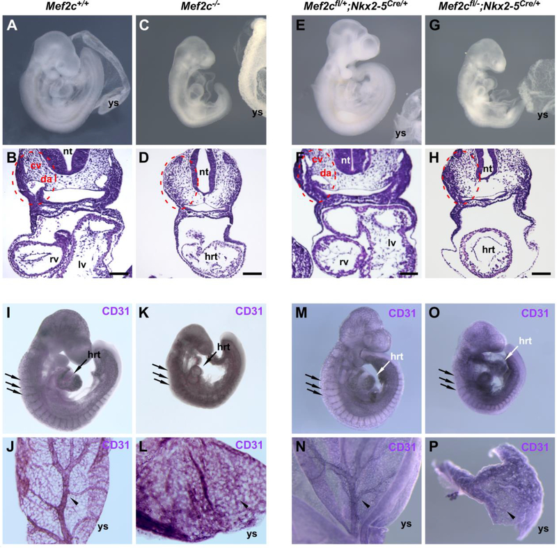
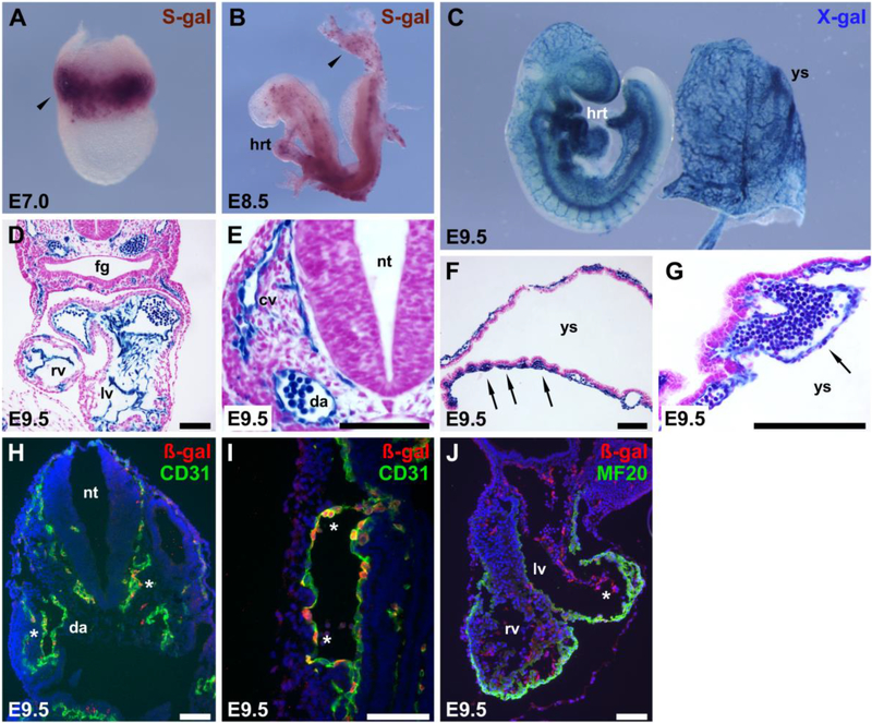
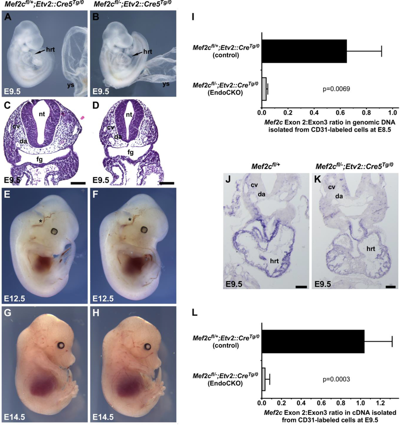
MEF2C is a member of the highly conserved MEF2 family of transcription factors and is a key regulator of cardiovascular development. In mice, Mef2c is expressed in the developing heart and vasculature, including the endothelium. Loss of Mef2c function in germline knockout mice leads to early embryonic demise and profound developmental abnormalities in the cardiovascular system. Previous attempts to uncover the cause of embryonic lethality by specifically disrupting Mef2c function in the heart or vasculature failed to recapitulate the global Mef2c knockout phenotype and instead resulted in relatively minor defects that did not compromise viability or result in significant cardiovascular defects. However, previous studies examined the requirement of Mef2c in the myocardial and endothelial lineages using Cre lines that begin to be expressed after the expression of Mef2c has already commenced. Here, we tested the requirement of Mef2c in the myocardial and endothelial lineages using conditional knockout approaches in mice with Cre lines that deleted Mef2c prior to onset of its expression in embryonic development. We found that deletion of Mef2c in the early myocardial lineage using Nkx2-5Cre resulted in cardiac and vascular abnormalities that were indistinguishable from the defects in the global Mef2c knockout. In contrast, early deletion of Mef2c in the vascular endothelium using an Etv2::Cre line active prior to the onset of Mef2c expression resulted in viable offspring that were indistinguishable from wild type controls with no overt defects in vascular development, despite nearly complete early deletion of Mef2c in the vascular endothelium. Thus, these studies support the idea that the requirement of MEF2C for vascular development is secondary to its requirement in the heart and suggest that the observed failure in vascular remodeling in Mef2c knockout mice results from defective heart function.
Keywords: Endothelial cells; Endothelium; Heart development; MEF2C; Morphogenesis; Mouse; Vascular development; Vascular remodeling.
Copyright © 2018 Elsevier Inc. All rights reserved.
Conflict of interest statement
Competing interests
The authors declare that they have no competing interests.
Figures
Similar articles
-
Requirement of the MADS-box transcription factor MEF2C for vascular development.Development. 1998 Nov;125(22):4565-74. doi: 10.1242/dev.125.22.4565. Development. 1998. PMID: 9778514
-
Generation of conditional Mef2cloxP/loxP mice for temporal- and tissue-specific analyses.Genesis. 2005 Sep;43(1):43-8. doi: 10.1002/gene.20152. Genesis. 2005. PMID: 16106363
-
MEF2C regulates outflow tract alignment and transcriptional control of Tdgf1.Development. 2016 Mar 1;143(5):774-9. doi: 10.1242/dev.126383. Epub 2016 Jan 25. Development. 2016. PMID: 26811383 Free PMC article.
-
Differential chamber-specific expression and regulation of long non-coding RNAs during cardiac development.Biochim Biophys Acta Gene Regul Mech. 2019 Oct;1862(10):194435. doi: 10.1016/j.bbagrm.2019.194435. Epub 2019 Nov 1. Biochim Biophys Acta Gene Regul Mech. 2019. PMID: 31678627 Review.
-
Role of the vascular endothelial growth factor isoforms in retinal angiogenesis and DiGeorge syndrome.Verh K Acad Geneeskd Belg. 2005;67(4):229-76. Verh K Acad Geneeskd Belg. 2005. PMID: 16334858 Review.
Cited by
-
microRNA Temporal-Specific Expression Profiles Reveal longissimus dorsi Muscle Development in Tianzhu White Yak.Int J Mol Sci. 2024 Sep 21;25(18):10151. doi: 10.3390/ijms251810151. Int J Mol Sci. 2024. PMID: 39337635 Free PMC article.
-
Role of MEF2C in the Endothelial Cells Derived from Human Induced Pluripotent Stem Cells.Stem Cells. 2023 Apr 25;41(4):341-353. doi: 10.1093/stmcls/sxad005. Stem Cells. 2023. PMID: 36639926 Free PMC article.
-
Promoting cardiomyocyte proliferation for myocardial regeneration in large mammals.J Mol Cell Cardiol. 2024 Mar;188:52-60. doi: 10.1016/j.yjmcc.2024.01.005. Epub 2024 Feb 9. J Mol Cell Cardiol. 2024. PMID: 38340541 Free PMC article. Review.
-
Optimizing Cardiomyocyte Differentiation: Comparative Analysis of Bone Marrow and Adipose-Derived Mesenchymal Stem Cells in Rats Using 5-Azacytidine and Low-Dose FGF and IGF Treatment.Biomedicines. 2024 Aug 22;12(8):1923. doi: 10.3390/biomedicines12081923. Biomedicines. 2024. PMID: 39200387 Free PMC article.
-
The benign nature and rare occurrence of cardiac myxoma as a possible consequence of the limited cardiac proliferative/ regenerative potential: a systematic review.BMC Cancer. 2023 Dec 18;23(1):1245. doi: 10.1186/s12885-023-11723-3. BMC Cancer. 2023. PMID: 38110859 Free PMC article.
References
Publication types
MeSH terms
Substances
Grants and funding
LinkOut - more resources
Full Text Sources
Molecular Biology Databases
