

Evaluation of the theoretical optimal angle of the tibial tunnel in transtibial anatomic posterior cruciate ligament reconstruction by computed tomography
- PMID: 30522472
- PMCID: PMC6284300
- DOI: 10.1186/s12891-018-2348-4
Evaluation of the theoretical optimal angle of the tibial tunnel in transtibial anatomic posterior cruciate ligament reconstruction by computed tomography
Abstract
Background: "Killer turn" effect is a critical explanation for the recurrent posterior laxity following transtibial posterior cruciate ligament (PCL) reconstruction, which affected by the angle of the tibial tunnel. Meanwhile, excessive tunnel angle would have an adverse impact on the healing of tendon to bone. The purpose was to evaluate the theoretical optimal angle of the tibial tunnel in transtibial anatomic PCL reconstruction.
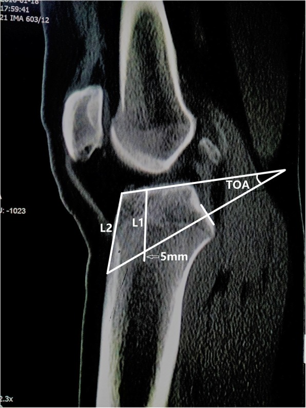
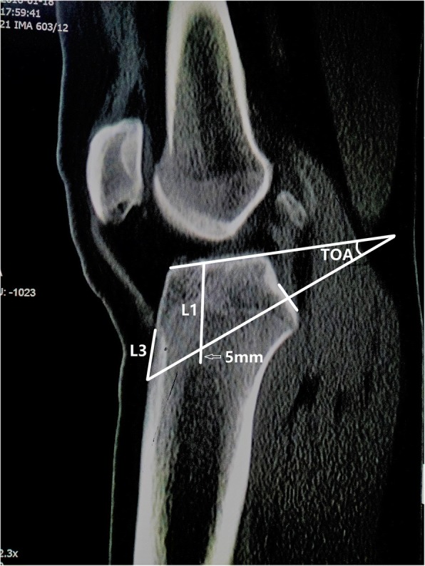
Methods: The measurements were performed on CT sagittal plane, including the thickness of cancellous bone (L1), the theoretical optimal angle of the tibial tunnel (TOA, which was measured between tibial plateau and the extension cord connecting the center of PCL insertion site with a point 5 mm superior from marrow cavity vertex), L2 - the distance from anterior tunnel aperture to anterior end of tibial plateau, L3 - the distance from anterior tunnel aperture to tibial tuberosity (lowest edge of patellar ligament attachment).
Results: The value of TOA and L3 were 35.4 ± 7.9 ° and 26.8 ± 11.4 mm, respectively. L1 and L2 were higher in males than females (L1, P = 0.002; L2, P = 0.046). Regarding age, L1, TOA, L2 and L3 were higher in the 46-60 years group than 31-45 years group (P = 0.02, P = 0.001, P = 0.038, P = 0.032, respectively). With regard to height, L1 was lower in group I - < 1.66 m than group II - 1.66 to 1.75 m and group III - > 1.75 m (I v II, P = 0.015, I v III, P = 0.026). L2 was also lower in group I than group II and group III (I v II, P = 0.026, I v III, P = 0.006). TOA and L3 showed no significant differences among sex and height groups (P > 0.05).
Conclusions: TOA (35.4 ° ± 7.9 °) and L3 (26.8 ± 11.4 mm) could be used as a reference for ideal tibial tunnel placement in transtibial anatomic PCL reconstruction, so as to prevent recurrent PCL laxity and ensure good graft healing. However, further clinical validation is needed.
Keywords: Computed tomography; Posterior cruciate ligament; Reconstruction; Tunnel angle.
Conflict of interest statement
Ethics approval and consent to participate
Ethics approval for this study was obtained from the Institutional Review Board of the Lanzhou University Second Hospital, Lanzhou, Gansu Province, China. The informed consent was waived by the Ethics Committee as the study was performed using the data and images without the patients’ identification information and this is a retrospective study.
Consent for publication
Not applicable.
Competing interests
The authors declare that they have no competing interests.
Publisher’s Note
Springer Nature remains neutral with regard to jurisdictional claims in published maps and institutional affiliations.
Figures
References
-
- Ochiai S, Hagino T, Senga S, Yamashita T, Haro H. Treatment outcome of reconstruction for isolated posterior cruciate injury: subjective and objective evaluations. J Knee Surg. 2018. 10.1055/s-0038-1653947. - PubMed
-
- Chahla J, Moatshe G, Cinque ME, Dornan GJ, Mitchell JJ, Ridley TJ, Single-Bundle LPRF. Double-bundle posterior cruciate ligament reconstructions: a systematic review and meta-analysis of 441 patients at a minimum 2 Years’ follow-up. Arthroscopy. 2017;33(11):2066–2080. - PubMed
Publication types
MeSH terms
Grants and funding
LinkOut - more resources
Full Text Sources
Medical
