Review
. 2018 Dec 6;475(23):3707-3723.
doi: 10.1042/BCJ20170726.

The split protein phosphatase system

Affiliations
Review

The split protein phosphatase system

Anne Bertolotti. Biochem J. .

Abstract

Reversible phosphorylation of proteins is a post-translational modification that regulates all aspect of life through the antagonistic action of kinases and phosphatases. Protein kinases are well characterized, but protein phosphatases have been relatively neglected. Protein phosphatase 1 (PP1) catalyzes the dephosphorylation of a major fraction of phospho-serines and phospho-threonines in cells and thereby controls a broad range of cellular processes. In this review, I will discuss how phosphatases were discovered, how the view that they were unselective emerged and how recent findings have revealed their exquisite selectivity. Unlike kinases, PP1 phosphatases are obligatory heteromers composed of a catalytic subunit bound to one (or two) non-catalytic subunit(s). Based on an in-depth study of two holophosphatases, I propose the following: selective dephosphorylation depends on the assembly of two components, the catalytic subunit and the non-catalytic subunit, which serves as a high-affinity substrate receptor. Because functional complementation of the two modules is required to produce a selective holophosphatase, one can consider that they are split enzymes. The non-catalytic subunit was often referred to as a regulatory subunit, but it is, in fact, an essential component of the holoenzyme. In this model, a phosphatase and its array of mostly orphan substrate receptors constitute the split protein phosphatase system. The set of potentially generalizable principles outlined in this review may facilitate the study of these poorly understood enzymes and the identification of their physiological substrates.

Keywords: biochemical techniques and resources; intracellular signaling; protein phosphatases.


Conflict of interest statement

A.B. is a founder and Chief Scientific Officer of a small company, CamPhos Therapeutics, which develops phosphatase inhibitors.

Figures
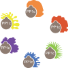
Figure 1.
Figure 1.. PP1 phosphatases are obligatory heteromers composed of shared catalytic subunits PP1c (gray) bound to one (or two) of many diverse non-catalytic subunits (colored).
It is unclear how many PP1 holoenzymes exist, but it is estimated that there may be several hundred.
Figure 2.
Figure 2.. Regulatory subunits as inhibitors of PP1c.
PP1c alone dephosphorylates phosphorylase a. When bound to an inhibitor, it fails to do so.
Figure 3.
Figure 3.. Targeting PP1c to glycogen.
The G subunit (green) binds to both glycogen (light blue polymer) and PP1c, thereby targeting PP1c to glycogen. Phosphorylase a (Phos a, red), glycogen synthase (GS, navy blue) and phosphorylase kinase (Phos K, orange) are PP1c substrates. How PP1 recognizes these substrates is unknown.
Figure 4.
Figure 4.. Summary of how the smooth muscle myosin regulatory subunits M130 and M20 change the substrate specificity of PP1c.
In vitro, PP1c dephosphorylates a broad range of substrates: phosphorylase a, phosphorylase kinase, glycogen synthase. When PP1c is bound to M130 and M20, dephosphorylation of phosphorylase a, phosphorylase kinase, glycogen synthase is suppressed while dephosphorylation of the myosin light and heavy chains is enhanced.
Figure 5.
Figure 5.. Trypsin converts myosin phosphatase into PP1c.
Low concentrations of trypsin digest M130-M20, but spare PP1c. This assay has been used to remove non-catalytic subunits of PP1c, thereby generating an enzyme of broad substrate selectivity.
Figure 6.
Figure 6.. An assay with recombinant proteins recapitulates the function and selectivity of PPP1R15–PP1c holophosphatases.
PPP1R15A: R15A. PPP1R15B: R15B. At physiological concentrations, PP1c does not dephosphorylate eIF2α. However, R15A–PP1c and R15B–PP1c are active and selective eIF2α phosphatases: they dephosphorylate eIF2α but not phosphorylase a. An unrelated holoenzyme, R3A-PP1c, does not dephosphorylate eIF2α. Thus, the function and selectivity of these holoenzymes can be recapitulated in vitro with purified proteins.
Figure 7.
Figure 7.. Complexes composed of carboxy-terminal fragments of PPP1R15s are capable of recruiting PP1c but are not functional.
Holoenzymes assembled with large fragments of PPP1R15s (R15A or R15B) are functional. The carboxy-terminal regions of R15s bind PP1c, but the resulting complexes are not functional.
Figure 8.
Figure 8.. Understanding the selectivity of PP1 holophosphatases and the substrate specifier functions of non-catalytic subunits.
PP1c has low affinity for phosphorylated eIF2α, explaining why at physiological concentrations it is unable to dephosphorylate this substrate. At high concentration, binding can occur, enabling dephosphorylation. PPP1R15s (R15A/B) provides a high-affinity receptor for eIF2α, enabling dephosphorylation at physiological concentrations of PP1c. The unrelated subunit R3A prevents PP1c binding to eIF2α and its dephosphorylation.
Figure 9.
Figure 9.. Assays to enable identification of selective phosphatase inhibitors.
PPP1R1 (R1) and PPP1R2 (R2) are non-catalytic subunits. An SPR screen enables the identification of molecules binding selectively to a holophosphatase of interest, R1-PP1c, and a counter-screen with a different phosphatase, R2-PP1c, filters out nonselective binders. Cell-based assays consisting of monitoring increased phosphorylation of the substrate of the R1-PP1 phosphatase or downstream signaling events select for compounds capable of inhibiting their target in cells. The same assays performed in cells knockedout for R1 identifies on-target compounds. Biochemical dephosphorylation assays validate the mechanism of action and selectivity of the inhibitors.
Figure 10.
Figure 10.. The split protein phosphatase system (SPS).
PP1 holoenzymes are composed of a catalytic subunit PP1c bound to a non-catalytic subunit which is composed of at least two modules: a substrate receptor module and a PP1c-binding module. Additional modules can be added such as a glycogen-targeting module or other to target the complex to a specific subcellular location.
Figure 11.
Figure 11.. The split phosphatase system explains the dual function of non-catalytic subunits.
R1 (PPP1R1) is the substrate receptor of the holophosphatase that recruits a specific substrate S1 and PP1c to enable selective dephosphorylation. Likewise, R2 (PPP1R2) is the substrate receptor for S2. R1 is inhibitory towards S2 and R2 is inhibitory towards S1.

References

    1. Manning G., Whyte D.B., Martinez R., Hunter T. and Sudarsanam S. (2002) The protein kinase complement of the human genome. Science 298, 1912–1934 10.1126/science.1075762 - DOI - PubMed
    1. Chen M.J., Dixon J.E. and Manning G. (2017) Genomics and evolution of protein phosphatases. Sci. Signal. 10, eaag1796 10.1126/scisignal.aag1796 - DOI - PubMed
    1. Brandt H., Killilea S.D. and Lee E.Y.C. (1974) Activation of phosphorylase phosphatase by a novel procedure: evidence for a regulatory mechanism involving the release of a catalytic subunit from enzyme-inhibitor complex(es) of higher molecular weight. Biochem. Biophys. Res. Commun. 61, 598–604 10.1016/0006-291X(74)90999-1 - DOI - PubMed
    1. Verbinnen I., Ferreira M. and Bollen M. (2017) Biogenesis and activity regulation of protein phosphatase 1. Biochem. Soc. Trans. 45, 89–99 10.1042/BST20160154 - DOI - PubMed
    1. Cori G.T. and Green A.A. (1943) Crystalline muscle phosphorylase II. Prosthetic group. J. Biol. Chem. 151, 31–38

Publication types

MeSH terms