. 2019 Jan 4;363(6422):88-91.
doi: 10.1126/science.aav7271. Epub 2018 Dec 6.

Functionally diverse type V CRISPR-Cas systems

Affiliations

Functionally diverse type V CRISPR-Cas systems

Winston X Yan et al. Science. .

Abstract

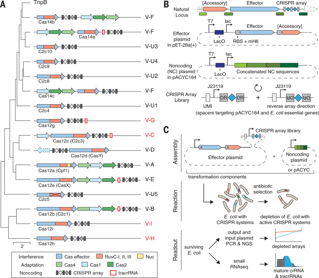
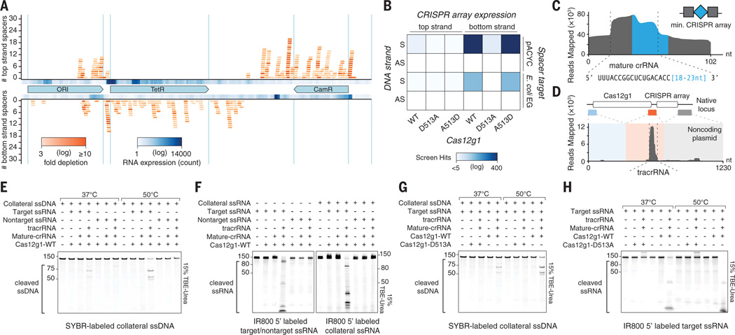
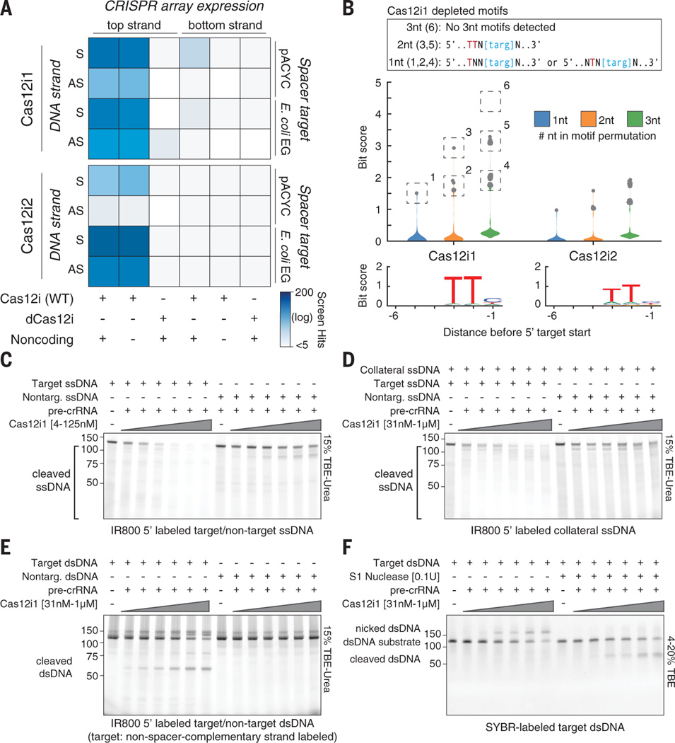
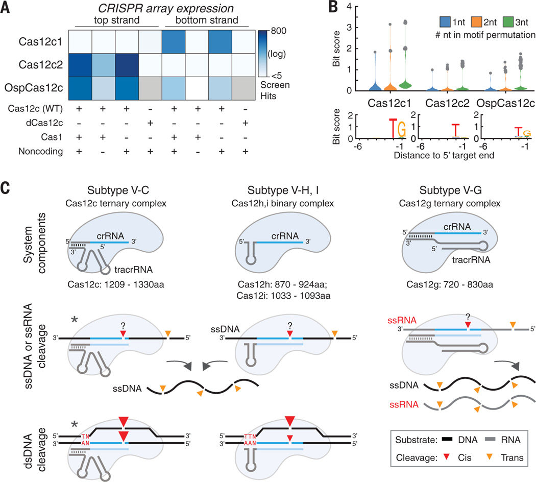
Type V CRISPR-Cas systems are distinguished by a single RNA-guided RuvC domain-containing effector, Cas12. Although effectors of subtypes V-A (Cas12a) and V-B (Cas12b) have been studied in detail, the distinct domain architectures and diverged RuvC sequences of uncharacterized Cas12 proteins suggest unexplored functional diversity. Here, we identify and characterize Cas12c, -g, -h, and -i. Cas12c, -h, and -i demonstrate RNA-guided double-stranded DNA (dsDNA) interference activity. Cas12i exhibits markedly different efficiencies of CRISPR RNA spacer complementary and noncomplementary strand cleavage resulting in predominant dsDNA nicking. Cas12g is an RNA-guided ribonuclease (RNase) with collateral RNase and single-strand DNase activities. Our study reveals the functional diversity emerging along different routes of type V CRISPR-Cas evolution and expands the CRISPR toolbox.


Figures

Fig. 1.
Fig. 1.. Discovery and screening of type V CRISPR-Cas diversity.
(A) Classification tree of type V effectors (Cas12 proteins) with the corresponding CRISPR-Cas loci organization shown for each branch. Cas12 proteins analyzed in this work are highlighted in red. (B) Design of in vivo screen effector and noncoding plasmids. CRISPR array libraries were designed with spacers uniquely and uniformly sampled from both strands of pACYC184 or E. coli essential genes, then flanked by two DRs and transcribed by a J23119 promoter. (C) Workflow schematic of the in vivo E. coli screen.
Fig. 2.
Fig. 2.. Cas12g displays RNA-activated target cleavage of RNA and collateral trans-cleavage of RNA and ssDNA.
(A) Strongly depleted CRISPR arrays from in vivo screening of Cas12g1 and its noncoding plasmid mapped to pACYC184. (B) Heatmap showing strongly depleted CRISPR arrays (screen hits) to evaluate RuvC and substrate strand dependencies of Cas12g1 (S, sense; AS, antisense; EG, essential genes). A513D was cloned from the D513A construct to rescue its activity. Strongly depleted CRISPR arrays in negative control screens without the effector were subtracted from this and similar analyses. (C and D) Mature crRNA (C) and tracrRNA (D) identified from small RNA sequencing of in vivo screen samples containing Cas12g1 and noncoding plasmid. The schematic above tracrRNA shows construction of noncoding plasmid from native locus sequences. (E and F) Target ssRNA activated collateral ssDNA cleavage at 37°C and 50°C (E) and target and collateral ssRNA cleavage at 37°C (F). (G and H) Cleavage assays targeting collateral ssDNA (G) and ssRNA (H) with purified RuvC mutant dCas12g1 D513A.
Fig. 3.
Fig. 3.. In vivo and in vitro activity of Cas12i.
(A) Evaluation of a minimal active system for Cas12i, with heatmaps showing strongly depleted CRISPR arrays from in vivo screening in different Cas12i system compositions (S, sense; AS, antisense; EG, essential genes). (B) (Top) Distribution of bit scores for all permutations of 1- to 3-nucleotide (nt) motifs within the target and 15-nt flanking sequences corresponding to strongly depleted in vivo arrays, calculated as described in (19). The box above describes motif analysis for Cas12i1 as an example. (Bottom) Web logos from target-flanking sequences. (C to E) Titration of a Cas12i1 binary complex on target and nontarget ssDNA (C), collateral ssDNA with target and nontarget ssDNA (D), and target and nontarget dsDNA (E). (F) S1 nuclease treatment to resolve dsDNA nicks (induced by Cas12i1) into dsDNA breaks.
Fig. 4.
Fig. 4.. In vivo dsDNA interference by Cas12c.
(A) Evaluation of a minimal active system for Cas12c, with heatmaps showing strongly depleted CRISPR arrays from in vivo screening in different Cas12c system compositions. Gray boxes indicate data not available. (B) (Top) Distribution of bit scores for all permutations of 1- to 3-nt motifs within the target and 15-nt flanking sequences corresponding to strongly depleted arrays. (Bottom) Web logos from target-flanking sequences. (C) Overview of minimal components and interference mechanisms of Cas12g, -h, -i, and -c. Asterisks denote putative mechanisms subject to additional validation.

Comment in

References

    1. Burstein D. et al., Nature 542, 237–241 (2017). - PMC - PubMed
    1. Doron S. et al., Science 359, eaar4120 (2018).
    1. Koonin EV, Makarova KS, Wolf YI, Annu. Rev. Microbiol 71, 233–261 (2017). - PMC - PubMed
    1. Barrangou R, Horvath P, Nat. Microbiol 2, 17092 (2017). - PubMed
    1. Barrangou R. et al., Science 315, 1709–1712 (2007). - PubMed

Publication types