. 2018 Dec 6;9(1):5224.
doi: 10.1038/s41467-018-07598-9.

GPCR-specific autoantibody signatures are associated with physiological and pathological immune homeostasis

Affiliations

GPCR-specific autoantibody signatures are associated with physiological and pathological immune homeostasis

Otavio Cabral-Marques et al. Nat Commun. .

Abstract

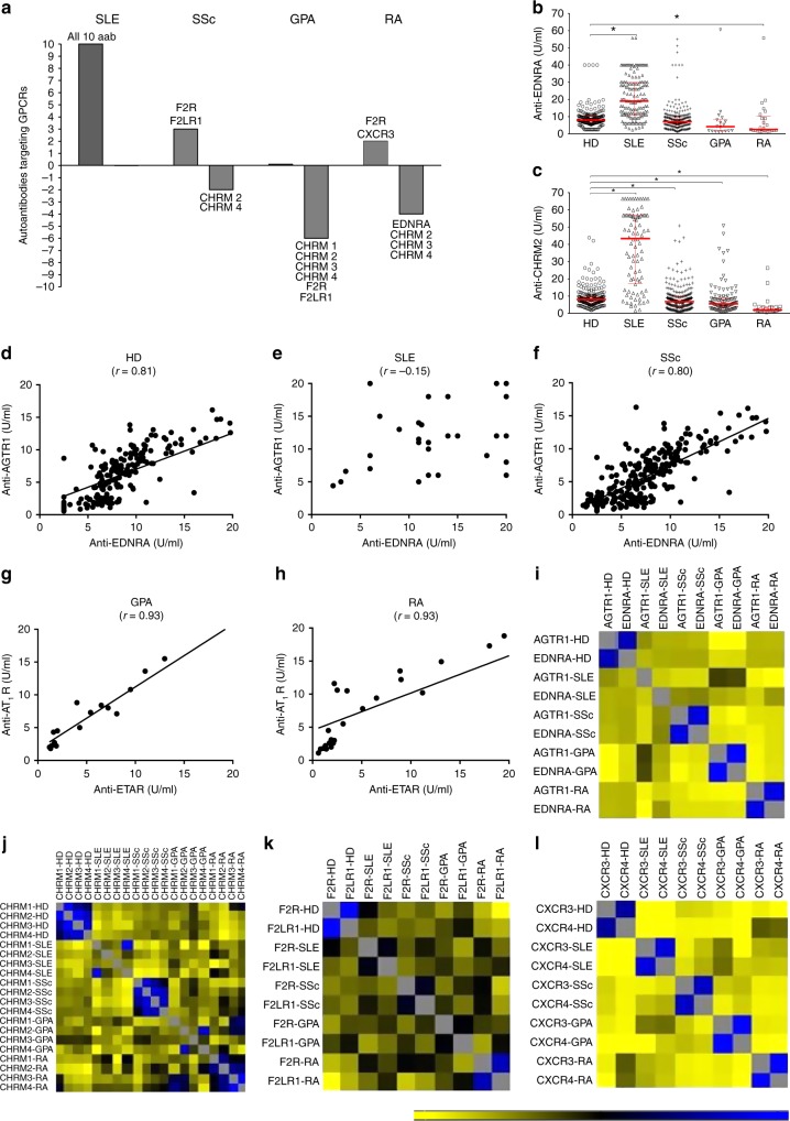
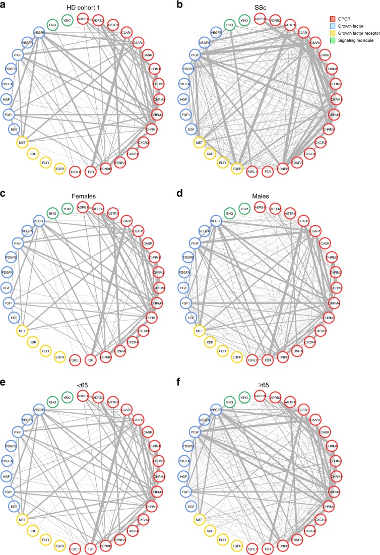
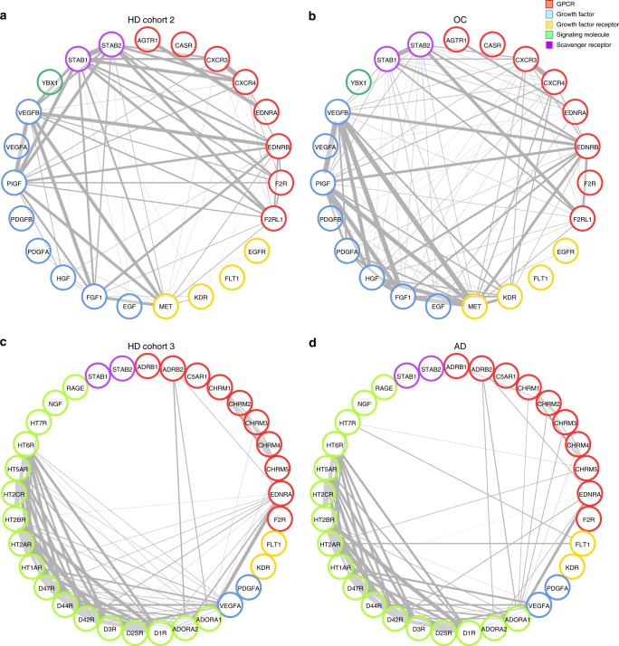
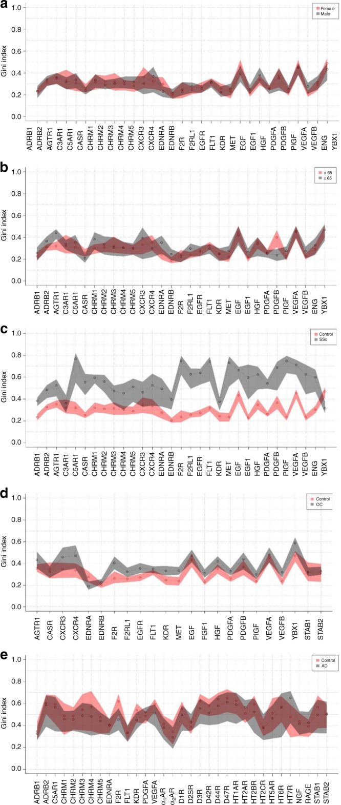
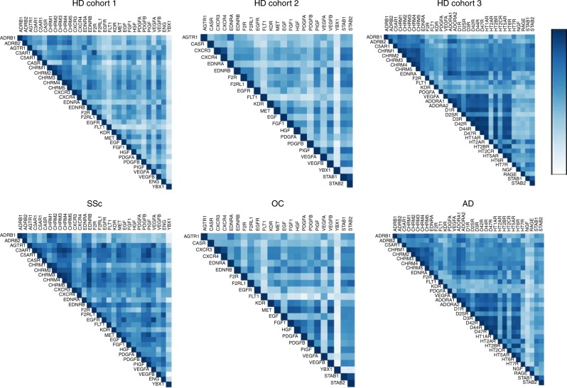
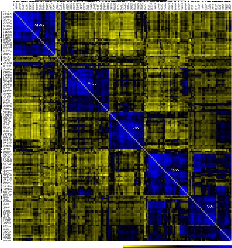
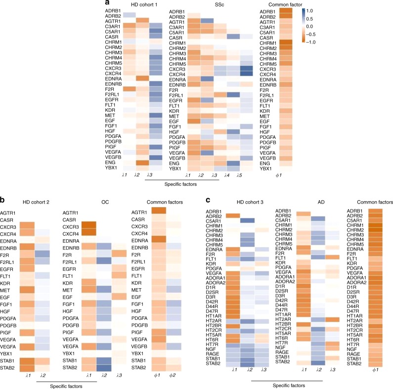
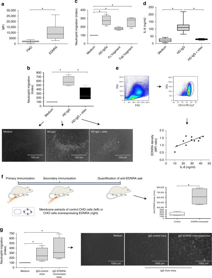
Autoantibodies have been associated with autoimmune diseases. However, studies have identified autoantibodies in healthy donors (HD) who do not develop autoimmune disorders. Here we provide evidence of a network of immunoglobulin G (IgG) autoantibodies targeting G protein-coupled receptors (GPCR) in HD compared to patients with systemic sclerosis, Alzheimer's disease, and ovarian cancer. Sex, age and pathological conditions affect autoantibody correlation and hierarchical clustering signatures, yet many of the correlations are shared across all groups, indicating alterations to homeostasis. Furthermore, we identify relationships between autoantibodies targeting structurally and functionally related molecules, such as vascular, neuronal or chemokine receptors. Finally, autoantibodies targeting the endothelin receptor type A (EDNRA) exhibit chemotactic activity, as demonstrated by neutrophil migration toward HD-IgG in an EDNRA-dependent manner and in the direction of IgG from EDNRA-immunized mice. Our data characterizing the in vivo signatures of anti-GPCR autoantibodies thus suggest that they are a physiological part of the immune system.


Conflict of interest statement

The authors declare that H.H. and K.S.F. are CellTrend managing directors and that Gabriela Riemekasten is an advisor of the company CellTrend and earned an honorarium for her advice between 2011 and 2015. The other authors declare no competing interests.

Figures

Fig. 1
Fig. 1
Relationships among autoantibodies in health and autoimmune diseases. a The graphic summarizes anti-GPCR autoantibodies (aab) in healthy donors (HD), which showed significantly increased or decreased concentrations when compared to those in the disease cohorts (systemic lupus erythematosus or SLE, systemic sclerosis or SSc, granulomatosis with polyangiitis or GPA, and rheumatoid arthritis or RA). Further details are shown in Supplementary Figure 1. The x-axis represents healthy controls. Graphics display concentrations of aab directed against b EDNRA and c CHRM2. The median with interquartile range is shown in red. *p ≤ 0.05 (Mann–Whitney test). Linear regression graphics exhibit the correlation between anti-EDNRA and anti-AGTR1 aab in sera from d HD, e SLE, f SSc, g GPA, and h RA. Heatmaps of aab vs. aab correlations demonstrate the spectrum of relationships among aab targeting i EDNRA and AGTR1; j CHRMs; k F2R and FLR1; and l CXCR3 and CXCR4 (for nomenclature, see Supplementary Table 2, aab dataset 1). The bar ranging from yellow to blue (−0.3 to 1) represents negative to positive correlations, respectively. In the correlation matrix, each small square represents a pairwise correlation between aab, as exemplified by dh. The correlation matrices used to perform the hierarchical correlograms shown in Fig. 1i–l are provided as source data
Fig. 2
Fig. 2
Effects of gender, age, and systemic sclerosis on autoantibody correlations. We analyzed the relationships among the different autoantibodies (aab) in sera from healthy donors (HD) and the effects of gender and age, as shown by circular networks based on Spearman’s rank correlation coefficients for aab. Circle plots show the correlation matrix of aab comparing each condition: a all HD evaluated, b patients with systemic sclerosis (SSc; Supplementary Table 1, cohort 1; Supplementary Table 2, aab dataset 1); subgroups of HD (cohort 1) analyzed according to c, d gender and e, f age (< and ≥65 years)
Fig. 3
Fig. 3
Effects of ovarian cancer and Alzheimer’s disease on autoantibody correlations. Graphics display the comparisons of a healthy donors (HD) and patients with b ovarian cancer (OC; Supplementary Table 1, cohort 2; Supplementary Table 2, aab dataset 2) and c HD and patients with d Alzheimer’s disease (AD; Supplementary Table 1, cohort 3; Supplementary Table 2, aab dataset 3). The nodes in the graphs represent variables (each aab), and a line between two nodes indicates the Spearman’s rank correlation coefficient. The line width indicates the strength of the association, with stronger correlations indicated by thicker lines. Only correlations >0.6 are shown. Multiple connections of nodes indicate clustering
Fig. 4
Fig. 4
Autoantibody relationships reflect their concentration distribution patterns. Gini index confidence intervals were obtained by bootstrap analysis. The red and gray shadows represent confidence intervals, and each small circle indicates the Gini index value. The graphics exhibit comparisons of a HD females and males and b HD above and below 65 years of age (Supplementary Table 1, cohort 1; Supplementary Table 2, aab dataset 1) and comparisons between c HD and patients with systemic sclerosis (SSc, Supplementary Table 1, cohort 1; Supplementary Table 2, aab dataset 1, d HD and patients with ovarian cancer (OC, Supplementary Table 1, cohort 2; Supplementary Table 2, aab dataset 2) or e HD and patients with Alzheimer’s disease (AD, Supplementary Table 1, cohort 3; Supplementary Table 2, aab dataset 3)
Fig. 5
Fig. 5
Heatmaps of autoantibody correlations. The images show Spearman’s correlation of autoantibody datasets (Supplementary Table 2) a 1, b 2, and c 3 in sera from healthy donor (HD) cohorts 1–3 and patients (SSc systemic sclerosis, OC ovarian cancer, and AD Alzheimer’s disease), respectively (Supplementary Table 1). The color scale bar (0 to 1) corresponds to weak and strong correlations, respectively
Fig. 6
Fig. 6
Hierarchical clustering of the autoantibody correlation signature. Correlogram matrix displays clusters (modules) of autoantibody (aab) correlations in all healthy donors (HD) according to gender and age (< and ≥65 years old) compared with patients with systemic sclerosis (SSc; Supplementary Table 1, cohort 1; Supplementary Table 2, aab dataset 1). Clusters of the correlations among aab are displayed in dendrograms on the top and side of the correlation matrix. The bar ranging from yellow to blue (−0.6 to 0.9) represents negative to positive correlations, respectively. In the heat map matrix, each small square represents the pairwise correlations between aab. The correlation matrix used to perform the hierarchical correlogram of SSc is provided as source data
Fig. 7
Fig. 7
Multistudy factor analysis of autoantibodies. The multistudy factor analysis (MSFA) was performed to analyze autoantibodies (aab) from healthy donors (HD) compared with patients with a systemic sclerosis (SSc), b ovarian cancer (OC), and c Alzheimer’s disease (AD). Supplementary Tables 1 and 2 provide further details about the HD and patient groups, as well as the aab datasets analyzed. The images are heatmaps of estimated factor loadings of common and specific latent factors. The color scale bar ranging from orange (−1 to 1) to blue corresponds to negative and positive factor loadings. Loadings close to −1 or 1 indicate aab that strongly influence factors in opposite directions
Fig. 8
Fig. 8
Effect of HD-IgG and anti-EDNRA autoantibodies on neutrophil migration. a Expression of endothelin receptor type A (EDNRA) by human neutrophils (n = 16). The fluorescence minus one control (FMO) was analyzed as shown in Supplementary Figure 5. b Neutrophil chemotaxis toward 0.5 mg/ml IgG from healthy donors (HD-IgG) in the presence or absence of the EDNRA antagonist sitaxsentan (sitax; n = 3). A representative image of neutrophils (white dots in the figure) on the bottom surface of transwell plates is shown. c Neutrophil migration toward intact human IgG, antigen-binding fragment (Fab), and the crystallized fragment (Fc) region. The results are representative of three independent experiments (n = 3). d HD-IgG-induced IL-8 production by peripheral blood mononuclear cells (PBMCs) in the absence (n = 13) or presence of sitax (n = 4). e IL-8 spontaneously released into the culture supernatants correlates with the level of EDNRA expression on CD14+ monocytes (n = 11). f Concentrations of anti-EDNRA autoantibodies (aab) in mouse sera were assessed after secondary immunization with membrane extracts from control Chinese hamster ovary (CHO) cells (n = 5) or CHO cells overexpressing human EDNRA (n = 4). Mouse images as well as syringe and membrane cartoons were adapted from Motifolio Drawing Toolkits (www.motifolio.com). EDNRA immunization was carried out by administration into footpads of 0.2 mg of membrane extracts prepared from CHO cells overexpressing human EDNRA (Celltrend, Germany). Three weeks after the primary immunization, mice were boosted with the same amount of membrane emulsified with incomplete Freund’s adjuvant (IFA, Sigma-Aldrich, USA). In the control group, mice were treated with the same amount of membrane extract from untransfected CHO cells. Six weeks after the booster immunization, all mice were sacrificed for sample collection and quantification of anti-EDNRA aab. g The migration of human neutrophils (white dots in the figure) toward IgG from control and EDNRA-immunized mice (n = 3). Error bars denote SD. *p ≤ 0.05 (unpaired t test)

References

    1. Ehrlich P. Croonian Lecture: on immunity with special reference to cell life. Proc. R. Soc. Lond. 1899;66:424–448. doi: 10.1098/rspl.1899.0121. - DOI
    1. Silverstein AM. Autoimmunity versus horror autotoxicus: the struggle for recognition. Nat. Immunol. 2001;2:279–281. doi: 10.1038/86280. - DOI - PubMed
    1. Nagele EP, et al. Natural IgG autoantibodies are abundant and ubiquitous in human sera, and their number is influenced by age, gender, and disease. PLoS. ONE. 2013;8:e60726. doi: 10.1371/journal.pone.0060726. - DOI - PMC - PubMed
    1. Plotz PH. The autoantibody repertoire: searching for order. Nat. Rev. Immunol. 2003;3:73–78. doi: 10.1038/nri976. - DOI - PubMed
    1. Meyer S, et al. AIRE-deficient patients harbor unique high-affinity disease-ameliorating autoantibodies. Cell. 2016;166:582–595. doi: 10.1016/j.cell.2016.06.024. - DOI - PMC - PubMed

Publication types

MeSH terms