Review
. 2018 Dec 6;4(1):50.
doi: 10.1038/s41572-018-0047-y.

Polycystic kidney disease

Affiliations
Review

Polycystic kidney disease

Carsten Bergmann et al. Nat Rev Dis Primers. .

Abstract

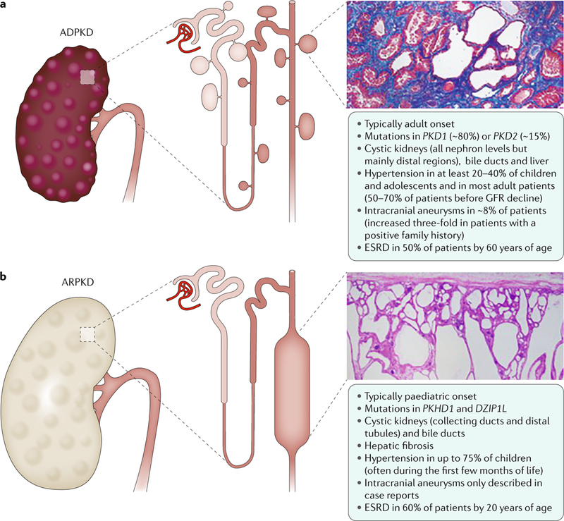
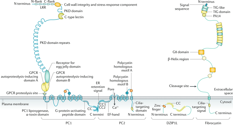
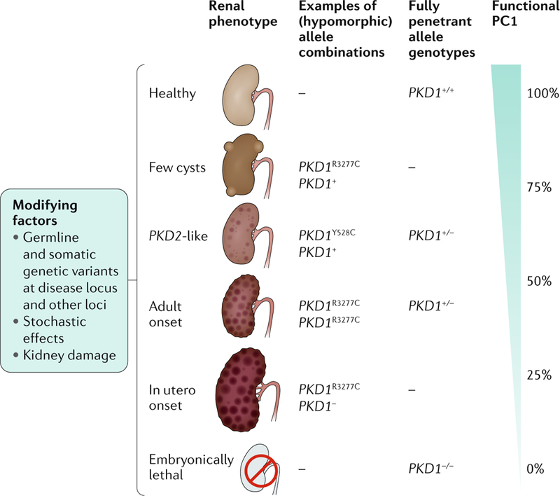
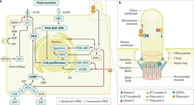
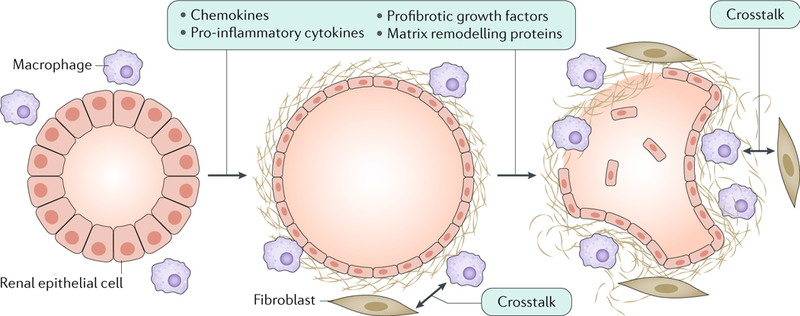
Cystic kidneys are common causes of end-stage renal disease, both in children and in adults. Autosomal dominant polycystic kidney disease (ADPKD) and autosomal recessive polycystic kidney disease (ARPKD) are cilia-related disorders and the two main forms of monogenic cystic kidney diseases. ADPKD is a common disease that mostly presents in adults, whereas ARPKD is a rarer and often more severe form of polycystic kidney disease (PKD) that usually presents perinatally or in early childhood. Cell biological and clinical research approaches have expanded our knowledge of the pathogenesis of ADPKD and ARPKD and revealed some mechanistic overlap between them. A reduced 'dosage' of PKD proteins is thought to disturb cell homeostasis and converging signalling pathways, such as Ca2+, cAMP, mechanistic target of rapamycin, WNT, vascular endothelial growth factor and Hippo signalling, and could explain the more severe clinical course in some patients with PKD. Genetic diagnosis might benefit families and improve the clinical management of patients, which might be enhanced even further with emerging therapeutic options. However, many important questions about the pathogenesis of PKD remain. In this Primer, we provide an overview of the current knowledge of PKD and its treatment.


Figures

Fig. 1 |
Fig. 1 |. Renal and extrarenal manifestations in polycystic kidney disease.
a | Autosomal dominant polycystic kidney disease (ADPKD) is the most common form of polycystic kidney disease (PKD) and is mainly caused by mutations in PKD1 and PKD2, which encode polycystin 1 and polycystin 2, respectively. ADPKD is usually an adult-onset disease that is characterized by the formation of fluid-filled cysts in various locations in the kidneys, but mostly in the distal regions. The histology image is of the kidney (stained with Malloy trichrome stain) of a 49-year-old patient with end-stage ADPKD. Small cysts and extensive fibrosis (blue) are visible. b | Autosomal recessive PKD (ARPKD) is rarer and more severe than ADPKD and is caused by mutations in polycystic kidney and hepatic disease 1 (PKHD1; which encodes fibrocystin) and DZIP1L (which encodes DAZ-interacting protein 1-like protein (DZIP1L)). ARPKD usually presents in utero, perinatally or in infancy and is characterized by the formation of cysts from the renal distal tubules and collecting ducts. Microscopically, cysts are fusiform dilatations of the aforementioned distal parts of the nephron, which are lined by a columnar or cuboidal epithelium. Respective dilated collecting ducts run perpendicular to the renal capsule (renal section is stained with haematoxylin and eosin). Both diseases often progress to end-stage renal disease (ESRD) that requires renal replacement therapy. GFR,glomerular filtration rate.
Fig. 2 |
Fig. 2 |. Domain organization of proteins implicated in polycystic kidney disease.
The structure of polycystin 1 (PC1), polycystin 2 (PC2), the membrane-bound form of fibrocystin and DAZ-interacting protein 1-like protein (DZIP1L) are depicted (not to scale). PC1 and PC2 are multispan membrane proteins that form a complex that is localized to multiple subcellular locations, including the primary cilium. Fibrocystin is also localized to the primary cilium and is subject to Notch-like proteolytic processing, resulting in release of the carboxy-terminal tail, which can translocate to the nucleus and may regulate gene expression. DZIP1L is a soluble zinc-finger protein that is localized to the centrioles and basal bodies at the ciliary transition zone. CC, coiled coil; C flank, carboxyl flank; C terminus, carboxyl terminus; ER, endoplasmic reticulum; GPCR, G protein-coupled receptor; LRR, leucine-rich repeat; N flank, amino flank; N terminus, amino terminus; PKD, polycystic kidney disease. Adapted from REF., Springer Nature Limited.
Fig. 3 |
Fig. 3 |. The dosage model of cystogenesis in autosomal dominant polycystic kidney disease.
The level of functional polycystin 1 (PC1; encoded by PKD1) directly influences the renal phenotype in patients with autosomal dominant polycystic kidney disease (ADPKD) — an ~50% reduction in PC1 levels (for example, from haploinsufficiency due to a single inactivating allele) is associated with adult-onset PKD, whereas the complete absence of PC1 is lethal. Furthermore, incompletely penetrant (hypomorphic) PKD1 alleles of different strengths and combinations also influence the renal phenotype. For example, the PKD1Y528C allele results in a phenotype similar to that in patients with mutations in PKD2, whereas the PKD1R3277C allele can result in a phenotype that ranges in severity from just a few cysts to adult-onset disease to early-onset disease, depending on which PKD1 allele is present in trans. Additional mutations and/or variants of the disease-causing locus and somatic and germline mutations at other loci, as well as chance and environmental factors, influence the disease course by determining the frequency of cyst development and their progression. Adapted with permission from REF., Elsevier.
Fig. 4 |
Fig. 4 |. Mechanisms of cyst formation and expansion.
Polycystin 1 (PC1) and PC2 form a multimeric protein complex that is localized to several cellular compartments, including cell–cell junctions, cell–matrix interfaces and in the primary cilium (part a; the ciliary localization of the polycystins is shown in part b). The polycystin proteins are also post-translationally modified, which regulates their transport, localization and/or function,,,. In addition, many proteins have been reported to bind directly to polycystin proteins; for example, PC1 and fibrocystin bind to PC2 and modulate its channel activity,,. Furthermore, DAZ-interacting protein 1-like protein (DZIP1L) interacts with septin 2 (SEPT2; in the septin ring), a protein implicated in maintenance of the periciliary diffusion barrier at the ciliary transition zone. Consistent with a defect in the diffusion barrier, the localization of PC1 and PC2 to the ciliary membrane is compromised in DZIP1L-mutant cells, suggesting that DZIP1L is required for regulating the integrity of the transition zone. How PC1, PC2, fibrocystin and DZIP1L directly affect cellular signalling is not known with certainty. However, these proteins modulate several signalling pathways, which in turn control essential cellular functions, such as proliferation, apoptosis, cell adhesion and differentiation. Reduced Ca2+ influx, increased cAMP levels and aberrant activation of RAS–RAF–ERK signalling in renal epithelial cells are important mediators of cyst growth. Other proposed mechanisms that regulate cystogenesis and/or cyst growth include altered signalling mediated by G proteins, mechanistic target of rapamycin (mTOR), phosphoinositide 3-kinase (PI3K)–AKT, AMP-activated protein kinase (AMPK), Janus kinase 2 (JAK2)–signal transducer and activator of transcription 1 (STAT1; or STAT3 or STAT6), nuclear factor of activated T cells (NFAT) and nuclear factor-κB. In addition, cyst expansion is accompanied by changes in cellular metabolism, such as a switch to aerobic glycolysis as well as impaired fatty acid oxidation. AC6, adenylyl cyclase 6; AVP, arginine vasopressin; CFTR, cystic fibrosis transmembrane conductance regulator; EGFR, epidermal growth factor receptor; ER, endoplasmic reticulum; IFT, intraflagellar transport; PDE1, phosphodiesterase 1; PKA, protein kinase A; PKD, polycystic kidney disease; SST, somatostatin; SSTR, somatostatin receptor; V2R, vasopressin V2 receptor.Part a is adapted from REF., Springer Nature Limited. Part b is adapted from REF., Springer Nature Limited.
Fig. 5 |
Fig. 5 |. Renal fibrosis in autosomal dominant polycystic kidney disease.
Altered expression of cystic proteins and dysregulated signalling in renal epithelial cells affect cell–extracellular matrix interactions and induce the production of chemokines (such as monocyte chemotactic protein 1 (MCP1), CC-chemokine ligand 6 (CCL6), CCL28, CXC-chemokine ligand 1 (CXCL1), CXCL8 and CX3C-chemokine ligand 1 (CX3CL1)), pro-inflammatory cytokines (such as tumour necrosis factor (TNF), IL-1, IL-2, IL-6, IL-8, macrophage colony-stimulating factor 1 (CSF1) and macrophage migration inhibitory factor (MIF)), profibrotic growth factors (such as transforming growth factor-β (TGFβ), TGFα, epidermal growth factor (EGF), fibroblast growth factors (FGFs) and platelet-derived growth factor (PDGF)) and proteins involved in matrix remodelling (such as matrix metalloproteinases (MMPs) and tissue inhibitors of metalloproteinases (TIMPs)),. These changes result in the accumulation of extracellular matrix and the recruitment of inflammatory cells, which is already observed in the early stages of the disease. In particular, M1 and M2 macrophages are important contributors to disease progression. Macrophages and interstitial fibroblasts also produce cytokines, and abnormal cytokine-mediated crosstalk between renal epithelial cells and inflammatory cells results in a positive feedback loop of increasing fibrosis.
Fig. 6 |
Fig. 6 |. Hepatobiliary lesions in hepatorenal disease.
a | Hepatobiliary lesions result from an architectural defect in the developing biliary tree. The normal ramifications of the portal venous system and the lattice-like network of associated biliary ducts (left) are disrupted owing to ductal plate malformation (DPM) (right), likely owing to a defect in terminal differentiation of cholangiocytes. b | The DPM results in marked cystic and fusiform dilatation of the intrahepatic biliary system (coronal T2-weighted image of the abdomen), nephromegaly with small cysts (arrowhead), cystic biliary disease (arrow) and marked splenomegaly (asterisk). c | The histopathological manifestation of the DPM is congenital hepatic fibrosis (section stained with haematoxylin and eosin), which is characterized by extensive fibrosis of the portal area (asterisk), ectatic, tortuous bile ducts (arrows) and hypoplasia of the portal vein (arrowhead). Magnification is 40×. Part a is reprinted, with permission, from Marchal G J, Desmet V J, Proesmans W C, et al. Caroli disease: high-frequency US and pathologic findings.
Fig. 7 |
Fig. 7 |. Diagnosis of autosomal dominant polycystic kidney disease using different imaging techniques.
a–b | Comparison of MRI (T2; part a) and CT (without contrast; part b) scans of a 35-year-old woman with autosomal dominant polycystic kidney disease (ADPKD), showing widespread kidney cysts and a few liver cysts. c–e | T2 MRI images of patients with ADPKD who have a truncating mutation in PKD1 (part c; 41-year-old man), a non-truncating mutation in PKD1 (part d, 40-year-old man) or a splicing mutation in PKD2 (part e; 41-year-old man). Patients with truncating mutations in PKD1 typically have more cysts, whereas patients with non-truncating mutations in PKD1 have an intermediate number of cysts and patients with splicing mutations in PKD2 have the fewest cysts.
Fig. 8 |
Fig. 8 |. Diagnosis of autosomal recessive polycystic kidney disease using MRI.
MRI can be used to detect renal and extrarenal manifestations of autosomal recessive polycystic kidney disease (ARPKD). In this coronal, T2-weighted MRI scan of a 32-week-old fetus, the kidneys (arrow) are massively enlarged and are abnormally bright with innumerable tiny cysts, most of which cannot be individually resolved. Cystic biliary disease (arrowhead) and extremely low lung volumes (asterisk) in patients with ARPKD are due to oligohydramnios (deficiency of amniotic fluid).

References

    1. Bergmann C ARPKD and early manifestations of ADPKD: the original polycystic kidney disease and phenocopies. Pediatr. Nephrol. 30, 15–30 (2015). - PMC - PubMed
    1. Torres VE, Harris PC & Pirson Y Autosomal dominant polycystic kidney disease. Lancet 369, 1287–1301 (2007). - PubMed
    1. Bergmann C & Weiskirchen R It’s not all in the cilium, but on the road to it: genetic interaction network in polycystic kidney and liver diseases and how trafficking and quality control matter. J. Hepatol.56, 1201–1203 (2012). - PubMed
    1. Gerdes JM, Davis EE & Katsanis N The vertebrate primary cilium in development, homeostasis, and disease. Cell 137, 32–45 (2009). - PMC - PubMed
    1. Hildebrandt F, Benzing T & Katsanis N Ciliopathies. N. Engl. J. Med. 364, 1533–1543 (2011). - PMC - PubMed

Publication types

MeSH terms