Review
. 2019 Mar;20(3):137-155.
doi: 10.1038/s41580-018-0085-z.

Dynamics and functions of lipid droplets

Affiliations
Review

Dynamics and functions of lipid droplets

James A Olzmann et al. Nat Rev Mol Cell Biol. 2019 Mar.

Abstract

Lipid droplets are storage organelles at the centre of lipid and energy homeostasis. They have a unique architecture consisting of a hydrophobic core of neutral lipids, which is enclosed by a phospholipid monolayer that is decorated by a specific set of proteins. Originating from the endoplasmic reticulum, lipid droplets can associate with most other cellular organelles through membrane contact sites. It is becoming apparent that these contacts between lipid droplets and other organelles are highly dynamic and coupled to the cycles of lipid droplet expansion and shrinkage. Importantly, lipid droplet biogenesis and degradation, as well as their interactions with other organelles, are tightly coupled to cellular metabolism and are critical to buffer the levels of toxic lipid species. Thus, lipid droplets facilitate the coordination and communication between different organelles and act as vital hubs of cellular metabolism.


Figures

Fig. 1 |
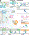
Fig. 1 |. Steps in lipid droplet biogenesis.
Lipid droplets emerge from the endoplasmic reticulum (ER). The correct shape and composition of the ER membranes, which are likely affected by the fat storage-inducing transmembrane 2 (FIT2) protein and other ER-resident proteins, are important determinants of organized lipid droplet biogenesis. Step 1: triacylglycerol (TAG) synthesis (see inset) and cholesterol ester synthesis enzymes deposit neutral lipids in between the leaflets of the ER bilayer. Beyond a certain concentration, the neutral lipids demix and coalesce into a lens. Step 2: seipin and other lipid droplet biogenesis factors are recruited to the lens structure and facilitate the growth of the nascent lipid droplet. The emergence of the lipid droplet into the cytosol is affected by differences in surface tension of the luminal and cytosolic leaflets of the ER bilayer, likely determined by asymmetrical protein binding and phospholipid composition (shown in inset). Step 3: in some mammalian cells, lipid droplets bud from the ER and grow through fusion or local lipid synthesis. AGPAT, acylglycerolphosphate acyltransferase; DAG, diacylglycerol; DGAT, acyl-CoA:diacylglycerol acyltransferase; G3P, glycerol-3-phosphate; GPAT, glycerol-3 phosphate acyltransferase; LPA, lysophosphatidic acid; PA, phosphatidic acid; PAP, phosphatidic acid phosphatase; PLIN, perilipin. Yeast orthologues of mammalian proteins are given in parentheses.
Fig. 2 |
Fig. 2 |. Mechanisms of lipid droplet protein targeting and degradation.
a | Class I lipid droplet proteins insert into the endoplasmic reticulum (ER) and laterally diffuse within the membrane entering the forming lipid droplets. In at least one case, the insertion of a class I protein is mediated through the association with farnesylated peroxisomal biogenesis factor 19 (PEX19) and its ER receptor PEX3. Most class I lipid droplet proteins contain a hydrophobic hairpin, through which they associate with the lipid monolayer. The asymmetrical distribution of class I proteins between the ER and the lipid droplets may be facilitated by targeting some proteins to ER-associated protein degradation (ERAD). b | Class II lipid droplet proteins insert directly from the cytosol into the lipid droplet via amphipathic helices that recognize packing defects in the membrane (inset) or through fatty acid modifications. In the absence of lipid droplets, these proteins may be degraded by the ubiquitin–proteasome system. These proteins may also be susceptible to degradation by the proteasome if they are forced off the surface of the lipid droplet, for example, by molecular crowding during lipid droplet lipolysis. The heat shock cognate 71 kDa protein (HSC70) can extract select proteins bearing a characteristic pentapeptide consensus sequence directing proteins for chaperone-mediated autophagy (CMA) from the lipid droplet for degradation in the lysosome. CMA of lipid-droplet-associated proteins may potentially occur at lipid droplet–lysosome contacts. L AMP2A, lysosome-associated membrane protein 2A; UBXD8, UBX domain-containing protein 8; VCP, transitional endoplasmic reticulum ATPase.
Fig. 3 |
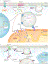
Fig. 3 |. Lipid droplet–organelle contacts.
Lipid droplets interact with nearly all organelles in the cell. The molecular basis for many of these contacts remains poorly understood. Select proteins implicated in tethering or organization of lipid droplet contact sites are shown, with lipid droplet proteins in blue and all other proteins in green. Although the organelle contacts are depicted as distinct and spatially separate, it is likely that individual lipid droplets participate in contacts with multiple organelles simultaneously. Tethering mechanisms that are thus far undefined are indicated by a question mark. AUP1, ancient ubiquitous protein 1; CIDEA, cell death-inducing DFFA-like effector A; DGAT2, diacylglycerol acyltransferase 2 protein; ER, endoplasmic reticulum; FATP1, fatty acid transport protein 1; HSC70, heat shock cognate 71 kDa protein; Ice2, inheritance of cortical ER protein 2; L AMP2A, lysosome-associated membrane protein 2A; Mdm1, structural protein MDM1; MFN2, mitofusin 2; NVJ, nuclear ER–vacuole junction; NRZ, NAG–RINT1–ZW10 complex; Nvj1, nuclear vacuolar junction protein 1; PLIN, perilipin; Vac8, vacuolar protein 8.
Fig. 4 |
Fig. 4 |. Lipid droplets as buffers against stress.
a | Fatty acids (FAs) are important sources of cellular energy. FAs derived from the LipoLysis of lipid droplets are converted at the mitochondrial membrane into acylcarnitine (AC), which is then taken up by mitochondria and broken down via β-oxidation. However, increased FA levels can be toxic to cells, and sequestration of FAs by lipid droplets provides a buffering capacity that prevents lipotoxicity. In adipocytes, induction of lipolysis by external stimuli (such as noradrenergic signalling) releases an enormous amount of FAs, many of which are immediately re-esterified and packaged as triacylglycerol (TAG) into new diacylglycerol acyltransferase 1 (DGAT1)-dependent lipid droplets. When lipid droplet biogenesis is impaired, lipolytic release of FAs from the existing lipid droplets results in endoplasmic reticulum (ER) stress, leading to the activation of the unfolded protein response (UPR) through inositol-requiring protein 1 (IRE1) and protein kinase RNA (PKR)-like ER kinase (PERK). UPR activation may involve alterations in the ER lipid composition, which could directly activate IRE1 and PERK. Large amounts of FAs are also released during starvation owing to the breakdown of membranous organelles via starvation-induced autophagy. Loss of DGAT1-mediated lipid droplet formation under these conditions results in the flux of FAs into AC, which accumulates and disrupts mitochondrial integrity and function, likely by inducing mitochondrial membrane permeabilization. b | Lipid droplets have also been connected with ER protein homeostasis. In certain cases, lipid droplets may facilitate removal of ER-associated protein degradation (ERAD) substrates. ERAD substrates may be targeted to the proteasome from lipid droplets or lipid-droplet-associated ER subdomains. It has also been proposed that ERAD substrates associated with the lipid droplet may be degraded through microlipophagy.
None

References

    1. Schaffer JE Lipotoxicity: when tissues overeat. Curr. Opin. Lipidol. 14, 281–287 (2003). - PubMed
    1. Fawcett DW An Atlas of Fine Structure: The Cell, Its Organelles, and Inclusions (Saunders, 1966).
    1. Farese RV & Walther TC Lipid droplets finally get a little R-E-S-P-E-C-T. Cell 139, 855–860 (2009). - PMC - PubMed
    1. Sorger D, Athenstaedt K, Hrastnik C & Daum G A yeast strain lacking lipid particles bears a defect in ergosterol formation. J. Biol. Chem. 279, 31190–31196 (2004). - PubMed
    1. Sandager L et al. Storage lipid synthesis is non-essential in yeast. J. Biol. Chem. 277, 6478–6482 (2002). - PubMed

Publication types