Review
. 2019 Mar;76(6):1067-1080.
doi: 10.1007/s00018-018-2980-z. Epub 2018 Dec 6.

Stem cells within the shoot apical meristem: identity, arrangement and communication

Affiliations
Review

Stem cells within the shoot apical meristem: identity, arrangement and communication

Naoyuki Uchida et al. Cell Mol Life Sci. 2019 Mar.

Abstract

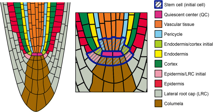
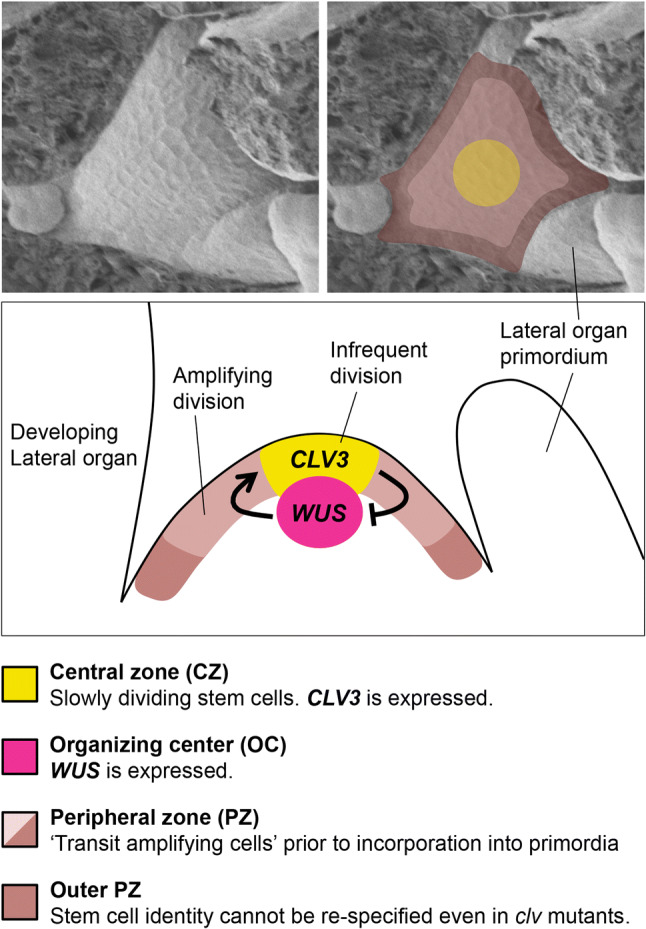
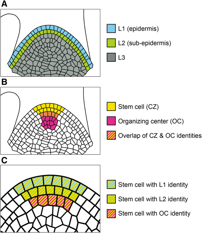
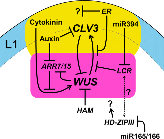
Stem cells are specific cells that renew themselves and also provide daughter cells for organ formation. In plants, primary stem cell populations are nurtured within shoot and root apical meristems (SAM and RAM) for the production of aerial and underground parts, respectively. This review article summarizes recent progress on control of stem cells in the SAM from studies of the model plant Arabidopsis thaliana. To that end, a brief overview of the RAM is provided first to emphasize similarities and differences between the two apical meristems, which would help in better understanding of stem cells in the SAM. Subsequently, we will discuss in depth how stem cells are arranged in an organized manner in the SAM, how dynamically the stem cell identity is regulated, what factors participate in stem cell control, and how intercellular communication by mobile signals modulates stem cell behaviors within the SAM. Remaining questions and perspectives are also presented for future studies.

Keywords: Central zone; Organizing center; Peripheral zone; Shoot apical meristem; Stem cell; Tissue layer.


Figures

Fig. 1
Fig. 1
Stem cell niche in the RAM. The stereotypical arrangement of cells at the Arabidopsis root tip is illustrated. Stem cells (also called ‘initial’ cells), which are shown as hatched cells, are located around the quiescent center (QC), and the stem cell region is surrounded by blue lines. Differentiation potency of each stem cell is strictly limited according to its position. Basically, each stem cell contributes to the formation of a specialized cell file shown in the same color. Exceptions are endodermis/cortex initial and epidermis/lateral root cap initial; the former gives rise to endodermis cell files and cortex cell files, and the later produces epidermis cell files and lateral root cap cells
Fig. 2
Fig. 2
Stem cell niche in the SAM. Photos are top-view images of the Arabidopsis SAM at the vegetative stage by electron scanning microscope. The left is the original image and the right is overlaid by the following zones with different colors. Yellow indicates the central zone (CZ) composed of stem cells expressing CLV3. Beige and dark beige indicate peripheral zone (PZ) composed of transit amplifying cells. The dark beige region shows the outer PZ. Magenta indicates the organizing center (OC) defined by WUS expression. Please see main text for further details
Fig. 3
Fig. 3
Layered arrangement of stem cell subpopulations in the SAM. a The SAM is composed of three tissue layers: the epidermal L1 (light blue), the sub-epidermal L2 layers (green) and the inner L3 tissue (grey). L1 and L2 cells divide anticlinally, resulting in clonal cell layers. L3 cells divide both anticlinally and periclinally, giving rise to internal tissues. b Stem cells consisting the CZ (yellow) spread between all the layers. The OC (magenta), which is defined by WUS expression, is located within the L3 tissue. The lowermost layer of the CZ overlaps with the uppermost layer of the OC (striped pattern of yellow and magenta). c There are three stem cell subpopulations; stem cells with the L1 identity (striped pattern of yellow and light blue), those with the L2 identity (striped pattern of yellow and green) and those with the OC identity (striped pattern of yellow and magenta)
Fig. 4
Fig. 4
Pathways involved in intercellular communication between tissue layers in the SAM. Yellow indicates the central zone composed of stem cells expressing CLV3. Magenta indicates the organizing center defined by WUS expression. Light blue shows the epidermal L1 layer. Please see main text for further details about each of the factors and pathways

References

    1. Heidstra R, Sabatini S. Plant and animal stem cells: similar yet different. Nat Rev Mol Cell Biol. 2014;15:301–312. doi: 10.1038/nrm3790. - DOI - PubMed
    1. Gaillochet C, Lohmann JU. The never-ending story: from pluripotency to plant developmental plasticity. Development. 2015;142:2237–2249. doi: 10.1242/dev.117614. - DOI - PMC - PubMed
    1. Greb T, Lohmann JU. Plant stem cells. Curr Biol. 2016;26:R816–R821. doi: 10.1016/j.cub.2016.07.070. - DOI - PubMed
    1. Rahni R, Efroni I, Birnbaum KD. A case for distributed control of local stem cell behavior in plants. Dev Cell. 2016;38:635–642. doi: 10.1016/j.devcel.2016.08.015. - DOI - PMC - PubMed
    1. Van Norman JM. Asymmetry and cell polarity in root development. Dev Biol. 2016;419:165–174. doi: 10.1016/j.ydbio.2016.07.009. - DOI - PubMed

Substances