

Pericytes for Therapeutic Bone Repair
- PMID: 30523587
- PMCID: PMC6503313
- DOI: 10.1007/978-3-030-02601-1_3
Pericytes for Therapeutic Bone Repair
Abstract
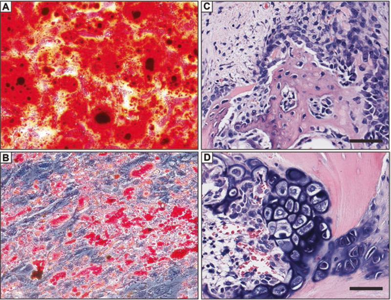
Besides seminal functions in angiogenesis and blood pressure regulation, microvascular pericytes possess a latent tissue regenerative potential that can be revealed in culture following transition into mesenchymal stem cells. Endowed with robust osteogenic potential, pericytes and other related perivascular cells extracted from adipose tissue represent a potent and abundant cell source for refined bone tissue engineering and improved cell therapies of fractures and other bone defects. The use of diverse bone formation assays in vivo, which include mouse muscle pocket osteogenesis and calvaria replenishment, rat and dog spine fusion, and rat non-union fracture healing, has confirmed the superiority of purified perivascular cells for skeletal (re)generation. As a surprising observation though, despite strong endogenous bone-forming potential, perivascular cells drive bone regeneration essentially indirectly, via recruitment by secreted factors of local osteo-progenitors.
Keywords: Blood vessel; Bone; Mesenchymal stem cell; Non-union; Osteogenesis; Pericyte; Perivascular cell; Spinal fusion; Stem cell; Tunica adventitia.
Figures
References
-
- Zimmermann KW (1923) Der feinere Bau der Blutkapillaren. Z Anat 68:29–109
-
- Gerhardt H, Betsholtz C (2003) Endothelial-pericyte interactions in angiogenesis. Cell Tissue Res 314(1):15–23 Epub 2003 Jul 22 - PubMed
Publication types
MeSH terms
Grants and funding
LinkOut - more resources
Full Text Sources
Research Materials
