. 2019 Jan 20;37(3):190-201.
doi: 10.1200/JCO.18.01583. Epub 2018 Dec 3.

Double-Hit Gene Expression Signature Defines a Distinct Subgroup of Germinal Center B-Cell-Like Diffuse Large B-Cell Lymphoma

Affiliations

Double-Hit Gene Expression Signature Defines a Distinct Subgroup of Germinal Center B-Cell-Like Diffuse Large B-Cell Lymphoma

Daisuke Ennishi et al. J Clin Oncol. .

Abstract

Purpose: High-grade B-cell lymphoma with MYC and BCL2 and/or BCL6 rearrangements (HGBL-DH/TH) has a poor outcome after standard chemoimmunotherapy. We sought to understand the biologic underpinnings of HGBL-DH/TH with BCL2 rearrangements (HGBL-DH/TH- BCL2) and diffuse large B-cell lymphoma (DLBCL) morphology through examination of gene expression.

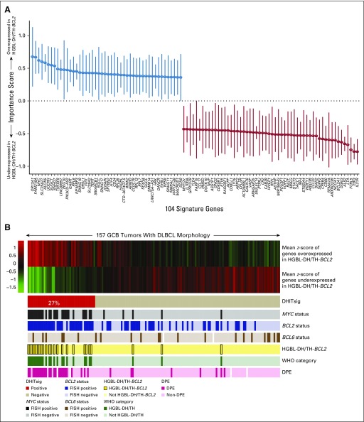
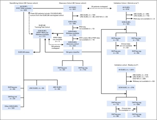
Patients and methods: We analyzed RNA sequencing data from 157 de novo germinal center B-cell-like (GCB)-DLBCLs, including 25 with HGBL-DH/TH- BCL2, to define a gene expression signature that distinguishes HGBL-DH/TH- BCL2 from other GCB-DLBCLs. To assess the genetic, molecular, and phenotypic features associated with this signature, we analyzed targeted resequencing, whole-exome sequencing, RNA sequencing, and immunohistochemistry data.

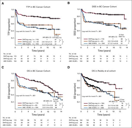
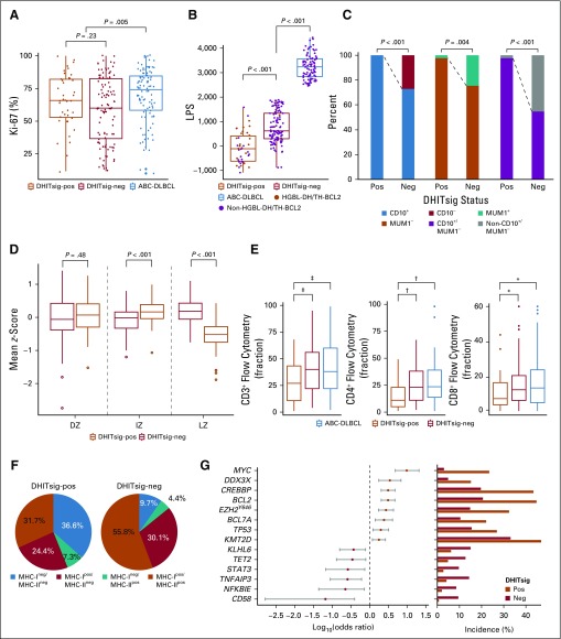
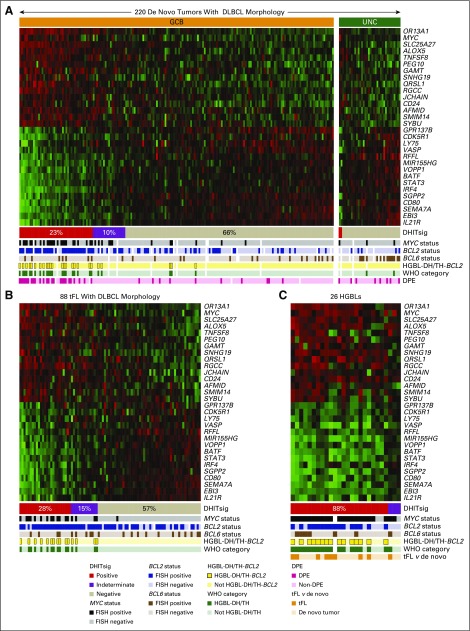
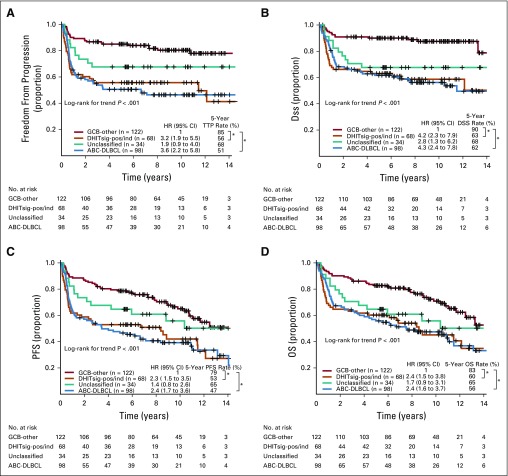
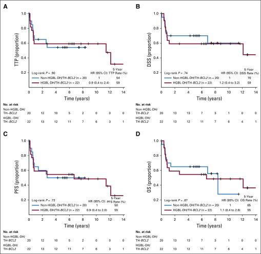
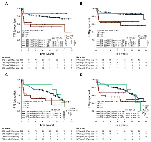
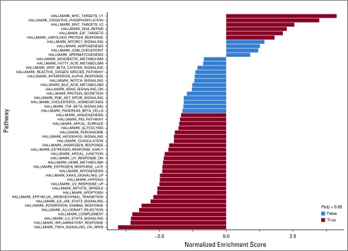
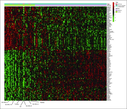
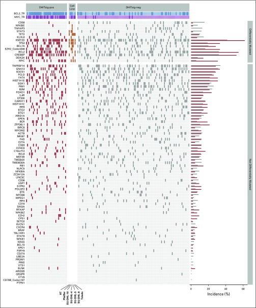
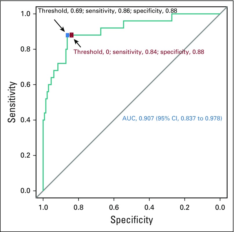
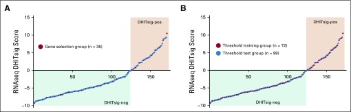
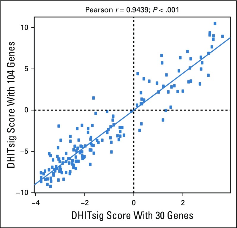
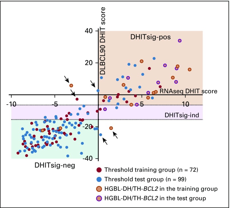
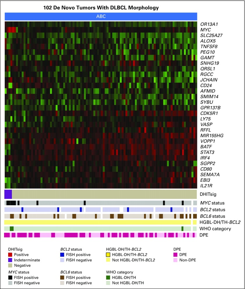
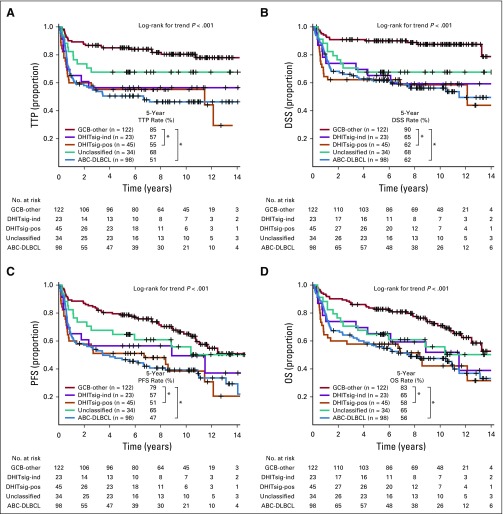
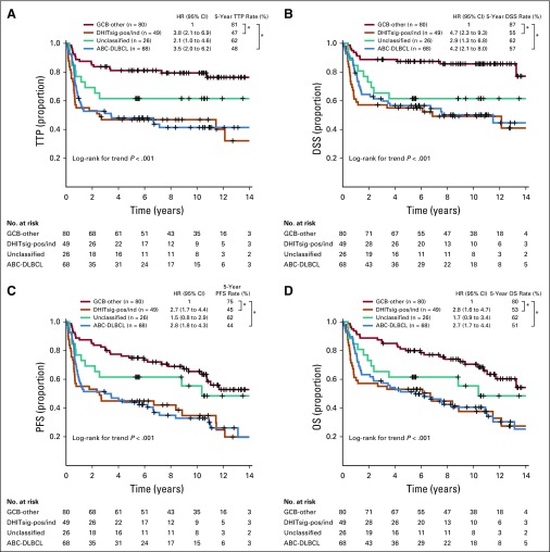
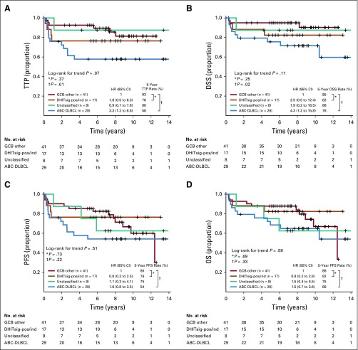
Results: We developed a 104-gene double-hit signature (DHITsig) that assigned 27% of GCB-DLBCLs to the DHITsig-positive group, with only one half harboring MYC and BCL2 rearrangements (HGBL-DH/TH- BCL2). DHITsig-positive patients had inferior outcomes after rituximab plus cyclophosphamide, doxorubicin, vincristine, and prednisone immunochemotherapy compared with DHITsig-negative patients (5-year time to progression rate, 57% and 81%, respectively; P < .001), irrespective of HGBL-DH/TH- BCL2 status. The prognostic value of DHITsig was confirmed in an independent validation cohort. DHITsig-positive tumors are biologically characterized by a putative non-light zone germinal center cell of origin and a distinct mutational landscape that comprises genes associated with chromatin modification. A new NanoString assay (DLBCL90) recapitulated the prognostic significance and RNA sequencing assignments. Validating the association with HGBL-DH/TH- BCL2, 11 of 25 DHITsig-positive-transformed follicular lymphomas were classified as HGBL-DH/TH- BCL2 compared with zero of 50 in the DHITsig-negative group. Furthermore, the DHITsig was shared with the majority of B-cell lymphomas with high-grade morphology tested.

Conclusion: We have defined a clinically and biologically distinct subgroup of tumors within GCB-DLBCL characterized by a gene expression signature of HGBL-DH/TH- BCL2. This knowledge has been translated into an assay applicable to routinely available biopsy samples, which enables exploration of its utility to guide patient management.


Figures

FIG 1.
FIG 1.
The gene expression–based model of 104 genes on the basis of high-grade B-cell lymphoma with MYC and BCL2 and/or BCL6 rearrangements with BCL2 translocations (HGBL-DH/TH-BCL2) status. (A) Importance score with 95% CI of the 104 most significantly differentially expressed genes between HGBL-DH/TH-BCL2 and germinal center B-cell-like (GCB) diffuse large B-cell lymphoma (DLBCL). Genes with blue and red bars are over- and underexpressed in HGBL-DH/TH-BCL2, respectively. (B) Mean z-score of genes over- or underexpressed in HGBL-DH/TH-BCL2 is shown in the form of a heat map, with the 157 patient biopsy samples shown as columns. Double-hit signature (DHITsig) groups identified by the signature are shown below the heat map. The status of MYC, BCL2, and BCL6 genetic alterations; HGBL-DH/TH-BCL2; WHO categories; and MYC/BCL2 dual protein expression (DPE) status are displayed beneath the heat map. FISH, fluorescent in situ hybridization.
FIG 2.
FIG 2.
Prognostic association of double-hit signature (DHITsig) in patients with diffuse large B-cell lymphoma (DLBCL) treated with rituximab plus cyclophosphamide, doxorubicin, vincristine, and prednisone. Kaplan-Meier curves of the DHITsig-positive (DHITsig-pos) germinal center B-cell-like DLBCL v DHITsig-negative (DHITsig-neg) GCB-DLBCL v activated B-cell-like (ABC)-DLBCL for (A) time to progression (TTP), (B) disease-specific survival (DSS) and (C) overall survival (OS) in the BC Cancer cohort and (D) OS in the Reddy et al validation cohort. *P < .001, †P = .011. HR, hazard ratio; PFS, progression-free survival.
FIG 3.
FIG 3.
Genetic, molecular, and phenotypic features of the double-hit signature (DHITsig). (A) Comparison of protein encoded by the MKI67 gene (Ki-67) staining by immunohistochemistry between DHITsig-positive (DHITsig-pos), DHITsig-negative (DHITsig-neg) germinal center B-cell-like (GCB) diffuse large B-cell lymphoma (DLBCL) and activated B-cell-like (ABC) DLBCL. (B) Comparison of linear predictor score (LPS) provided by the Lymph2Cx assay between DHITsig-pos, DHITsig-neg GCB-DLBCL and ABC-DLBCL. Dark orange dots represent the high-grade B-cell lymphoma with MYC and BCL2 and/or BCL6 rearrangements with BCL2 translocations (HGBL-DH/TH-BCL2) tumors. (C) Comparison of immunohistochemistry staining pattern of CD10 (membrane metallo-endopeptidase) and MUM1 (IRF4) between DHITsig-pos and DHITsig-neg GCB-DLBCL tumors. (D) Comparison of mean z-scores of dark zone (DZ), intermediate zone (IZ), and light zone (LZ) signature genes (20 genes each) between DHITsig-pos and DHITsig-neg groups. (E) Comparison of fraction of tumor-infiltrating T cells (CD3+, CD4+, and CD8+ T cells) measured by flow cytometry between DHITsig-pos, DHITsig-neg GCB-DLBCL and ABC-DLBCL. (F) Frequencies of MHC class I (MHC-I) and MHC-II double-negative, isolated MHC-II negative, isolated MHC-I negative, and MHC-I and -II double-positive cases in DHITsig-pos and DHITsig-neg cases. (G) Forest plots summarize the results of Fisher’s exact tests that compare the frequency of mutations that affect individual genes in DHITsig-neg (dark orange dots) and DHITsig-pos (red dots) GCB-DLBCL tumors. Significantly enriched genes in either DHITsig-pos or DHITsig-neg cases (false discovery rate < .10) are represented. Log10 odds ratios and 95% CIs are shown. Bar plots represent the frequency of mutations in either DHITsig-pos or DHITsig-neg groups. *P < .10, †P < .05, ‡P <.01.
FIG 4.
FIG 4.
The gene expression–based model for the double-hit signature (DHITsig). The DLBCL90 assay is shown in the form of a heat map, with the 30 informative genes shown as rows, and the tumors shown as columns, separated into (A) 220 germinal center B-cell-like (GCB) and unclassified (UNC) diffuse large B-cell lymphomas (DLBCLs). (B) Eighty-eight transformed follicular lymphomas (tFLs) with DLBCL morphology. (C) Twenty-six high-grade B-cell lymphomas (HGBLs). The tumors are arrayed from highest DHITsig score on the left to lowest DHITsig score on the right. DHITsig groups identified by the signature are shown below the heat map. The status of MYC, BCL2, and BCL6 genetic alterations; HGBL with MYC and BCL2 and/or BCL6 rearrangements with BCL2 translocations (HGBL-DH/TH-BCL2) status; and WHO categories also are shown. White bars indicate data that are not available or assignments that could not be made on the basis of the available data. DPE, dual protein expression; FISH, fluorescent in situ hybridization.
FIG 5.
FIG 5.
Prognostic association of DLBCL90 in patients with diffuse large B-cell lymphoma (DLBCL) treated with rituximab plus cyclophosphamide, doxorubicin, vincristine, and prednisone. Kaplan-Meier curves of the germinal center B-cell-like (GCB) DLBCL v double-hit signature (DHITsig)-positive (DHITsig-pos) and DHITsig-indeterminate (DHITsig-ind) v unclassified v activated B-cell-like (ABC) DLBCL for (A) time to progression (TTP), (B) disease-free survival (DSS), (C) progression-free survival (PFS), and (D) overall survival (OS) in 322 patients with de novo tumors of DLBCL morphology treated with rituximab plus cyclophosphamide, doxorubicin, vincristine, and prednisone. The same curves with separation of DHITsig-pos and DHITsig-ind are shown in Appendix Figure A13 (online only). *P < .001. HR, hazard ratio.
FIG A1.
FIG A1.
Patient flow for the discovery, two independent validation cohorts, and NanoString cohort. ABC, activated B-cell-like; COO, cell of origin; DHITsig, double-hit signature; DLBCL, diffuse large B-cell lymphoma; FFPET, formalin-fixed paraffin-embedded tissue; FISH, fluorescent in situ hybridization; GCB, germinal center B-cell-like; ind, indeterminate; neg, negative; pos, positive; RNAseq, RNA sequencing; UNC, unclassified.
FIG A2.
FIG A2.
Kaplan-Meier curves of the patients with high-grade B-cell lymphoma with MYC and BCL2 and/or BCL6 rearrangements with BCL2 translocations (HGBL-DH/TH-BCL2) v non-HGBL-DH/TH-BCL2 within double-hit signature–positive (DHITsig-pos) germinal center B-cell-like (GCB) diffuse large B-cell lymphoma (DLBCL) for (A) time to progression (TTP), (B) disease-specific survival (DSS), (C) progression-free survival (PFS), and (D) overall survival (OS). HR, hazard ratio.
FIG A3.
FIG A3.
Kaplan-Meier curves of patients stratified by double-hit signature (DHITsig) combined with dual protein expression (DPE) status in germinal center B-cell-like (GCB) diffuse large B-cell lymphoma (DLBCL) for (A) time to progression (TTP). (B) Disease-specific survival (DSS). (C) Progression-free survival (PFS). (D) Overall survival (OS). DPE-negative (DPE-neg)/DHITsig-positive (DHIT-pos) compared with DPE-positive (DPE-pos)/DHITsig-pos: TTP hazard ratio (HR), 1.4 (95% CI, 0.5 to 3.5; log-rank P = .51); DSS HR, 1.0 (95% CI, 0.4 to 2.8; log-rank P = .99); PFS HR, 1.2 (95% CI, 0.5 to 2.8; log-rank P = .67); and OS HR, 0.9 (95% CI, 0.4 to 2.2; log-rank P = .79).
FIG A4.
FIG A4.
Bar plot of the gene set enrichment analysis. This analysis includes differential expression genes between double-hit signature–positive (DHITsig-pos) and DHITsig-negative (DHITsig-neg) groups with false discovery rate < 0.1 and log2 fold change greater than an absolute value of 0.5.
FIG A5.
FIG A5.
Heat map of primary samples with germinal center B-cell-like (GCB) diffuse large B-cell lymphoma (DLBCL) along with seven GCB-DLBCL cell lines arrayed according to the double-hit signature (DHITsig) score.
FIG A6.
FIG A6.
Mutations detected by targeted sequencing of the discovery cohort and seven diffuse large B-cell lymphoma (DLBCL) cell lines are shown with germinal center B-cell-like (GCB) cases classified as double-hit signature–positive (DHITsig-pos) shown on the left. Genes with significantly different mutation abundance between groups are at the top. The incidence of mutations in each of the genes for the two groups is shown on the right. ind, indeterminate; neg, negative.
FIG A7.
FIG A7.
Receiver operating characteristic curve for the RNA sequencing double-hit signature model score for high-grade B-cell lymphoma with MYC and BCL2 and/or BCL6 rearrangements with BCL2 translocations. The threshold of 0 is indicated by the red dot, whereas the blue dot represents the threshold selected by the Youden index. AUC, area under the curve.
FIG A8.
FIG A8.
RNA sequencing (RNAseq) double-hit signature (DHITsig) scores from 171 germinal center B-cell-like (GCB) diffuse large B-cell lymphomas (DLBCLs) used to train and test the DLBCL90 assay. The tumors are arrayed from left to right with increasing DHITsig scores, with tumors with a score < 0 being designated DHITsig-negative (DHITsig-neg) and > 0 being DHITsig-positive (DHITsig-pos). (A) Tumors highlighted in red had digital expression performed using a code set that contained all 104 genes in the RNAseq model. (B) Tumors highlighted in red were used to train the threshold for the DLBCL90 assay. Note that all of the tumors highlighted in (A) are also highlighted in (B).
FIG A9.
FIG A9.
Plot that shows the association between the double-hit signature (DHITsig) scores from RNA sequencing using the full 104-gene model and the model using the 30 genes selected for the DLBCL90 model.
FIG A10.
FIG A10.
Plot of the double-hit signature (DHITsig) score from the RNA sequencing (RNAseq) model (x-axis) against the DHIT score from the DLBCL90 assay in 171 samples of germinal center B-cell-like (GCB) diffuse large B-cell lymphoma (DLBCL). The 72 biopsy samples highlighted in red were used to establish the thresholds for the assay. Arrows point to the five tumors (3%) that were frankly misclassified. HGBL-DH/TH-BCL2, high-grade B-cell lymphoma with MYC and BCL2 and/or BCL6 rearrangements with BCL2 translocations; ind, indeterminate; neg, negative; pos, positive.
FIG A11.
FIG A11.
Comparisons between the linear predictor score (LPS) from the Lymph2Cx and the DLBCL90 assay. (A) The uncalibrated DLBCL90 LPS scores. (B) The calibrated DLBCL90 LPS scores where 116.6 points were removed from the uncalibrated scores. The six tumors (2%) that moved from a definitive category to unclassified (or vice versa) are highlighted in red. ABC, activated B-cell-like; GCB, germinal center B-cell-like.
FIG A12.
FIG A12.
Heat map that shows the results of the DLBCL90 assignment of double-hit signature (DHITsig) in 102 activated B-cell-like (ABC) diffuse large B-cell lymphoma (DLBCL) tumors. The columns, which represent the tumors, are arrayed from highest scores on the left to lowest on the right. Arrayed below the heat map are pathology characteristics of the tumors. DPE, dual protein expression; FISH, fluorescent in situ hybridization; HGBL-DH/TH-BCL2, high-grade B-cell lymphoma with MYC and BCL2 and/or BCL6 rearrangements with BCL2 translocations.
FIG A13.
FIG A13.
Kaplan-Meier curves of the outcomes in 322 patients with de novo diffuse large B-cell lymphoma treated with curative intent with rituximab plus cyclophosphamide, doxorubicin, vincristine, and prednisone in the BC Cancer cohort grouped according to double-hit signature (DHITsig) status and cell of origin. (A) Time to progression (TTP). (B) Progression-free survival (PFS). (C) Disease-specific survival (DSS). (D) Overall survival (OS). *P < .001. ABC, activated B-cell-like; GCB, germinal center B-cell-like; ind, indeterminate; pos, positive.
FIG A14.
FIG A14.
Kaplan-Meier curves of the outcomes in 223 patients with de novo diffuse large B-cell lymphoma (DLBCL) with advanced-stage disease treated with curative intent with rituximab plus cyclophosphamide, doxorubicin, vincristine, and prednisone in the BC Cancer cohort grouped according to double-hit signature (DHITsig) status and cell of origin. (A) Time to progression (TTP). (B) Disease-specific survival (DSS). (C) Progression-free survival (PFS). (D) Overall survival (OS). *P < .001. ABC, activated B-cell-like; GCB, germinal center B-cell-like; HR, hazard ratio; ind, indeterminate; pos, positive.
FIG A15.
FIG A15.
Kaplan-Meier curves of the outcomes in 95 patients with de novo diffuse large B-cell lymphoma (DLBCL) with limited-stage disease treated with curative intent with rituximab plus cyclophosphamide, doxorubicin, vincristine, and prednisone in the BC Cancer cohort grouped according to double-hit signature (DHITsig) status and cell of origin. (A) Time to progression (TTP). (B) Disease-specific survival (DSS). (C) Progression-free survival (PFS). (D) Overall survival (OS). ABC, activated B-cell-like; GCB, germinal center B-cell-like; HR, hazard ratio; ind, indeterminate; pos, positive.

Comment in

References

    1. Lenz G, Wright G, Dave SS, et al. Stromal gene signatures in large-B-cell lymphomas. N Engl J Med. 2008;359:2313–2323. - PMC - PubMed
    1. Shipp MA, Ross KN, Tamayo P, et al. Diffuse large B-cell lymphoma outcome prediction by gene-expression profiling and supervised machine learning. Nat Med. 2002;8:68–74. - PubMed
    1. Alizadeh AA, Eisen MB, Davis RE, et al. Distinct types of diffuse large B-cell lymphoma identified by gene expression profiling. Nature. 2000;403:503–511. - PubMed
    1. Swerdlow SH, Campo E, Pileri SA, et al. The 2016 revision of the World Health Organization classification of lymphoid neoplasms. Blood. 2016;127:2375–2390. - PMC - PubMed
    1. Scott DW, King RL, Staiger AM, et al. High-grade B-cell lymphoma with MYC and BCL2 and/or BCL6 rearrangements with diffuse large B-cell lymphoma morphology. Blood. 2018;131:2060–2064. - PMC - PubMed

Publication types

MeSH terms