

A Pipeline for Volume Electron Microscopy of the Caenorhabditis elegans Nervous System
- PMID: 30524248
- PMCID: PMC6262311
- DOI: 10.3389/fncir.2018.00094
A Pipeline for Volume Electron Microscopy of the Caenorhabditis elegans Nervous System
Erratum in
-
Corrigendum: A Pipeline for Volume Electron Microscopy of the Caenorhabditis elegans Nervous System.Front Neural Circuits. 2019 Mar 20;13:16. doi: 10.3389/fncir.2019.00016. eCollection 2019. Front Neural Circuits. 2019. PMID: 30949033 Free PMC article.
Abstract
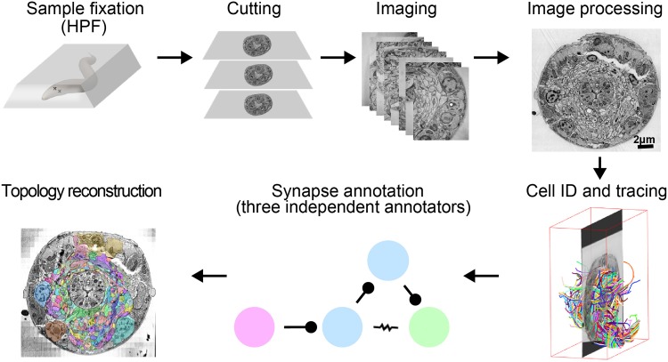
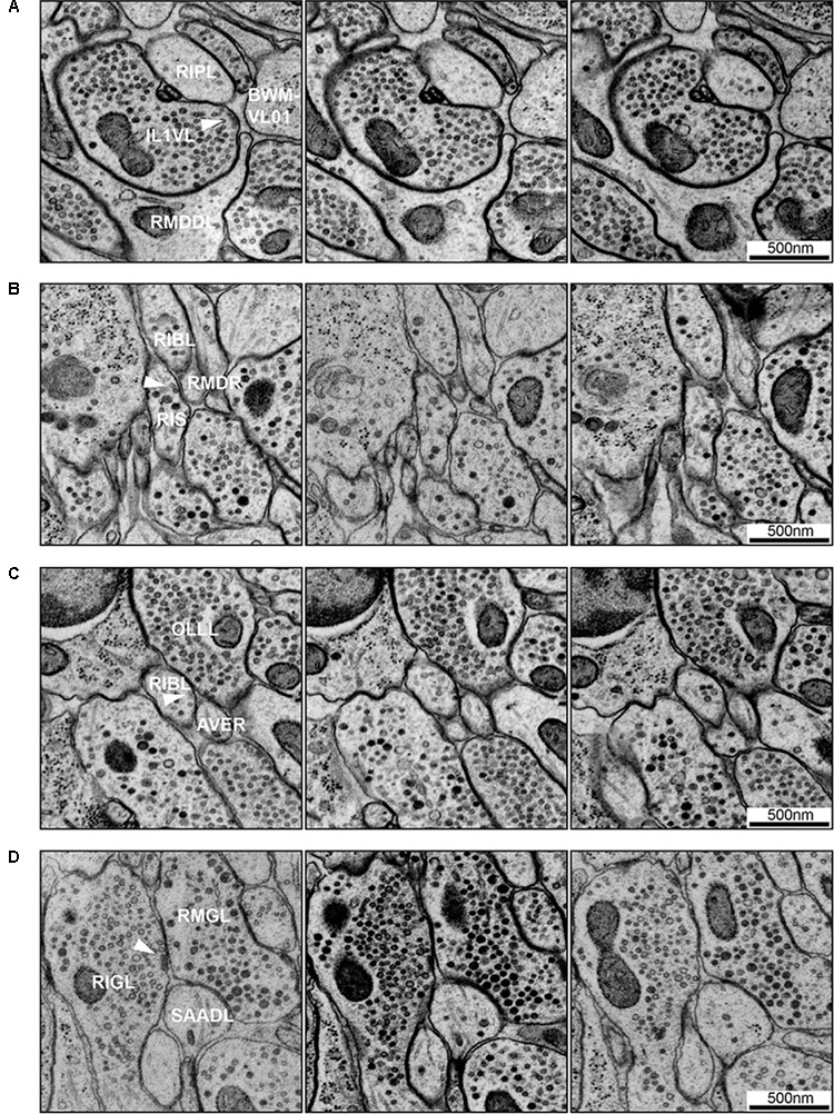
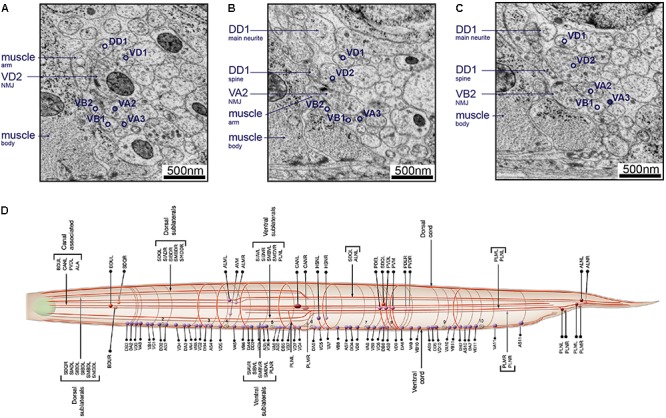
The "connectome," a comprehensive wiring diagram of synaptic connectivity, is achieved through volume electron microscopy (vEM) analysis of an entire nervous system and all associated non-neuronal tissues. White et al. (1986) pioneered the fully manual reconstruction of a connectome using Caenorhabditis elegans. Recent advances in vEM allow mapping new C. elegans connectomes with increased throughput, and reduced subjectivity. Current vEM studies aim to not only fill the remaining gaps in the original connectome, but also address fundamental questions including how the connectome changes during development, the nature of individuality, sexual dimorphism, and how genetic and environmental factors regulate connectivity. Here we describe our current vEM pipeline and projected improvements for the study of the C. elegans nervous system and beyond.
Keywords: C. elegans; connectome; high-pressure freezing; nervous system; volume electron microscopy.
Figures
References
-
- Altun Z. F., Herndon L. A., Wolkow C. A., Crocker C., Lints R., Hall D. H. (2002–2018). WormAtlas. Available at: http://www.wormatlas.org.