. 2018 Nov 22:9:685.
doi: 10.3389/fendo.2018.00685. eCollection 2018.

GnRH Neurons Provide Direct Input to Hypothalamic Tyrosine Hydroxylase Immunoreactive Neurons Which Is Maintained During Lactation

Affiliations

GnRH Neurons Provide Direct Input to Hypothalamic Tyrosine Hydroxylase Immunoreactive Neurons Which Is Maintained During Lactation

Zsuzsanna Bardóczi et al. Front Endocrinol (Lausanne). .

Abstract

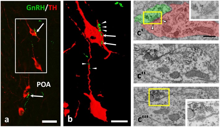
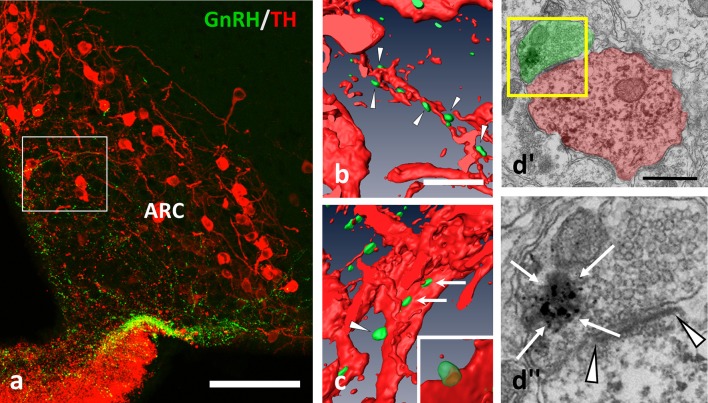
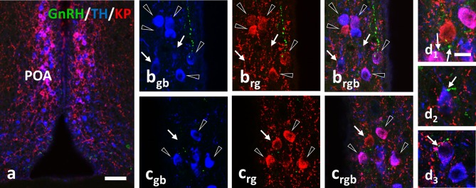
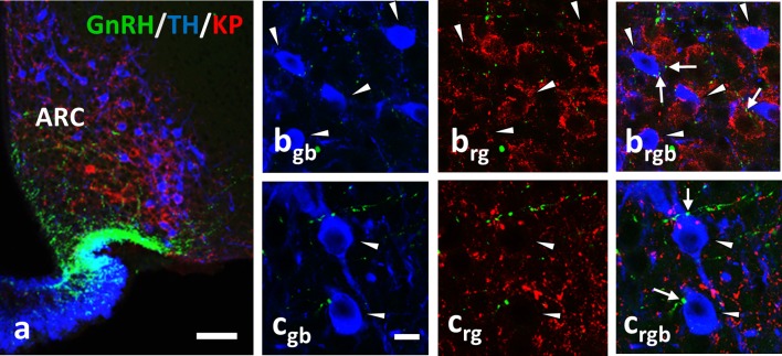
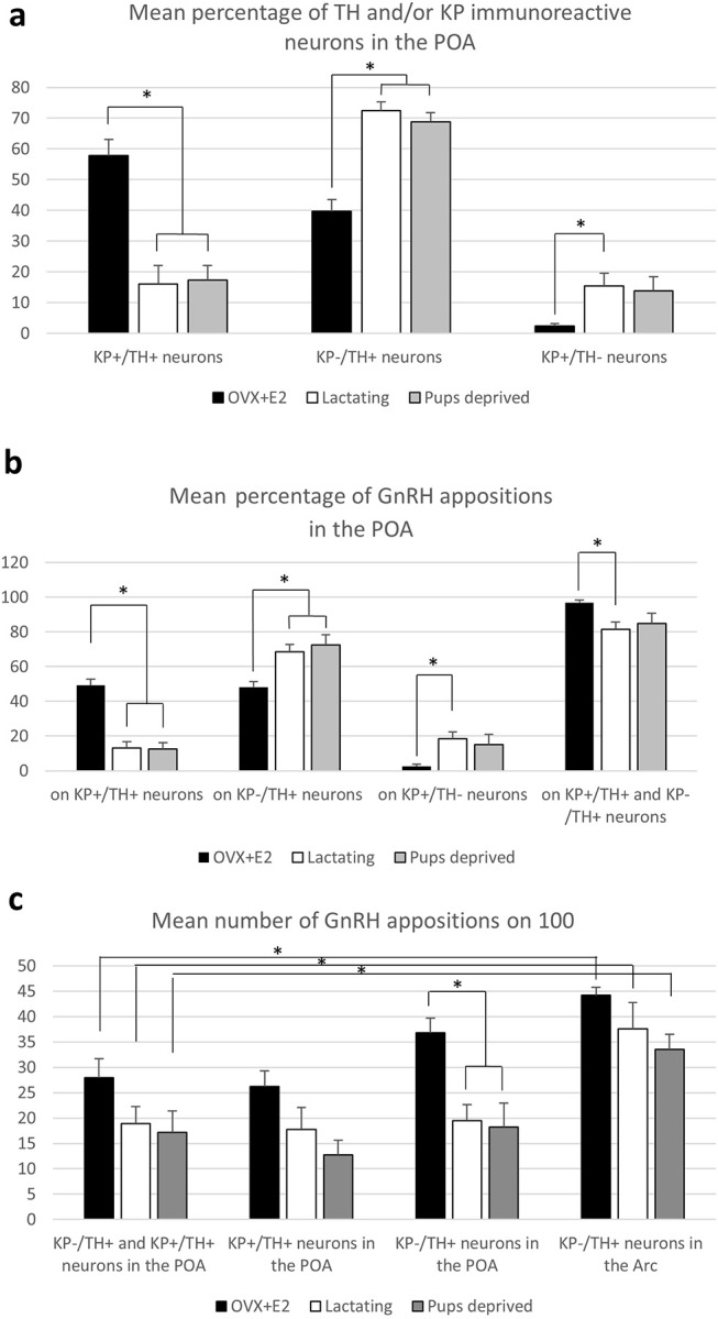
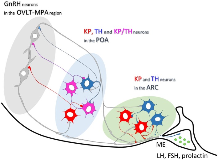
Gonadotropin releasing hormone (GnRH) neurons provide neuronal input to the preoptic area (POA) and the arcuate nucleus (Arc), two regions involved critically in the regulation of neuroendocrine functions and associated behaviors. These areas contain tyrosine hydroxylase immunoreactive (TH-IR) neurons, which play location-specific roles in the neuroendocrine control of both the luteinizing hormone and prolactin secretion, as well as, sexually motivated behaviors. Concerning changes in the activity of GnRH neurons and the secretion pattern of GnRH seen under the influence of rising serum estrogen levels and during lactation, we tested the hypothesis that the functional state of GnRH neurons is mediated via direct synaptic connections to TH-IR neurons in the POA and Arc. In addition, we examined putative changes of these inputs in lactating mice and in mothers separated from their pups. Confocal microscopic and pre-embedding immunohistochemical studies on ovariectomized mice treated with 17β-estradiol (OVX+E2) provided evidence for direct appositions and asymmetric synapses between GnRH-IR fiber varicosities and TH-IR neurons in the POA and the Arc. As TH co-localizes with kisspeptin (KP) in the POA, confocal microscopic analysis was continued on sections additionally labeled for KP. The TH-IR neurons showed a lower level of co-labeling for KP in lactating mice compared to OVX+E2 mice (16.1 ± 5% vs. 57.8 ± 4.3%). Removing the pups for 24 h did not alter significantly the KP production in TH-IR neurons (17.3 ± 4.6%). The mean number of GnRH-IR varicosities on preoptic and arcuate TH cells did not differ in the three animal models investigated. This study shows evidence that GnRH neurons provide direct synaptic inputs to POA and Arc dopaminergic neurons. The scale of anatomical connectivity with these target cells was unaltered during lactation indicating a maintained GnRH input, inspite of the altered hormonal condition.

Keywords: arcuate nucleus; asymmetric synapses; estrogen; kisspeptin; lactation; preoptic area; tyrosine hydroxylase.


Figures

Figure 1
Figure 1
GnRH-immunoreactive (IR) beaded axons contacting processes of TH-IR neurons in the preoptic area of the mouse brain. (a,b) Double immunofluorescent labeling for GnRH (green) and TH (red) shows varicose GnRH-IR fibers in apposition to TH-IR cell bodies (arrows) and dendrites (arrowheads); the gap free connection was validated by reconstructing the image of the immunolabeled profiles in 3D (b). Using medium electron dense DAB to label TH-IR neurons and highly electron dense SGI-NiDAB to mark GnRH-IR axons in pre-embedding immuno-electron microscopy, (c) asymmetric synapses (arrowheads) were identified. The presence of silver grains and the asymmetric character of the synapse were validated in consecutive ultrathin sections (c'–c)”'. Insets in c' and c”' demonstrate the boxed areas at higher power and a reduced contrast. Scale bar in (a) is 25 μm; in (b) it is 10 μm and in (c'–c)”' 500 nm.
Figure 2
Figure 2
GnRH-IR varicosities (green) establish multiple appositions on TH-IR neurons (red) also in the Arc. (a–c) Axo-dendritic and axo-somatic appositions are indicated by arrowheads and arrows; (b,c) are snapshots from the 3D reconstructed and rotated image of the boxed area in (a). The presence of asymmetric synapses (d; arrowheads) were confirmed also by the arcuate appositions. (d)” The GnRH-IR axon terminal contained both dense-core granules (arrows, heavily labeled with SGI-NiDAB) and round-shaped, small, clear vesicles, as shown in the high power image of the boxed area of (d)'. Scale bar in (a) is 100 μm; in (b) and (c) 10 μm; in (d') it is 500 nm.
Figure 3
Figure 3
Immunohistochemical triple labeling for GnRH (green), TH (blue) and KP (red) in the POA, where the majority of TH neurons are also immunoreactive for KP (a; co-localization in purple). Besides double-labeled neurons (arrowheads), single-labeled for TH (arrows in b) or KP (arrow in c) are also present. The GnRH-IR varicosities form appositions (arrows) on all three phenotypes of neurons (d1–d3). Scale bar in (a) is 50 μm; in (bgb–d3) 10 μm.
Figure 4
Figure 4
Immunohistochemical triple labeling for GnRH (green), TH (blue) and KP (red) in the Arc, where TH neurons are not immunoreactive for KP (a); TH- and KP-IR neurons form two separate populations. The TH-IR neurons (arrowheads) receive axo-somatic and axo-dendritic appositions from GnRH-IR fibers (arrows; b,c). rgb; red-green-blue channels. Scale bar in (a) is 50 μm; in (bgb-crgb) 10 μm.
Figure 5
Figure 5
Characterization of preoptic and arcuate TH-IR neurons for kisspeptin expression and GnRH-IR afferentation. (a) Percentage of neurons immunoreactive (IR) for tyrosine hydroxylase (TH) and/or kisspeptin (KP) in the preoptic area (POA). (b) Percentage of the GnRH appositions on each of this neuronal phenotype in the POA. (c) The mean number of GnRH appositions on 100 TH-IR neurons in the POA and arcuate nucleus (Arc). Asterisks mark significant changes.
Figure 6
Figure 6
Summary scheme illustrating neuronal interactions between GnRH and dopaminergic neurons in mice. In the mouse preoptic area, about two-thirds of TH-IR neurons are also immunoreactive for KP. One-third of them, similarly to the Arc dopaminergic neurons, are distinct from KP-IR neurons. Axonal branches of GnRH processes establish synaptic connections with the TH- and KP-IR cell populations in both the preoptic area and arcuate nucleus.

References

    1. Chen WP, Witkin JW, Silverman AJ. beta-Endorphin and gonadotropin-releasing hormone synaptic input to gonadotropin-releasing hormone neurosecretory cells in the male rat. J Comp Neurol. (1989) 286:85–95. 10.1002/cne.902860106 - DOI - PubMed
    1. Leranth C, Segura LM, Palkovits M, MacLusky NJ, Shanabrough M, Naftolin F. The LH-RH-containing neuronal network in the preoptic area of the rat: demonstration of LH-RH-containing nerve terminals in synaptic contact with LH-RH neurons. Brain Res. (1985) 345:332–6. 10.1016/0006-8993(85)91011-X - DOI - PubMed
    1. Thind KK, Goldsmith PC. Infundibular gonadotropin-releasing hormone neurons are inhibited by direct opioid and autoregulatory synapses in juvenile monkeys. Neuroendocrinology (1988) 47:203–16. 10.1159/000124914 - DOI - PubMed
    1. Kallo I, Vida B, Bardoczi Z, Szilvasy-Szabo A, Rabi F, Molnar T, et al. . Gonadotropin-releasing hormone neurones innervate kisspeptin neurones in the female mouse brain. Neuroendocrinology (2013) 98:281–9. 10.1159/000355623 - DOI - PubMed
    1. Herbison AE, Hubbard JI, Sirett NE. LH-RH in picomole concentrations evokes excitation and inhibition of rat arcuate neurones in vitro. Neurosci Lett. (1984) 46:311–5. 10.1016/0304-3940(84)90117-4 - DOI - PubMed