. 2018 Nov 19:9:1661.
doi: 10.3389/fpls.2018.01661. eCollection 2018.

Rice Calcineurin B-Like Protein-Interacting Protein Kinase 31 (OsCIPK31) Is Involved in the Development of Panicle Apical Spikelets

Affiliations

Rice Calcineurin B-Like Protein-Interacting Protein Kinase 31 (OsCIPK31) Is Involved in the Development of Panicle Apical Spikelets

Yongbin Peng et al. Front Plant Sci. .

Abstract

Panicle apical abortion (PAA) causes severe yield losses in rice production, but details about its development and molecular basis remain elusive. Herein, a PAA mutant, paa1019, was identified among the progeny of an elite indica maintainer rice line Yixiang 1B (YXB) mutagenized population obtained using ethyl methyl sulfonate. The abortion rate of spikelets in paa1019 was observed up to 60%. Genetic mapping combined with Mutmap analysis revealed that LOC_Os03g20380 harbored a single-bp substitution (C to T) that altered its transcript length. This gene encodes calcineurin B-like protein-interacting protein kinase 31 (OsCIPK31) localized into the cytoplasm, and is preferentially expressed in transport tissues of rice. Complementation of paa1019 by transferring the open reading frame of LOC_Os03g20380 from YXB reversed the mutant phenotype, and conversely, gene editing by knocking out of OsCIPK31 in YXB results in PAA phenotype. Our results support that OsCIPK31 plays an important role in panicle development. We found that dysregulation is caused by the disruption of OsCIPK31 function due to excessive accumulation of ROS, which ultimately leads to cell death in rice panicle. OsCIPK31 and MAPK pathway might have a synergistic effect to lead ROS accumulation in response to stresses. Meanwhile the PAA distribution is related to IAA hormone accumulation in the panicle. Our study provides an understanding of the role of OsCIPK31 in panicle development by responding to various stresses and phytohormones.

Keywords: CIPK; IAA; MAPK signaling; ROS; apical dominance; auxin; panicle apical abortion.


Figures

FIGURE 1
FIGURE 1
Phenotypes of the paa1019 mutant. Phenotypes, microscopy analysis, and agronomic traits of wild-type (WT) and paa1019 mutant plants. (A) Phenotype of the heading stage. (B) Phenotype of seeds. (C) Phenotype of panicles. (D) Microscopy analysis of the seed surface. (E) Quantification of plant height. (F) Main panicle length. (G) 1000-grain weight. (H). Grain number per panicle. (I) Primary branch number. Statistical analysis was performed using Student’s t-tests. p < 0.05, ∗∗p < 0.01. Scale bar in (D) = 400 μm.
FIGURE 2
FIGURE 2
Genetic and physical maps of the OsCIPK31 gene. (A) The OsCIPK31 gene is located on chromosome 3 between Os3-50.8 and RM 5748. (B) Manhattan plot of chromosome 3. (C) Gene structure of OsCIPK31. The splicing pattern is altered in the mutant, resulting in the deletion of the second exon. (D) Phenotype of WT, paa1019, and complementation lines. CE1 – CE3 represent the three complementation transgenic lines. (E) Phenotype of WT, paa1019, and knock-out lines. KO1 – KO3 represent three knock-out transgenic lines. (F) Abortion rate analysis of lines corresponding to (E). Statistical analysis was performed using Student’s t-tests, ∗∗p < 0.01.
FIGURE 3
FIGURE 3
Characteristics of degraded spikelets. (A–D) Scanning electron microscopy (SEM) of shoot apical meristems (SAMs). Branch and spikelet primordia of WT (A,C) and paa1019 (B,D) plants. PB, primary branches; SB, second branches. (E–J) Panicle development at different growth stages. Panicles were observed when the length was 4, 10, and 15 cm for WT (E,G,I) and paa1019 (F,H,J) plants. (K) Phenotype of primary branches from the bottom to the top of the panicle in paa1019. (L) Statistics for the spikelet number correspond to (K). (M) Model of apical abortion distribution in paa1019. (N,O) Transverse sections of pedicels. (O) Secondary pedicel in PAA. (N) Corresponding parts in WT plants. VB, vascular bundle; GC, green cells; SC, sclerenchymatous cells; T, vessel; P, phloem. (P) Concentration of indole-3-acetic acid (IAA/auxin). PBA, panicles before abortion; PFA, panicles following abortion. Scale bar = 100 μm (A–D,N,O) and 2 cm (E–J). p < 0.05, ∗∗p < 0.01.
FIGURE 4
FIGURE 4
ROS accumulation leads to plasma membrane damage in paa1019. (A) Trypan blue staining. (B) Evans blue staining. (C) DAB staining at different growth stages. Primordium differentiation stage (1) = 20 days before flowering (DBF); (2) = 15 DBF; (3) = 10 DBF; (4) = 5 DBF; (5) = heading stage; (6) differences between WT and paa1019. (D) Concentration of H2O2 in panicles. (E–G) Relative expression of CAT1, CAT2, and CAT3 isozymes. Rice ACTIN was used as an internal control. Data are presented as mean ± SE (n = 3). (H–M) Transverse sections of spikelets at booting stages (H,I) and heading stages (J,K). (L,M) Enlargement of stamens in (H,I). Scale bar = 0.2 c (C) and 100 μm (H–M). p < 0.05, ∗∗p < 0.01.
FIGURE 5
FIGURE 5
paa1019 plants are more sensitive to cold and salt stresses. (A) Phenotype following cold treatment. (B) Relative water concentration in leaves. (C–F) Relative expression of OsCOLD1 and OsLTG. (G) Phenotype following salt treatment. (H) Relative water concentration in leaves. (I–L) Relative expression of CAT and P5CS. p < 0.05, ∗∗p < 0.01.
FIGURE 6
FIGURE 6
Analysis of OsCIPK31 expression. (A–J) Detection of GUS activity in OsCIPK31 promoter:GUS transgenic plants. GUS staining was performed using more than three independent transgenic lines with similar patterns. Primary roots (A), stems (B,C), center of young leaves (D), adult leaves (E), young stem nodes (F), stamens (G), embryos of seeds (H), germinating seeds (I), seedlings (J). (K–M) In situ hybridization of an OsCIPK31-specific probe with a longitudinal section of young panicle (K), and cross-sections of spikelets (L) and stems (M). (N) Subcellular location of the OsCIPK31 protein. Scale bar = 0.5 cm (A–J), 500 μm (K–M) and 10 μm (N).
FIGURE 7
FIGURE 7
Differentially expressed genes (DEGs) in panicles of WT and paa1019 plants. (A) Number of DEGs in host tissues. Green and red bars indicated down- and upregulated genes, respectively. a, b, c, and d represent WT-2 cm vs. WT-6 cm, paa1019-2 cm vs. paa1019-6 cm, WT-2 cm vs. paa1019-2 cm, and WT-6 cm vs. paa1019-6 cm, respectively. 765 significantly dysregulated genes with a fold change (FC) > 2 and a p-value less than 0.05 was identified. Among these, 421 were upregulated and 344 were downregulated in mutant panicles before degradation. By contrast, 1995 significantly dysregulated genes, including 787 upregulated and 1208 downregulated genes, were identified between WT and mutant panicles after degradation. A total of 2657 genes, including 2139 upregulated and 518 downregulated genes, were observed in WT groups, and 1862 genes, including 1290 upregulated and 572 downregulated genes, were identified in mutant groups. (B) Venn diagram showing genes common to WT-1, WT-2, paa1019-1, and paa1019-2 groups (represented by WT-2 cm, WT-6 cm, paa1019-2 cm, and paa1019-6 cm, respectively). (C) Analysis of the expression of common genes.
FIGURE 8
FIGURE 8
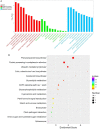
Functional enrichment in response to OsCIPK31 mutation. (A) Comparison of GO enrichment analysis. The top 10 significantly enriched GO terms for biological process (BP, cellular component (CC) and molecular function (MF) in PAA relative to normal panicle development in WT plants are shown in red, green, and blue, respectively. (B) Comparison of Kyoto Encyclopedia of Genes and Genomes (KEGG) pathway enrichment. The top 20 significantly enriched KEGG pathways in PAA relative to normal panicle development in WT plants are shown. The x-axis represents the enrichment factor for each pathway, and the y-axis shows the KEGG pathway names. The size of the circle represents the number of genes involved in each significant pathway. The color represents the significance level of the -log (p-value), from red (most significant) to green (least significant).
FIGURE 9
FIGURE 9
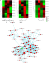
Interaction networks involving OsCIPK31 and the most significantly enriched pathways. (A) Comparison of DEGs in different pathways between WT and paa1019 plants. (B) Protein-protein interaction (PPI) networks involving OsCIPK31 and the most significantly enriched pathways (the MAPK signaling pathway, plant hormone signal transduction, peroxisome, and alpha-linolenic acid metabolism).
FIGURE 10
FIGURE 10
Putative regulation of panicle development by OsCIPK31. OsCIPK31 perceives the response against stresses and regulates ROS accumulation, which might be collaboration with MAPK cascades. Meanwhile OsCIPK31 regulates IAA phytohormone distribution in the panicle, and excessive IAA might lead to the ROS accumulation in the apical spikelet, and ROS regulates PCD of the panicle.

References

    1. Albrecht V., Ritz O., Linder S., Harter K., Kudla J. (2001). The NAF domain defines a novel protein–protein interaction module conserved in Ca2+-regulated kinases. EMBO J. 20 1051–1063. 10.1093/emboj/20.5.1051 - DOI - PMC - PubMed
    1. Ansari T. H., Yamamoto Y., Yoshida T., Miyazaki A., Wang Y. (2003). Cultivar differences in the number of differentiated spikelets and percentage of degenerated spikelets as determinants of the spikelet number per panicle in relation to dry matter production and nitrogen absorption. Soil Sci. Plant Nutr. 49 433–444. 10.1080/00380768.2003.10410029 - DOI
    1. Ashikari M., Sakakibara H., Lin S., Yamamoto T., Takashi T., Nishimura A., et al. (2005). Cytokinin oxidase regulates rice grain production. Science 309 741–745. 10.1126/science.1113373 - DOI - PubMed
    1. Bai J., Zhu X., Wang Q., Zhang J., Chen H., Dong G., et al. (2015). Rice TUTOU1 encodes a suppressor of CAMP receptor-like protein that is important for actin organization and panicle development. Plant Physiol. 169 1179. 10.1104/pp.15.00229 - DOI - PMC - PubMed
    1. Batistiè O., Rehers M., Akerman A., Schl Cking K., Steinhorst L., Yalovsky S., et al. (2012). S-acylation-dependent association of the calcium sensor CBL2 with the vacuolar membrane is essential for proper abscisic acid responses. Cell Res. 22 1155–1168. 10.1038/cr.2012.71 - DOI - PMC - PubMed

LinkOut - more resources