

Isolated bilateral renal mucormycosis in apparently immunocompetent patients-a case series from India and review of the literature
- PMID: 30524710
- PMCID: PMC6275442
- DOI: 10.1093/ckj/sfy034
Isolated bilateral renal mucormycosis in apparently immunocompetent patients-a case series from India and review of the literature
Abstract
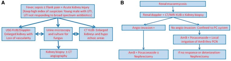
Background: Isolated renal mucormycosis (IRM) is a potentially fatal disease affecting immunocompromised hosts. IRM affecting apparently immunocompetent patients is rare, with few previous reports, mostly from India. We describe 10 cases of bilateral IRM with no underlying risk factors.
Methods: We performed a retrospective analysis of cases of IRM from our hospital information system admitted between 2009 and 2016. We analyzed the data of this cohort of IRM, including epidemiological characteristics, clinical presentation, diagnostic procedures, treatment details and outcome.
Results: In all, 10 cases of bilateral IRM were identified. All of them were males with a mean age of 24.7 years (range 10-42). Most patients were initially managed as acute bacterial pyelonephritis with acute kidney injury. A total of eight patients were diagnosed antemortem. Diagnostic clues include sepsis not controlled with broad-spectrum antibiotics and enlarged kidneys with or without hypodensities on ultrasound/computed tomography imaging. Three patients also gave a specific history of passing white flakes in their urine. Eight patients received specific antifungal therapy with amphotericin B with or without posaconazole. Three patients in whom the disease was apparently confined to the pelvicalyceal system underwent local irrigation with Amp-B. One patient underwent bilateral nephrectomy. Four patients succumbed to the disease while five patients were successfully treated. One patient was discharged against medical advice.
Conclusions: IRM is a rare, life-threatening disease associated with high mortality even in immunocompetent individuals. Typical clinical and radiological findings and a high index of suspicion may help in early diagnosis, but definitive diagnosis requires histopathological and/or microbiological confirmation. Early and rapid diagnosis along with aggressive multidisciplinary management including initiation of specific antifungal therapy with or without surgical debridement is vital for a successful outcome.
Keywords: amphotericin B; immunocompetent; isolated renal mucormycosis; nephrectomy; posaconazole.
Figures


Similar articles
-
Renal mucormycosis presenting during the COVID-19 pandemic: A series of 11 cases from a tertiary care center in India.Indian J Urol. 2022 Apr-Jun;38(2):115-120. doi: 10.4103/iju.iju_437_21. Epub 2022 Apr 1. Indian J Urol. 2022. PMID: 35400861 Free PMC article.
-
Isolated Renal Mucormycosis in Immunocompetent Hosts: Clinical Spectrum and Management Approach.Am J Trop Med Hyg. 2019 Apr;100(4):791-797. doi: 10.4269/ajtmh.18-0103. Am J Trop Med Hyg. 2019. PMID: 30652661 Free PMC article.
-
Intra-abdominal Mucormycosis in an Immunocompetent Host: A Rare Presentation and Literature Review.Cureus. 2025 Mar 17;17(3):e80730. doi: 10.7759/cureus.80730. eCollection 2025 Mar. Cureus. 2025. PMID: 40103914 Free PMC article.
-
Primary cutaneous mucormycosis in an immunocompetent host: report of a case.Surg Today. 2003;33(4):319-22. doi: 10.1007/s005950300073. Surg Today. 2003. PMID: 12707834 Review.
-
Gastrointestinal mucormycosis in apparently immunocompetent hosts-A review.Mycoses. 2018 Dec;61(12):898-908. doi: 10.1111/myc.12798. Epub 2018 Jun 20. Mycoses. 2018. PMID: 29855116 Review.
Cited by
-
Successful Treatment of Bilateral Renal Mucormycosis With Isavuconazole: A Case Report.Cureus. 2023 Jul 20;15(7):e42219. doi: 10.7759/cureus.42219. eCollection 2023 Jul. Cureus. 2023. PMID: 37605691 Free PMC article.
-
Renal mucormycosis presenting during the COVID-19 pandemic: A series of 11 cases from a tertiary care center in India.Indian J Urol. 2022 Apr-Jun;38(2):115-120. doi: 10.4103/iju.iju_437_21. Epub 2022 Apr 1. Indian J Urol. 2022. PMID: 35400861 Free PMC article.
-
Isolated renal mucormycosis presenting with bilateral renal artery thrombosis: a case report.Afr J Urol. 2021;27(1):86. doi: 10.1186/s12301-021-00193-3. Epub 2021 Jun 30. Afr J Urol. 2021. PMID: 34226814 Free PMC article.
-
Disseminated mucormycosis presenting as a renal mass in an human immunodeficiency virus-infected patient: A case report.S Afr J Infect Dis. 2021 Mar 2;36(1):202. doi: 10.4102/sajid.v36i1.202. eCollection 2021. S Afr J Infect Dis. 2021. PMID: 34485490 Free PMC article.
-
Global Epidemiology of Mucormycosis.J Fungi (Basel). 2019 Mar 21;5(1):26. doi: 10.3390/jof5010026. J Fungi (Basel). 2019. PMID: 30901907 Free PMC article. Review.
References
-
- Prabhu RM, Patel R.. Mucormycosis and entomophthoramycosis: a review of the clinical manifestations, diagnosis and treatment. Clin Microbiol Infect 2004; 10(Suppl 1): 31–47 - PubMed
-
- Dansky AS, Lynne CM, Politano VA.. Disseminated mucormycosis with renal involvement. J Urol 1978; 119: 275–277 - PubMed
-
- Ingram CW, Sennesh J, Cooper JN. et al. Disseminated zygomycosis: report of four cases and review. Rev Infect Dis 1989; 11: 741–754 - PubMed
-
- Levy E, Bia MJ.. Isolated renal mucormycosis: case report and review. J Am Soc Nephrol 1995; 5: 2014–2019 - PubMed
LinkOut - more resources
Full Text Sources
