. 2018 Sep 5;7(12):e1486353.
doi: 10.1080/2162402X.2018.1486353. eCollection 2018.

Upregulation of tryptophanyl-tRNA synthethase adapts human cancer cells to nutritional stress caused by tryptophan degradation

Affiliations

Upregulation of tryptophanyl-tRNA synthethase adapts human cancer cells to nutritional stress caused by tryptophan degradation

Isabell Adam et al. Oncoimmunology. .

Abstract

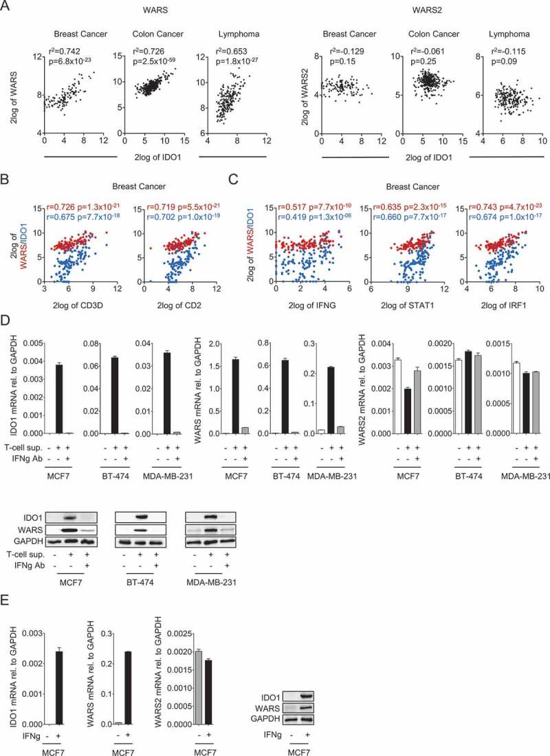
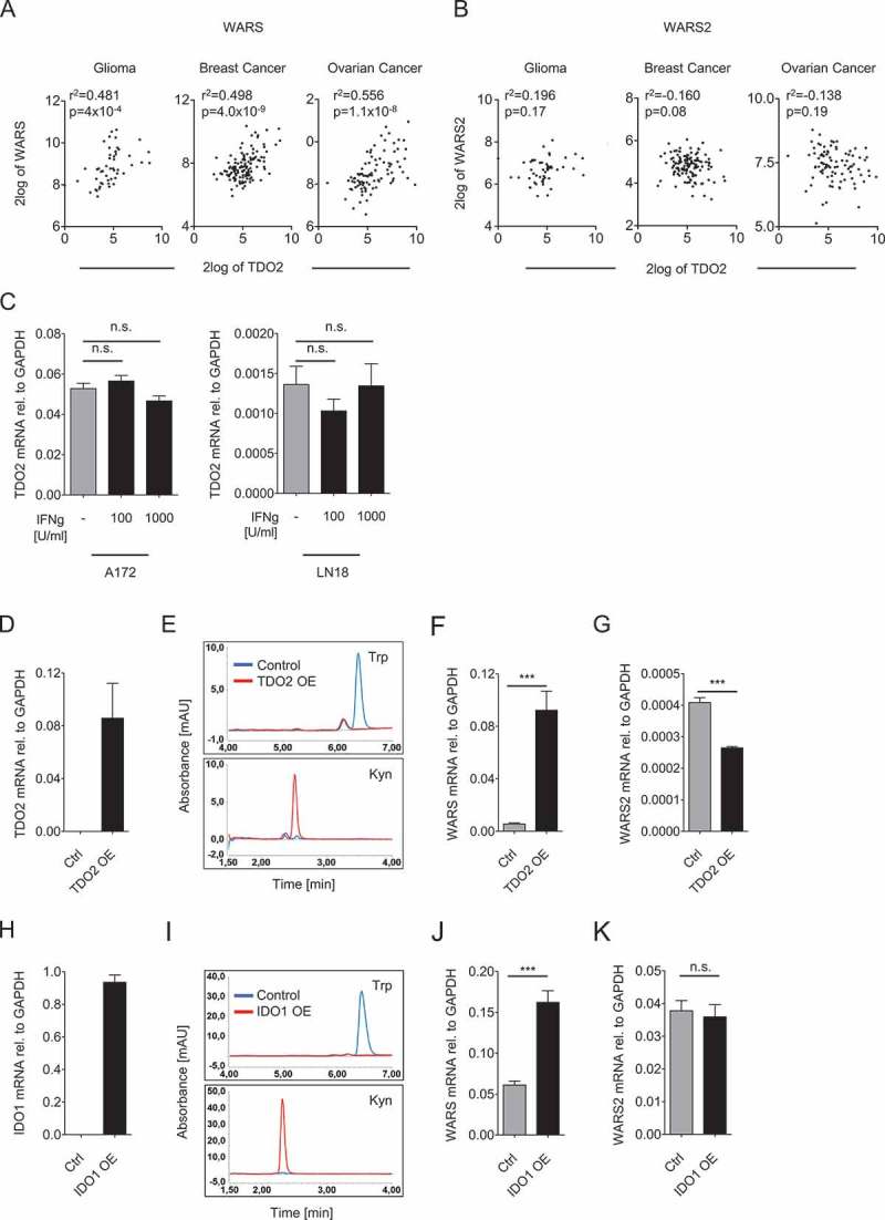
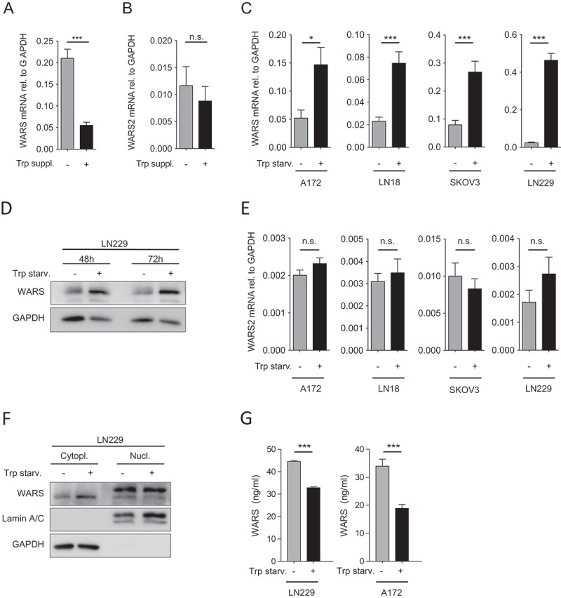
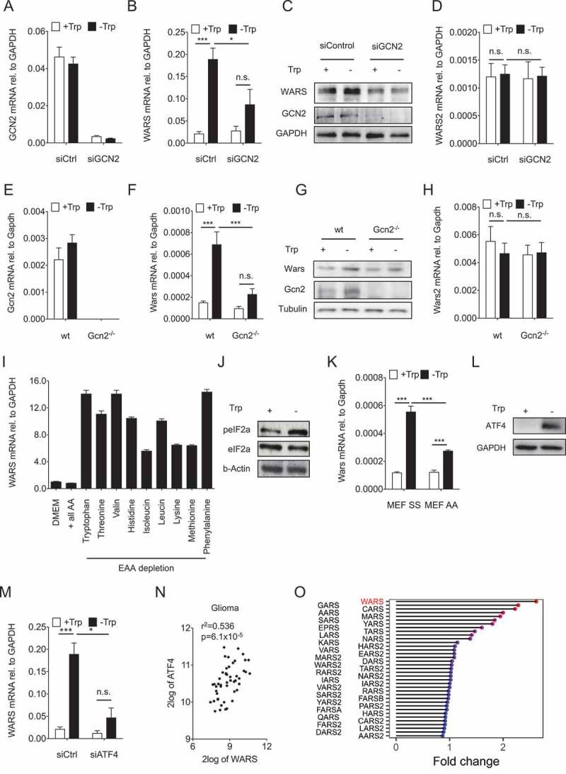
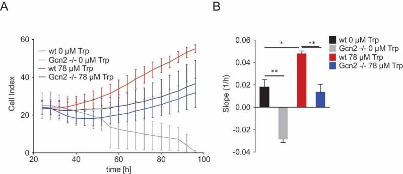
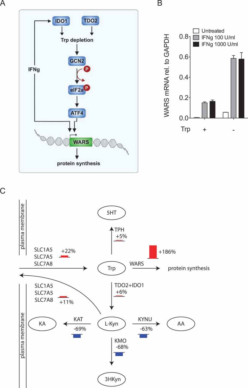
Tryptophan (Trp) metabolism is an important target in immuno-oncology as it represents a powerful immunosuppressive mechanism hijacked by tumors for protection against immune destruction. However, it remains unclear how tumor cells can proliferate while degrading the essential amino acid Trp. Trp is incorporated into proteins after it is attached to its tRNA by tryptophanyl-tRNA synthestases. As the tryptophanyl-tRNA synthestases compete for Trp with the Trp-catabolizing enzymes, the balance between these enzymes will determine whether Trp is used for protein synthesis or is degraded. In human cancers expression of the Trp-degrading enzymes indoleamine-2,3-dioxygenase-1 (IDO1) and tryptophan-2,3-dioxygenase (TDO2) was positively associated with the expression of the tryptophanyl-tRNA synthestase WARS. One mechanism underlying the association between IDO1 and WARS identified in this study is their joint induction by IFNγ released from tumor-infiltrating T cells. Moreover, we show here that IDO1- and TDO2-mediated Trp deprivation upregulates WARS expression by activating the general control non-derepressible-2 (GCN2) kinase, leading to phosphorylation of the eukaryotic translation initiation factor 2α (eIF2α) and induction of activating transcription factor 4 (ATF4). Trp deprivation induced cytoplasmic WARS expression but did not increase nuclear or extracellular WARS levels. GCN2 protected the cells against the effects of Trp starvation and enabled them to quickly make use of Trp for proliferation once it was replenished. Computational modeling of Trp metabolism revealed that Trp deficiency shifted Trp flux towards WARS and protein synthesis. Our data therefore suggest that the upregulation of WARS via IFNγ and/or GCN2-peIF2α-ATF4 signaling protects Trp-degrading cancer cells from excessive intracellular Trp depletion.

Keywords: 3-dioxygenase; Indoleamine-2, 3-dioxygenase; cancer metabolism; immunosuppression; immunosurveillance; inflammation and cancer; nutrients; proliferation; starvation; tRNA synthetase; tryptophan; tryptophan-2; tumor.


Figures

Figure 1.
Figure 1.
IDO1 and WARS correlate with each other and are both associated with the expression of T cell markers and with IFNγ signaling. (A) Left: Correlation between IDO1 and WARS expression in breast cancer, colon cancer and B-cell lymphoma. Right: IDO1 and WARS2 expression in breast cancer, colon cancer and B-cell lymphoma do not correlate with each other (= 124 for breast cancer, = 355 for colon cancer, = 215 for lymphoma, Spearman’s rank correlation). (B) WARS (red) and IDO1 (blue) expression are significantly correlated with the expression of the T cell markers CD3D and CD2, and (C) with the expression of IFNG, STAT1 and IRF1 in breast cancer (= 124, Spearman’s rank correlation). (D) IDO1, WARS and WARS2 mRNA levels measured by qRT-PCR and IDO1 and WARS protein measured by Western blot after treatment of the human breast cancer cell lines MCF7, BT-474 and MDA-MB-231 with supernatants of activated CD4 + T cells in the absence and presence of an IFNγ blocking antibody. (E) IDO1, WARS and WARS2 mRNA levels measured by qRT-PCR and IDO1 and WARS protein measured by Western blot after treatment of MCF7 cells with 1000 U/ml recombinant IFNγ. All data are expressed as mean ± s.e.m. Statistical significance is assumed at < 0.05 (*< 0.05, **< 0.01, ***P <0.001).
Figure 2.
Figure 2.
TDO2 and IDO1 activity upregulate WARS expression. (A) Correlation between TDO2 and WARS expression in human glioma, breast cancer, and ovarian cancer. (B) TDO2 and WARS2 expression in glioma, breast cancer, and ovarian cancer do not correlate with each other (= 50 for glioma, = 124 for breast cancer, = 90 for ovarian cancer, Spearman’s rank correlation). (C) Treatment of A172 and LN18 glioblastoma cells with 100 U/ml or 1000 U/ml IFNγ did not alter TDO2 mRNA expression measured by qRT-PCR. (D) TDO2 mRNA expression in LN229 glioblastoma cells after overexpression of TDO2. (E) HPLC chromatograms showing Trp (top) and Kyn (bottom) measured in the supernatants of control-transduced (blue) and TDO2-overexpressing (red) LN229 glioblastoma cells. (F) WARS mRNA expression measured by qRT-PCR in control-transduced and TDO2-overexpressing LN229 cells. (G) WARS2 mRNA expression measured by qRT-PCR in control-transduced and TDO2-overexpressing LN229 cells. (H) IDO1 mRNA expression in HEK 293 cells after overexpression of IDO1. (I) HPLC chromatograms showing Trp (top) and Kyn (bottom) measured in the supernatants of control-transfected (blue) and IDO1-overexpressing (red) HEK 293 cells. (J) WARS mRNA expression measured by qRT-PCR in control-transfected and IDO1-overexpressing HEK 293 cells. (K) WARS2 mRNA expression measured by qRT-PCR in control-transfected and IDO1-overexpressing HEK 293 cells. All data are expressed as mean ± s.e.m. Statistical significance is assumed at < 0.05 (*< 0.05, **< 0.01, ***P <0.001).
Figure 3.
Figure 3.
Trp depletion upregulates WARS expression. (A) WARS mRNA expression measured by qRT-PCR in A172 glioblastoma cells, which exhibit constitutive TDO2 activity, cultured for 120 h in normal DMEM or with additional supplementation of 78 µM Trp after 72 h. (B) WARS2 mRNA expression measured by qRT-PCR in A172 glioblastoma cells under the conditions described in (A). (C) WARS mRNA expression measured by qRT-PCR in glioblastoma (A172, LN18, LN229) and ovarian carcinoma cells (SKOV-3) after 24 h of cultivation in medium containing 78 µM versus no Trp. (D) WARS protein levels in LN229 glioblastoma cells detected by Western blot after 48 h or 72 h of cultivation in medium containing 78 µM or no Trp. GAPDH served as loading control (E) WARS2 mRNA expression measured by qRT-PCR was not altered by the conditions described in (C). (F) WARS protein levels in the cytoplasmic or nuclear fraction of LN229 glioblastoma cells detected by Western blot after 48 h of cultivation in medium containing 78 µM or no Trp. GAPDH served as loading control for the cytoplasmic fraction, lamin A/C as loading control for the nuclear fraction. (G) WARS protein concentrations measured by ELISA in the supernatants of LN229 and A172 glioblastoma cells cultured for 48 h in the presence or absence of Trp. All data are expressed as mean ± s.e.m. Statistical significance is assumed at < 0.05 (*< 0.05, **< 0.01, ***P <0.001).
Figure 4.
Figure 4.
GCN2-peIF2a-ATF4 signaling mediates the upregulation of WARS in response to Trp depletion. (A) GCN2 mRNA expression measured by qRT-PCR in LN229 cells after knockdown of GCN2 by siRNA cultured in the absence and presence of 78 µM Trp for 24 h. (B) WARS mRNA expression measured by qRT-PCR and (C) WARS and GCN2 protein expression detected by Western blot under the conditions described in (A). GAPDH served as loading control. (D) WARS2 mRNA expression measured by qRT-PCR under the conditions described in (A). (E) Gcn2 mRNA expression in wildtype (wt) and Gcn2 −/- MEFs cultured in the absence and presence of 78 µM Trp for 24 h. (F) Wars mRNA expression measured by qRT-PCR and (G) WARS and GCN2 protein expression detected by Western blot under the conditions described in (E). Tubulin served as loading control. (H) Wars2 mRNA expression measured by qRT-PCR under the conditions described in (E). (I) WARS mRNA expression measured by qRT-PCR in full DMEM medium (DMEM), in amino acid-free DMEM supplemented with all amino acids (+ all AA) and in amino acid-free DMEM supplemented with all but each of the essential amino acids (EAA). (J) peIF2a and eIF2a detected by Western blot in LN229 cells cultured in the presence (+) or absence (-) of 78 µM Trp for 24 h. (K) Wars mRNA expression measured by qRT-PCR in MEFs that express wildtype Eif2a (SS) or an Eif2a version, in which serine 51 is mutated to an alanine and cannot be phosphorylated by Gcn2 (AA). MEFs were cultured for 24 h in the presence or absence of 78 µM Trp. (L) ATF4 protein detected by Western blot in LN229 cells cultured for 24 h in the presence or absence of 78 µM Trp. GAPDH served as loading control. (M) WARS mRNA expression measured by qRT-PCR in LN229 cells after knockdown of ATF4 by siRNA cultured in the absence and presence of 78 µM Trp for 24 h. (N) Correlation between ATF4 and WARS expression in human glioma (= 50, Spearman’s rank correlation). All data are expressed as mean ± s.e.m. Statistical significance is assumed at < 0.05 (*< 0.05, **< 0.01, ***P <0.001). (O) Lolli-plot showing the absolute fold changes of all the tRNA synthetases in a microarray of U87-MG cells upon 24 h of Trp starvation.
Figure 5.
Figure 5.
Gcn2 protects cells against the effects of Trp shortage and enables them to optimally make use of Trp when it is replenished. (A) MEFs were cultured in the presence or absence of 78 µM Trp following a 24 h period of Trp starvation. Cell proliferation of wt and Gcn2-/- MEFs was monitored by the xCELLigence RTCA system. Displayed are the values measured every 4 h over a 3-day period. (B) Slopes of the proliferation curves depicted in (A). Data are expressed as mean ± s.e.m. Statistical significance is assumed at < 0.05 (*< 0.05, **< 0.01, ***P <0.001).
Figure 6.
Figure 6.
Trp shortage channels Trp towards WARS and protein synthesis. (A) Cartoon depicting the mechanisms underlying the regulation of WARS by Trp-degrading enzymes. (B) WARS mRNA expression measured by qRT-PCR of LN18 glioblastoma cells treated with 0, 100 and 1000 U/ml of IFNγ in the presence and absence of 78 µM Trp. (C) Scheme depicting the most prominent flux changes in Trp metabolism upon Trp depletion. Fluxes were calculated by integration of microarray data from U87-MG glioblastoma cells into a computational model of Trp metabolism. Bar heights represent relative change in percentage between cells growing in normal media and cells growing without Trp. All data are expressed as mean ± s.e.m.

References

    1. Munn DH, Mellor AL.. Indoleamine 2,3 dioxygenase and metabolic control of immune responses. Trends Immunol. 2013;34:137–143. doi: 10.1016/j.it.2012.10.001. - DOI - PMC - PubMed
    1. Opitz CA, Wick W, Steinman L, Platten M. Tryptophan degradation in autoimmune diseases. Cell Mol Life Sci. 2007;64:2542–2563. doi: 10.1007/s00018-007-7140-9. - DOI - PMC - PubMed
    1. Yuasa HJ, Mizuno K, Ball HJ. Low efficiency IDO2 enzymes are conserved in lower vertebrates, whereas higher efficiency IDO1 enzymes are dispensable. FEBS J. 2015;282:2735–2745. doi: 10.1111/febs.2015.282.issue-14. - DOI - PubMed
    1. Chon SY, Hassanain HH, Gupta SL. Cooperative role of interferon regulatory factor 1 and p91 (STAT1) response elements in interferon-gamma-inducible expression of human indoleamine 2,3-dioxygenase gene. J Biol Chem. 1996;271:17247–17252. doi: 10.1074/jbc.271.29.17247. - DOI - PubMed
    1. Chen W, Liang X, Peterson AJ, Munn DH, Blazar BR. The indoleamine 2,3-dioxygenase pathway is essential for human plasmacytoid dendritic cell-induced adaptive T regulatory cell generation. J Immunol. 2008;181:5396–5404. doi: 10.4049/jimmunol.181.8.5396. - DOI - PMC - PubMed

Publication types