. 2018 Nov 21:6:173.
doi: 10.3389/fbioe.2018.00173. eCollection 2018.

Transdifferentiating Astrocytes Into Neurons Using ASCL1 Functionalized With a Novel Intracellular Protein Delivery Technology

Affiliations

Transdifferentiating Astrocytes Into Neurons Using ASCL1 Functionalized With a Novel Intracellular Protein Delivery Technology

Meghan Robinson et al. Front Bioeng Biotechnol. .

Abstract

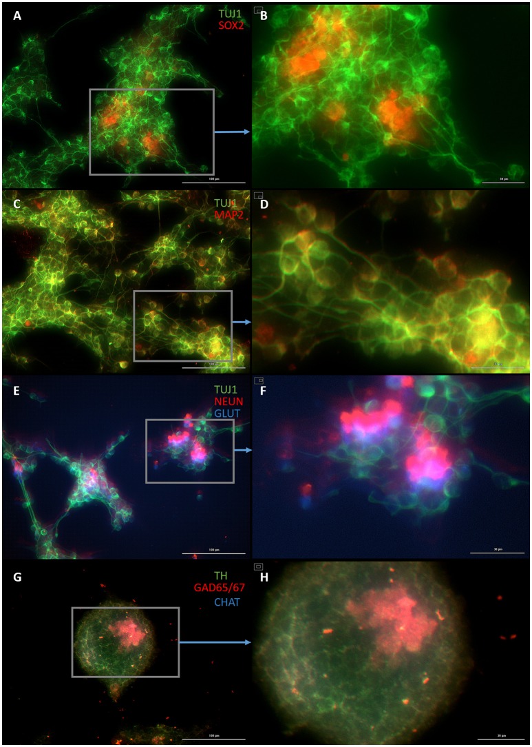
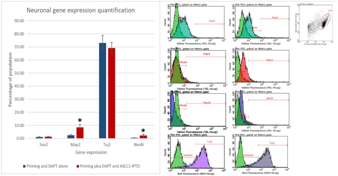
Cellular transdifferentiation changes mature cells from one phenotype into another by altering their gene expression patterns. Manipulating expression of transcription factors, proteins that bind to DNA promoter regions, regulates the levels of key developmental genes. Viral delivery of transcription factors can efficiently reprogram somatic cells, but this method possesses undesirable side effects, including mutations leading to oncogenesis. Using protein transduction domains (PTDs) fused to transcription factors to deliver exogenous transcription factors serves as an alternative strategy that avoids the issues associated with DNA integration into the host genome. However, lysosomal degradation and inefficient nuclear localization pose significant barriers when performing PTD-mediated reprogramming. Here, we investigate a novel PTD by placing a secretion signal sequence next to a cleavage inhibition sequence at the end of the target transcription factor-achaete scute homolog 1 (ASCL1), a powerful regulator of neurogenesis, resulting in superior stability and nuclear localization. A fusion protein consisting of the amino acid sequence of ASCL1 transcription factor with this novel PTD added can transdifferentiate cerebral cortex astrocytes into neurons. Additionally, we show that the synergistic action of certain small molecules improves the efficiency of the transdifferentiation process. This study serves as the first step toward developing a clinically relevant in vivo transdifferentiation strategy for converting astrocytes into neurons.

Keywords: drug delivery; neuroscience; reprogramming; small molecules; transcription factors.


Figures

Figure 1
Figure 1
Mouse embryonic fibroblasts (MEFs) express the skeletal myoblast gene myosin heavy chain 3 (MYH3) and lack the neuronal gene beta tubulin (TUJ1) after 14 days of exposure to ASCL1-IPTD and the cAMP activator forskolin, indicating that they have adopted a myogenic cell fate rather than a neuronal cell fate. (A) Control. Scale bar is 100 μm. (B) High magnification of (A). Scale bar is 10 μm. (C) Forskolin only. Scale bar is 100 μm. (D) High magnification of (C). Scale bar is 10 μm. (E) ASCL1-IPTD only. Scale bar is 100 μm. (F) High magnification of (E). Scale bar is 10 μm. (G) ASCL1-IPTD with forskolin. Scale bar is 100 μm. (H) High magnification of (H). Scale bar is 10 μm.
Figure 2
Figure 2
Small molecule screening of combinations of forskolin, ISX9, DAPT and CHIR99021 with ASCL1-IPTD show that DAPT is the most potent inducer of neural transdifferentiation. (A) Cells express beta tubulin (TUJ1), an early neuronal marker, and ASCL1 after 2 days of priming with LDN193189 and SB431542 and ASCL1-IPTD followed by 10 days of treatment with ASCL1-IPTD and DAPT. Scale bar is 100 μm. (B) High magnification of (A). Scale bar is 20 μm. (C) Cell survival varied in each condition, with the DAPT alone condition being the least toxic by far (n = 3). (D) Neurons counted from each small molecule combination on day 12 (n = 3). (E) Neuronal conversion percentages. (F) Protocol timeline: 2 days of priming with SMAD inhibitors LDN193189 and SB431542 followed by 10 days of small molecule combinations. All 12 days included the addition of ASCL1-IPTD. Statistical analysis was performed by a one-way ANOVA followed by a one-tailed Student's t-test with a 95% confidence level (α = 0.05). Results are presented as the mean ± standard deviation, with a * representing a statistically significant difference between that condition and the DAPT alone condition.
Figure 3
Figure 3
NGN2-IPTD in place of ASCL1-IPTD does not generate neurons, but does produce cells expressing doublecortin (DCX), a marker for neuroblasts. (A) Control. Scale bar is 100 μm. (B) Small molecules only. Scale bar is 100 μm. (C) NGN2-IPTD plus small molecules. Scale bar is 100 μm. (D) High magnification image of (C). Scale bar is 10 μm. Note that TUJ1 is weakly expressed in astrocytes in addition to neurons and can be regarded as background stain in these images.
Figure 4
Figure 4
Small molecule only group after 2 days of priming by LDN193189 and SB431542 followed by 10 days of treatment with DAPT. (A) Cells express the early neural marker TUJ1 and weakly express the neural stem cell marker SOX2. Scale bar is 100 μm. (B) High magnification of (A). Scale bar is 30 μm. (C) Cells are negative for the mature neural marker MAP2. Scale bar is 100 μm. (D) High magnification of (C). Scale bar is 10 μm (E) Cells are negative for the mature neural marker NEUN and the neurotransmitter glutamate expressed by excitatory interneurons GLUT. Scale bar is 100 μm. (F) High magnification of (E). Scale bar is 20 μm. (G) Cells are negative for the neurotransmitter tyrosine hydroxylase expressed by dopaminergic neurons TH, the neurotransmitter glutamic acid decarboxylase expressed by inhibitory interneurons GAD65/67, and the neurotransmitter choline acetyltransferase expressed by motor neurons CHAT. Scale bar is 100 μm. (H) High magnification of (G). Scale bar is 30 μm. Note that some fluorescence is picked up by all the cells producing background fluorescence which can be ignored.
Figure 5
Figure 5
Plastic astrocytes generate neurospheres and mature neurons after 12 days of exposure to ASCL1-IPTD, including 2 days of priming by LDN193189 and SB431542 followed by 10 days of DAPT. (A) Cells express the early neural marker TUJ1 and the neural stem cell marker SOX2. Scale bar is 100 μm. (B) High magnification of (A). Scale bar is 30 μm. (C) Cells express the mature neural marker MAP2. Scale bar is 100 μm. (D) High magnification of (C). Scale bar is 30 μm (E) Cells express the mature neural marker NEUN and the neurotransmitter glutamate expressed by excitatory interneurons GLUT. Scale bar is 100 μm. (F) High magnification of (E). Scale bar is 30 μm. (G) Cells are negative for the neurotransmitter tyrosine hydroxylase expressed by dopaminergic neurons TH, and negative for the neurotransmitter choline acetyltransferase expressed by motor neurons CHAT, but do express the neurotransmitter glutamic acid decarboxylase expressed by inhibitory interneurons GAD65/67. Scale bar is 100 μm. (H) High magnification of (G). Scale bar is 30μm. Note that some fluorescence is picked up by all the cells producing background fluorescence which can be ignored.
Figure 6
Figure 6
Quantification of neural gene expression for the immature neural marker TUJ1 and the mature neuronal markers MAP2 and NEUN by flow cytometry. Plastic astrocytes were exposed to 2 days of priming by LDN193189 and SB431542 followed by 10 days of exposure to DAPT, with or without ASCL1-IPTD. Results show that TUJ1 expression is similar in both groups, whereas MAP2 and NEUN expression are significantly upregulated in the ASCL1-IPTD group. Statistical analysis was performed by a one-way ANOVA followed by a one-tailed Student's t-test with a 95% confidence level (α = 0.05). Results are presented as the mean ± standard deviation, with a * representing a statistically significant difference between the ASCL1-IPTD group with the DAPT Alone group.

References

    1. Abe K. (2000). Therapeutic potential of neurotrophic factors and neural stem cells against ischemic brain injury. J. Cereb. Blood Flow Metab. 20, 1393–1408. 10.1097/00004647-200010000-00001 - DOI - PubMed
    1. Addis R. C., Hsu F.-C., Wright R. L., Dichter M. A., Coulter D. A., Gearhart J. D. (2011). Efficient conversion of astrocytes to functional midbrain dopaminergic neurons using a single polycistronic vector. PLoS ONE 6:e28719. 10.1371/journal.pone.0028719 - DOI - PMC - PubMed
    1. Agbay A., De La Vega L., Nixon G., Willerth S. (2018). Guggulsterone-releasing microspheres direct the differentiation of human induced pluripotent stem cells into neural phenotypes. Biomed. Mater. 13:034104. 10.1088/1748-605X/aaaa77 - DOI - PubMed
    1. Ambasudhan R., Talantova M., Coleman R., Yuan X., Zhu S., Lipton S. A., et al. . (2011). Direct reprogramming of adult human fibroblasts to functional neurons under defined conditions. Cell Stem Cell 9, 113–118. 10.1016/j.stem.2011.07.002 - DOI - PMC - PubMed
    1. Berninger B., Costa M. R., Koch U., Schroeder T., Sutor B., Grothe B., et al. . (2007). Functional properties of neurons derived from in vitro reprogrammed postnatal astroglia. J. Neurosci. 27, 8654–8664. 10.1523/JNEUROSCI.1615-07.2007 - DOI - PMC - PubMed

LinkOut - more resources