. 2018 Aug 24:9:58-70.
doi: 10.1016/j.reth.2018.06.003. eCollection 2018 Dec.

Application of induced pluripotent stem cells for cartilage regeneration in CLAWN miniature pig osteochondral replacement model

Affiliations

Application of induced pluripotent stem cells for cartilage regeneration in CLAWN miniature pig osteochondral replacement model

Sakura Uto et al. Regen Ther. .

Abstract

Introduction: Pluripotent stem cells have an advantage that they can proliferate without reduction of the quality, while they have risk of tumorigenesis. It is desirable that pluripotent stem cells can be utilized safely with minimal effort in cartilage regenerative medicine. To accomplish this, we examined the potential usefulness of induced pluripotent stem cells (iPS cells) after minimal treatment via cell isolation and hydrogel embedding for cartilage regeneration using a large animal model.

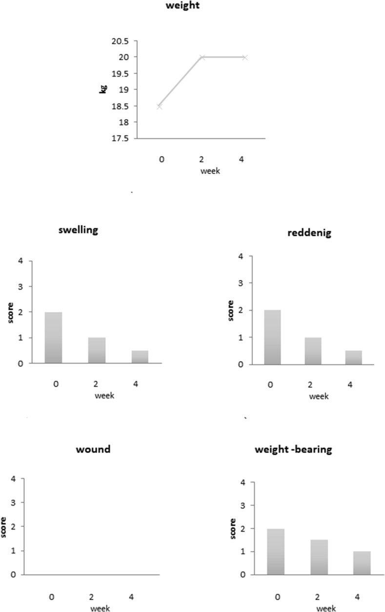
Methods: Porcine iPS-like cells were established from the CLAWN miniature pig. In vitro differentiation was examined for porcine iPS-like cells with minimal treatment. For the osteochondral replacement model, osteochondral defect was made in the quarters of the anteromedial sides of the proximal tibias in pigs. Porcine iPS-like cells and human iPS cells with minimal treatment were seeded on scaffold made of thermo-compression-bonded beta-TCP and poly-L-lactic acid and transplanted to the defect, and cartilage regeneration and tumorigenesis were evaluated.

Results: The in vitro analysis indicated that the minimal treatment was sufficient to weaken the pluripotency of the porcine iPS-like cells, while chondrogenic differentiation did not occur in vitro. When porcine iPS-like cells were transplanted into osteochondral replacement model after minimal treatment in vitro, cartilage regeneration was observed without tumor formation. Additionally, fluorescent in situ hybridization (FISH) indicated that the chondrocytes in the regenerative cartilage originated from transplanted porcine iPS-like cells. Transplantation of human iPS cells also showed the regeneration of cartilage in miniature pigs under immunosuppressive treatment.

Conclusion: Minimally-treated iPS cells will be a useful cell source for cartilage regenerative medicine.

Keywords: Cartilage regeneration; Minimal treatment; Osteochondral replacement model; iPS cells.


Figures

Fig. 1
Fig. 1
Experimental design of in vivo study. (A) Three-dimensional image of the bone and cartilage defect. (B, C) Three-dimensional images of the PLLA (B) and the beta-TCP (C) scaffolds formed to fit the defect in the knee joint. (D) Three-dimensional image of thermo-compression-bonded beta-TCP and PLLA. (E) A Scheme of the preparation of the transplant. Cells were singly isolated and embedded in collagen. The mixture of cells and collagen was administered to PLLA layer. Following administration, collagen was gelatinized. (F) Gross appearance of the transplant. (G) The exposed surgical site. (H) The defect formed at the anteromedial quarter of the tibial side of the knee joint. (I) The surgical site after transplantation. The transplant was placed at the defect and fixed with thin titan mesh plates.
Fig. 2
Fig. 2
Characterization of porcine iPS-like cells. (A) Alkaline phosphatase staining of colonies of porcine iPS-like cells. (B, C) Real-time RT-PCR analyses of pluripotent markers NANOG and OCT3/4 (B) and transgenes oct3/4, sox2, klf4, and c-myc (C) in porcine iPS-like cells and fibroblasts. (D) RT-PCR analyses of early mesodermal marker BRACHYURY in porcine iPS-like cells with or without induction of mesodermal differentiation (D).
Fig. 3
Fig. 3
Gene expression of the porcine cells cultured in 3D chondrogenic condition determined by real-time RT-PCR. MSCs (A) and porcine iPS-like cells (B) were three-dimensionally cultured in chondrogenic differentiation medium and mRNA from each cells was subjected to real time RT-PCR. All values are presented as mean plus standard deviation of 3 samples per group. Statistical analysis was done by Dunnett's test (*p < 0.05, **p < 0.01 versus plate).
Fig. 4
Fig. 4
Physiological findings of the pigs after transplantation. β-TCP: β-TCP scaffold only, MSC: scaffolds (β-TCP + PLLA) with porcine MSCs, iPS-like: scaffolds (β-TCP + PLLA) with porcine iPS-like cells. Values are means of the scores from 2 animals for each group.
Fig. 5
Fig. 5
Macroscopic findings of knee joints 8 weeks after transplantation with porcine cell-based transplants. Tibial (A, B, C, G, H, I) and femoral (D, E, F, J, K, L) sides of knee joints transplanted with the beta-TCP scaffold only (A, D, G, J), the beta-TCP/PLLA scaffold with MSCs (B, E, H, K) and the beta-TCP/PLLA scaffold with porcine iPS-like cells (C, F, I, L) and higher magnifications of each samples indicated by squares (A', B', C', D', E', F', G', H', I', J', K', L'). Granulations are indicated by arrows.
Fig. 6
Fig. 6
Histological findings of the transplantation sites. (A) Hematoxylin-eosin staining of the unaffected cartilage (a, e), site with the beta-TCP scaffold only (b, f, i), the beta-TCP/PLLA scaffold with MSCs (c, g, j) and the beta-TCP/PLLA scaffold with porcine iPS-like cells (d, h, k). Scale bars = 1 mm for upper panels and 100 μm for lower panels. (B) FISH analysis. HE staining (upper panels) and FISH (lower panels) for unaffected cartilage (l, m) and regenerated cartilages with of the transplant of the beta-TCP/PLLA scaffold with porcine iPS-like cells (n, o).
Fig. 7
Fig. 7
Physiological findings of the pigs after transplantation of human iPS cells. Scaffolds: (beta-TCP + PLLA). Values are means of the scores from 2 animals for each group.
Fig. 8
Fig. 8
Macroscopic and histological findings of knee joints 4 weeks after transplantation with human iPS cell-based transplants. Tibial (A) and femoral (B) sides of knee joint transplanted with beta-TCP/PLLA scaffold with human iPS cells. Granulation is indicated by an arrow. (C) Hematoxylin-Eosin staining of regenerative cartilage. (D) Magnification of squared are in figure D. (E) FISH analysis of the same area as figure E. Scale bars = 100 μm.

References

    1. Takahashi K., Tanabe K., Ohnuki M., Narita M., Ichisaka T., Tomoda K. Induction of pluripotent stem cells from adult human fibroblasts by defined factors. Cell. 2007;131:861–872. - PubMed
    1. Khoo M.L., Shen B., Tao H., Ma D.D. Long-term serial passage and neuronal differentiation capability of human bone marrow mesenchymal stem cells. Stem Cells Dev. 2008;17:883–896. - PubMed
    1. Tanaka Y., Yamaoka H., Nishizawa S., Nagata S., Ogasawara T., Asawa Y. The optimization of porous polymeric scaffolds for chondrocyte/atelocollagen based tissue-engineered cartilage. Biomaterials. 2010;31:4506–4516. - PubMed
    1. Yamaoka H., Nishizawa S., Asawa Y., Fujihara Y., Ogasawara T., Yamaoka K. Involvement of fibroblast growth factor 18 in dedifferentiation of cultured human chondrocytes. Cell Prolif. 2010;43:67–76. - PMC - PubMed
    1. Oldershaw R.A., Baxter M.A., Lowe E.T., Bates N., Grady L.M., Soncin F. Directed differentiation of human embryonic stem cells toward chondrocytes. Nat Biotechnol. 2010;28(11):1187–1194. - PubMed

LinkOut - more resources