Review
. 2019 Feb 15;199(4):433-445.
doi: 10.1164/rccm.201810-1944CI.

Role of Biologics in Asthma

Affiliations
Review

Role of Biologics in Asthma

Mary Clare McGregor et al. Am J Respir Crit Care Med. .

Abstract

Patients with severe uncontrolled asthma have disproportionally high morbidity and healthcare utilization as compared with their peers with well-controlled disease. Although treatment options for these patients were previously limited, with unacceptable side effects, the emergence of biologic therapies for the treatment of asthma has provided promising targeted therapy for these patients. Biologic therapies target specific inflammatory pathways involved in the pathogenesis of asthma, particularly in patients with an endotype driven by type 2 (T2) inflammation. In addition to anti-IgE therapy that has improved outcomes in allergic asthma for more than a decade, three anti-IL-5 biologics and one anti-IL-4R biologic have recently emerged as promising treatments for T2 asthma. These targeted therapies have been shown to reduce asthma exacerbations, improve lung function, reduce oral corticosteroid use, and improve quality of life in appropriately selected patients. In addition to the currently approved biologic agents, several biologics targeting upstream inflammatory mediators are in clinical trials, with possible approval on the horizon. This article reviews the mechanism of action, indications, expected benefits, and side effects of each of the currently approved biologics for severe uncontrolled asthma and discusses promising therapeutic targets for the future.

Keywords: asthma treatments; biologics; eosinophils; monoclonal antibodies; severe asthma.


Figures

Figure 1.
Figure 1.
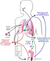
Schematic of the immunopathobiology of asthma with sites of the targeted treatments with approved and investigational monoclonal antibodies marked. In asthma, the interaction of genetic susceptibility and environmental exposures—such as with allergens, viruses, pollutants, and irritants—creates airway inflammation. In type 2 (T2) asthma, the interaction of environmental exposures with the airway epithelium leads to the release of the mediators IL-33, IL-25, and TSLP (thymic stromal lymphopoietin). In addition, allergens are taken up by dendritic cells and presented to naive T-helper (Th0) cells. A cascade of events as shown ensues that leads to production of the type 2 cytokines IL-4, IL-5, and IL-13; secretion of IgE by B cells; and chemoattraction of mast cells, eosinophils, and basophils. This process lends itself to numerous therapeutic targets that have already been approved by the U.S. Food and Drug Administration (outlined in red) and others that remain in investigation (outlined in green). CRTh2 = chemoattractant receptor–homologous molecule expressed on T2 cells; DP-1 = prostaglandin D2 receptor type 1; ILC2 = innate lymphoid cell type 2; M2 macrophage = alternatively activated macrophage; SM = smooth muscle. Modified by permission from Reference from Sanofi.
Figure 2.
Figure 2.
Effect of omalizumab on (A) rate of asthma exacerbations and (B) lung function: the EXTRA trial (28) and INNOVATE study (27). (A) In the EXTRA trial, when evaluated over 48 weeks, treatment with omalizumab reduced the rate of asthma exacerbations by 25% (95% confidence interval, 8‒39%). (B) In the INNOVATE study, when evaluated over a 28-week period, treatment with omalizumab improved lung function, as measured by the least squares (LS) mean change in FEV1, by 94 ml more than placebo (P = 0.043). *P < 0.05; **P < 0.01.
Figure 3.
Figure 3.
Effect of mepolizumab on (A) oral corticosteroid reduction, (B) asthma exacerbations, and (C) lung function: the SIRIUS trial. (43). (A) In the SIRIUS trial, the reduction in steroid dosing at 24 weeks was 50% in the group receiving mepolizumab compared with 0% in the placebo group (P = 0.007). (B) At 24 weeks, there was a relative reduction of 32% in the cumulative number of asthma exacerbations with mepolizumab (P = 0.04). (C) Based on the Asthma Control Questionnaire 5 (ACQ-5), mepolizumab resulted in improvement in asthma control at 2 weeks that was sustained through the 24-week trial (P = 0.004). Error bars show 95% confidence intervals around the least squares mean.
Figure 4.
Figure 4.
Effect of reslizumab on (A) rate of asthma exacerbations and (B) lung function: the BREATHE trials (48). (A) In two separate trials, reslizumab reduced the rate of asthma exacerbations with a pooled relative reduction in the asthma exacerbation rate of 54% (95% confidence interval, 42–63%) over 52 weeks. (B) Additionally, reslizumab improved lung function as measured by FEV1 by 110 ml (95% confidence interval, 67–150 ml) more than placebo. **P < 0.01; ***P < 0.001. LS = least squares; q4w = every 4 weeks.
Figure 5.
Figure 5.
Effect of benralizumab on (A) time to first asthma exacerbation and (B) oral corticosteroid reduction: the ZONDA trial (55). (A) In the ZONDA trial, benralizumab dosed every 4 weeks or every 8 weeks led to a median percentage reduction from baseline in steroid requirement of 75% compared with a 25% reduction with placebo (P < 0.001). (B) As shown using a Kaplan-Meier cumulative incidence curve, benralizumab was associated with a longer time to first asthma exacerbation when administered every 4 weeks (hazard ratio [HR], 0.39; 95% confidence interval [CI], 0.22–0.66) or every 8 weeks (HR, 0.32; CI, 0.17–0.57). Error bars show 95% confidence intervals around the least squares mean.
Figure 6.
Figure 6.
Schematic of airway (luminal) eosinophils and eosinophilopoeitic factors with anti–IL-5 therapy in severe eosinophilic asthma. In response to a variety of airway stimuli (such as allergens, microbes, pollutants, etc.), CD4 (classic T-helper cell type 2 [Th2] response) or non-CD4 cells (such as type 2 innate lymphoid cells [ILC2] in the airways, nonclassic T2 response), secrete eosinophilopoeitic cytokines such as IL-5 and IL-13. In patients with severe asthma who are on high doses of systemic corticosteroids, airway ILC2 cells may dominate over CD4 cells as the predominant source of IL-5 and IL-13. Although IL-13 can prime the migrational response of eosinophil progenitor cells from the bone marrow into the lung in response to SDF-1 (stromal-derived factor-1), locally derived IL-5 can promote their “in situ differentiation” into mature eosinophils. Pharmacokinetic data of airway levels of biologics have never been evaluated. Clinically relevant doses have been selected based on mathematical modeling of airway levels of drugs from studies in normal volunteers or subjects with mild asthma and from pharmacodynamics studies guided by absolute blood eosinophil levels. In order to suppress airway eosinophils, treatment options may include higher levels of anti–IL-5 neutralizing monoclonal antibodies such as reslizumab or mepolizumab, or inhibition of the migration of progenitor cells into the airways (e.g., anti–IL-4R or antialarmins such as anti-TSLP [thymic stromal lymphopoietin] or anti–IL-33), or depletion of both mature and immature eosinophils and possibly the ILC2 cells (those that express IL-5R) by the antibody-dependent cell–mediated cytotoxicity action of benralizumab. eos = eosinophils; GM-CSF = granulocyte–macrophage colony–stimulating factor. Illustration by Patricia Ferrer Beals.
Figure 7.
Figure 7.
Effect of dupilumab on (A) percentage reduction in oral corticosteroid dosing and (B) lung function: the VENTURE trial (72). (A) In the VENTURE trial, the least squares mean percentage reduction in oral corticosteroid dosing in patients receiving dupilumab at 24 weeks was −70.1% (SE of ±4.9%) compared with −41.9% (SE of ±4.5%) in the group treated with placebo (P < 0.001). (B) At 24 weeks, dupilumab treatment resulted in a 220-ml (95% confidence interval, 90–340 ml) improvement in FEV1 compared with placebo. Error bars show the SE.

Comment in

References

    1. To T, Stanojevic S, Moores G, Gershon AS, Bateman ED, Cruz AA, et al. Global asthma prevalence in adults: findings from the cross-sectional world health survey. BMC Public Health. 2012;12:204. - PMC - PubMed
    1. Moore WC, Bleecker ER, Curran-Everett D, Erzurum SC, Ameredes BT, Bacharier L, et al. National Heart, Lung, Blood Institute’s Severe Asthma Research Program. Characterization of the severe asthma phenotype by the National Heart, Lung, and Blood Institute’s Severe Asthma Research Program. J Allergy Clin Immunol. 2007;119:405–413. - PMC - PubMed
    1. Chung KF, Wenzel SE, Brozek JL, Bush A, Castro M, Sterk PJ, et al. International ERS/ATS guidelines on definition, evaluation and treatment of severe asthma Eur Respir J 201443343–373.[Published erratum appears in Eur Respir J 52:1352020.] - PubMed
    1. Antonicelli L, Bucca C, Neri M, De Benedetto F, Sabbatani P, Bonifazi F, et al. Asthma severity and medical resource utilisation. Eur Respir J. 2004;23:723–729. - PubMed
    1. Hyland ME, Whalley B, Jones RC, Masoli M. A qualitative study of the impact of severe asthma and its treatment showing that treatment burden is neglected in existing asthma assessment scales. Qual Life Res. 2015;24:631–639. - PubMed

MeSH terms