Review
. 2019 Jun 4:21:1-31.
doi: 10.1146/annurev-bioeng-060418-052130. Epub 2018 Dec 10.

Exploring Dynamics and Structure of Biomolecules, Cryoprotectants, and Water Using Molecular Dynamics Simulations: Implications for Biostabilization and Biopreservation

Affiliations
Review

Exploring Dynamics and Structure of Biomolecules, Cryoprotectants, and Water Using Molecular Dynamics Simulations: Implications for Biostabilization and Biopreservation

Lindong Weng et al. Annu Rev Biomed Eng. .

Abstract

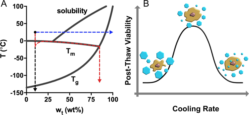
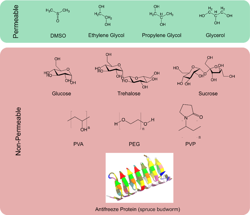
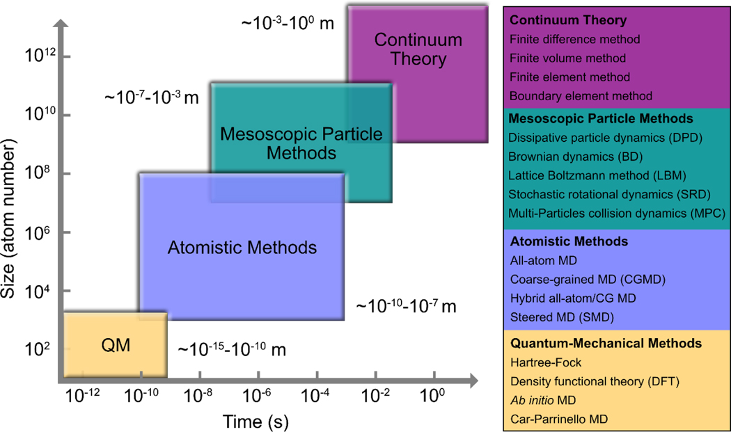
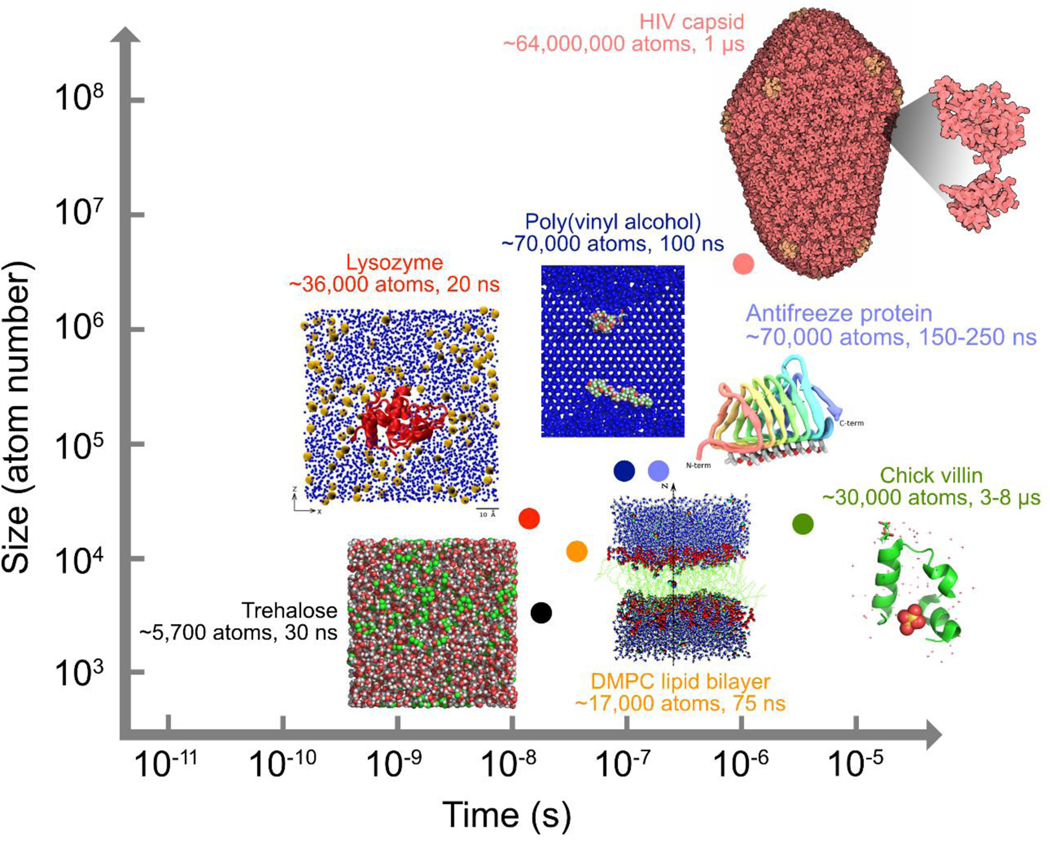
Successful stabilization and preservation of biological materials often utilize low temperatures and dehydration to arrest molecular motion. Cryoprotectants are routinely employed to help the biological entities survive the physicochemical and mechanical stresses induced by cold or dryness. Molecular interactions between biomolecules, cryoprotectants, and water fundamentally determine the outcomes of preservation. The optimization of assays using the empirical approach is often limited in structural and temporal resolution, whereas classical molecular dynamics simulations can provide a cost-effective glimpse into the atomic-level structure and interaction of individual molecules that dictate macroscopic behavior. Computational research on biomolecules, cryoprotectants, and water has provided invaluable insights into the development of new cryoprotectants and the optimization of preservation methods. We describe the rapidly evolving state of the art of molecular simulations of these complex systems, summarize the molecular-scale protective and stabilizing mechanisms, and discuss the challenges that motivate continued innovation in this field.

Keywords: anhydrobiosis; cryopreservation; hydrogen bond; molecular modeling; protein stabilization; trehalose.


Figures

Figure 1.
Figure 1.
Preservation pathways for a trehalose solution (initial condition: 10 wt% and 25 °C) and the two-factor hypothesis for slow-freezing of living cells. (A) In a slow-freezing method, ice crystallization drives the unfrozen trehalose solution to follow the liquidus curve until it crosses the Tg (red). Direct vitrification strategy brings the temperature of the solution below Tg ultra-rapidly in the absence of ice formation (black). Isothermal vitrification removes water from the solution until the concentration yields a Tg that is below 25 °C (blue). (B) High cooling rates may be associated with intracellular ice formation that causes mechanical disruption of membranes and organelles; Slow cooling rates may result in excessive cell dehydration and prolonged exposure of cells to a high electrolyte concentration. There is an optimal cooling rate at which the two mechanisms of damage are balanced, yielding the highest post-thaw viability.
Figure 2.
Figure 2.
Representative permeable or non-permeable cryoprotectants that have been widely employed for the stabilization and preservation of a variety of biologics.
Figure 3.
Figure 3.
The hierarchy of size and time scales covered by various computational modeling techniques
Figure 4.
Figure 4.
Size and time scales covered by representative MD simulations for biopreservation and some state-of-the-art biomolecular simulations beyond the field. Inset pictures were adapted from References [33], [64], [67], [106] and [114] under the Creative Commons Attribution license or with permission from American Chemical Society. Images of HIV capsid and chick villin were adapted from entries 3J3Q and 1YRF deposited at the Protein Data Bank (PDB).
Figure 5.
Figure 5.
Shares of “bound” and “free” water in trehalose-water mixtures at 295 K (upper panel); Snapshots of the MD simulation box showing the water or trehalose percolation at each of the trehalose–water concentrations under investigation (lower panel, 295 K). This figure was created using the raw data from Ref (33).
Figure 6.
Figure 6.
(A) Snapshot of a typical 1Me2SO–2H2O aggregate in which one Me2SO molecule acting as the hydrogen-bond acceptor forms hydrogen bonds with two water molecules. The 1Me2SO-2H2O structure was adapted with permission from Ref (45) Copyright (1992) American Chemical Society. (B) Snapshot of a 2Me2SO–1H2O aggregate which includes a central water molecule acting as the hydrogen-bond donor and two Me2SO molecules being the hydrogen-bond acceptors. The image was adapted from Ref (39), with the permission of AIP Publishing. (Color index: grey-carbon, orange-sulfur, red-oxygen, and white-hydrogen)
Figure 7.
Figure 7.
Snapshots of the simulation boxes of MgCl2–glycerol (left) and NaCl-glycerol (right) mixtures, respectively, at 560 K. The molar ratio of electrolyte to glycerol is 2. Only Mg2+ (green), Na+ (yellow), and Cl (grey) are shown. While the MgCl2–glycerol mixture is homogeneous, the NaCl–glycerol mixture has become heterogeneous with indication of NaCl crystallization. This figure was created using the raw data from Ref (63).
Figure 8.
Figure 8.
Role of PVA in inhibiting ice recrystallization. (A) An atactic PVA20 chain with a random arrangement of the −OH groups. (B) Illustration of the Gibbs–Thomson effect due to the PVA20–ice binding with two adjacent periodic images joined together. (C) Geometrical match between some of the −OH groups of PVA20 (indicated by the black arrows) and the lattice of ice front. Reprinted with permission from Ref (64). Copyright (2017) American Chemical Society.
Figure 9.
Figure 9.
Cartoon structure of lipid bilayers and chemical structures of three widely-studied phospholipids (POPC, DOPC, and DPPC). The liquid-crystalline phase features randomly oriented and fluid acyl chains. Below the phase transition temperature, lipid bilayers change into the gel phase in which acyl chains are fully extended and closely packed.
Figure 10.
Figure 10.
Distinct modes of action of Me2SO on phospholipid membranes (DPPC). Presented are side views of the final structures for the bilayer systems containing 0, 5, 10, and 40 mol% of Me2SO (lipid-free basis). Lipids are shown in cyan, water in red, and Me2SO in yellow. Reprinted with permission from Ref (103). Copyright (2007) American Chemical Society.
Figure 11.
Figure 11.
Hypothesized mechanisms about cryoprotectants protecting proteins against damages induced by freezing or dehydration. (Orange circles represent cryoprotectant molecules like trehalose and blue ones represent water molecules.)

References

    1. Zylberberg C, Matosevic S. 2016. Pharmaceutical liposomal drug delivery: a review of new delivery systems and a look at the regulatory landscape. Drug Deliv. 23:3319–29 - PubMed
    1. Leader B, Baca QJ, Golan DE. 2008. Protein therapeutics: a summary and pharmacological classification. Nat. Rev. Drug Discov 7:21–39 - PubMed
    1. Mazur P. 1970. Cryobiology: the freezing of biological systems. Science 168:939–49 - PubMed
    1. Acker JP, McGann LE. 2002. Innocuous intracellular ice improves survival of frozen cells. Cell Transplant. 11:563–71 - PubMed
    1. Rall WF, Fahy GM. 1985. Ice-free cryopreservation of mouse embryos at −196 C by vitrification. Nature 313:573–75 - PubMed

Supplementary concepts

LinkOut - more resources