

Contrast sensitivity deficits in patients with mutation-proven inherited retinal degenerations
- PMID: 30526558
- PMCID: PMC6286564
- DOI: 10.1186/s12886-018-0982-0
Contrast sensitivity deficits in patients with mutation-proven inherited retinal degenerations
Abstract
Background: Patients with retinal diseases frequently complain of poor visual function even when visual acuity is relatively unaffected. This clinical finding has been attributed to deficits in contrast sensitivity (CS). The purpose of our study was to evaluate the CS in patients with clinical and genetic diagnosis of inherited retinal degeneration (IRD) and relatively preserved visual acuity.
Methods: Seventeen patients (30 eyes) with IRD and visual acuity of 20/40 or better, and 18 controls (18 eyes) without any ocular condition underwent slit lamp examination, visual acuity testing via standard Snellen chart testing, CS testing via the Quick Contrast Sensitivity Function (QCSF), and Spectral Domain Optical Coherence Tomography (SD-OCT). CS were measured at 1.0, 1.5, 3.0, 6.0, 12.0, and 18.0 cycles per degree (cpd). T tests with general estimated equations were used to compare CS between groups. Wald chi square followed by pairwise comparisons was used to compare CS between multiple groups.
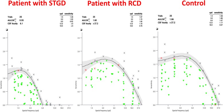
Results: We included 12 patients with rod-cone dystrophy (RCD), 3 patients with Stargardt disease (STGD) and 2 patients with Best disease. Patients with IRD had significantly worse CS than controls (p < 0.001) in all spatial frequencies. Patients with STGD had more marked deficits in CS than patients with Best disease (p < 0.001) and RCD (p < 0.001) despite having similar visual acuities.
Conclusion: Patients with IRD, especially patients with STGD with relatively preserved visual acuity have marked deficits in CS when measured across a range of spatial frequencies. We recommend that clinical trials for STGD incorporate CS measured over a range of spatial frequencies as a secondary clinical endpoint for monitoring visual function. CS may provide an explanation for complaints of visual dysfunction when visual acuity is not significantly altered.
Keywords: Best disease; Contrast sensitivity; Retinal dystrophy; Retinitis Pigmentosa; Stargardt disease.
Conflict of interest statement
Ethics approval and consent to participate
This study was approved by the University of Michigan Medical School IRB before any testing was done. The study was performed according to the tenets of the declaration of Helsinki, and the IRB number is HUM 12099. Written informed consent was obtained before all subjects participated.
Consent for publication
Not Applicable.
Competing interests
The authors declare that they have no competing interests.
Publisher’s Note
Springer Nature remains neutral with regard to jurisdictional claims in published maps and institutional affiliations.
Figures
References
-
- Bainbridge JW, Mehat MS, Sundaram V, Robbie SJ, Barker SE, Ripamonti C, Georgiadis A, Mowat FM, Beattie SG, Gardner PJ, Feathers KL, Luong VA, Yzer S, Balaggan K, Viswanathan A, de Ravel TJ, Casteels I, Holder GE, Tyler N, Fitzke FW, Weleber RG, Nardini M, Moore AT, Thompson DA, Petersen-Jones SM, Michaelides M, van den Born LI, Stockman A, Smith AJ, Rubin G and Ali RR (2015). “Long-term effect of gene therapy on Leber's congenital amaurosis.” N Engl J Med 372(20): 1887–1897.. - PMC - PubMed
MeSH terms
Grants and funding
LinkOut - more resources
Full Text Sources
