. 2018 Dec 6;103(6):1022-1029.
doi: 10.1016/j.ajhg.2018.10.023.

Aberrant Inclusion of a Poison Exon Causes Dravet Syndrome and Related SCN1A-Associated Genetic Epilepsies

Collaborators, Affiliations

Aberrant Inclusion of a Poison Exon Causes Dravet Syndrome and Related SCN1A-Associated Genetic Epilepsies

Gemma L Carvill et al. Am J Hum Genet. .

Abstract

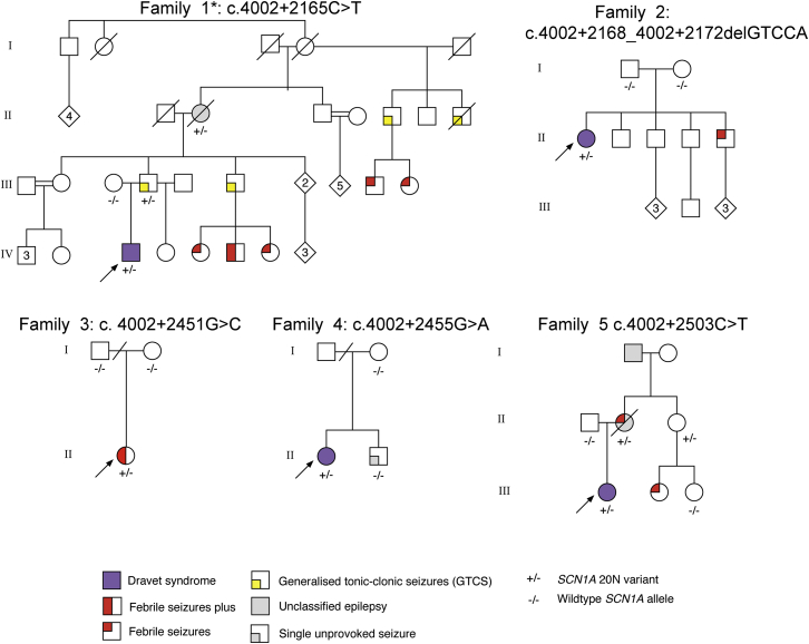
Developmental and epileptic encephalopathies (DEEs) are a group of severe epilepsies characterized by refractory seizures and developmental impairment. Sequencing approaches have identified causal genetic variants in only about 50% of individuals with DEEs.1-3 This suggests that unknown genetic etiologies exist, potentially in the ∼98% of human genomes not covered by exome sequencing (ES). Here we describe seven likely pathogenic variants in regions outside of the annotated coding exons of the most frequently implicated epilepsy gene, SCN1A, encoding the alpha-1 sodium channel subunit. We provide evidence that five of these variants promote inclusion of a "poison" exon that leads to reduced amounts of full-length SCN1A protein. This mechanism is likely to be broadly relevant to human disease; transcriptome studies have revealed hundreds of poison exons,4,5 including some present within genes encoding other sodium channels and in genes involved in neurodevelopment more broadly.6 Future research on the mechanisms that govern neuronal-specific splicing behavior might allow researchers to co-opt this system for RNA therapeutics.

Keywords: Dravet syndrome; SCN1A; alternative splicing; epilepsy; genome sequencing; noncoding; poison exon; variant interpretation.


Figures

Figure 1
Figure 1
Location of Intron 20 Variants and Impact on Poison Exon 20N Splicing (A) The region spanning SCN1A exon 20 through 21, highlighting the high level of conservation as illustrated by the GERP (Genomic Evolutionary Rate Profiling) score across the intron 20 region. Positive GERP scores indicate that the position is probably under evolutionary constraint, whereas negative GERP scores indicate that the site is probably evolving with neutral constraint. GERP scores are calculated based on conservation of 35 mammalian species., We selected three highly conserved regions for targeted resequencing (black bars = target regions); one of these regions (blue highlighted region) contains the 20N poison exon (purple). The level of conservation of this exon, and the genomic region surrounding it, is comparable to the known SCN1A coding exons (blue bars). The five intron 20 variants are shown in red, and the distance relative to exon 20N is shown below each proband number. SCN1A is transcribed in the reverse orientation in the human genome. This figure was modified from the UCSC Browser. (B) Three of the identified variants cause increased inclusion of the poison exon 20N, as measured via a splicing reporter in K562 cells. A splicing reporter containing SCN1A exon 20, intron 20, and exon 21 was generated with WT and each of the proband-specific variants. The reporter was transfected into K562 cells in triplicate and harvested after 24 hours. Then RTqPCR was used for measuring the amount of correctly spliced SCN1A (SCN1A exon 20:exon 21), 20N inclusion (SCN1A 20N:exon 21 and exon 20:20N), and GAPDH (a housekeeper). (Biological replicates n = 3, technical qPCR replicates n = 3). These data clearly show that the variants found in probands 1, 2, and 3 cause a significantly higher amount of 20N inclusion than the amount found in WT. Similar observations were made for cell line A549 (Figure S2). Data are shown as boxplots indicating median and interquartile ranges. (C) Putative mechanisms explaining the splicing reporter results seen from the variant in proband 1. At the upstream and downstream junctions, proband 1’s variant caused a large difference in 20N inclusion compared to the 20N inclusion seen in wild-type cells (Figures 1B and S2). This observation is highly reproducible across both cell lines used (Figure S2), and we suspect it might indicate a more complex mechanism than that proposed for the variants from probands 2 and 3; both of these variants show similar results on either side of 20N. One potential mechanism that could explain the muted signal for the junction between exon 20 and 20N might be mutual exclusivity of 20N and exon 20 in the presence of proband 1’s variant. If this were the case, we would also expect to see reduced signal for the junction between exon 20 and intron 20, as well as exon 20 to exon 21.
Figure 2
Figure 2
SCN1A Intron 20 Variants and Upstream Deletions in Individuals with Dravet Syndrome and Related Genetic Epilepsies Pedigrees and familial segregation of SCN1A intron 20 variants for the five probands. Genotypes are shown below each individual, and epilepsy phenotypes are color coded for each individual. Proband 1, diagnosed with DS, has an extensive family history of genetic epilepsy with febrile seizures plus (GEFS+). The SCN1A intron 20 variants segregate with several available affected relatives in family 1 and family 5. An asterisk indicates a consanguineous family of Turkish origin.

References

    1. Zhang Q., Li J., Zhao Y., Bao X., Wei L., Wang J. Gene mutation analysis of 175 Chinese patients with early-onset epileptic encephalopathy. Clin Genet. 2016;91:717–724. - PubMed
    1. Howell K.B., Eggers S., Dalziel K., Riseley J., Mandelstam S., Myers C.T., McMahon J.M., Schneider A., Carvill G.L., Mefford H.C., Victorian Severe Epilepsy of Infancy Study Group A population-based cost-effectiveness study of early genetic testing in severe epilepsies of infancy. Epilepsia. 2018;59:1177–1187. - PMC - PubMed
    1. Allen A.S., Berkovic S.F., Cossette P., Delanty N., Dlugos D., Eichler E.E., Epstein M.P., Glauser T., Goldstein D.B., Han Y., Epi4K Consortium. Epilepsy Phenome/Genome Project De novo mutations in epileptic encephalopathies. Nature. 2013;501:217–221. - PMC - PubMed
    1. Zhang X., Chen M.H., Wu X., Kodani A., Fan J., Doan R., Ozawa M., Ma J., Yoshida N., Reiter J.F. Cell-type-specific alternative splicing governs cell fate in the developing cerebral cortex. Cell. 2016;166:1147–1162.e15. - PMC - PubMed
    1. Yan Q., Weyn-Vanhentenryck S.M., Wu J., Sloan S.A., Zhang Y., Chen K., Wu J.Q., Barres B.A., Zhang C. Systematic discovery of regulated and conserved alternative exons in the mammalian brain reveals NMD modulating chromatin regulators. Proc. Natl. Acad. Sci. USA. 2015;112:3445–3450. - PMC - PubMed

Publication types

Substances