. 2019 Apr 15;85(8):667-678.
doi: 10.1016/j.biopsych.2018.10.009. Epub 2018 Oct 25.

The Influence of Microglial Elimination and Repopulation on Stress Sensitization Induced by Repeated Social Defeat

Affiliations

The Influence of Microglial Elimination and Repopulation on Stress Sensitization Induced by Repeated Social Defeat

Michael D Weber et al. Biol Psychiatry. .

Abstract

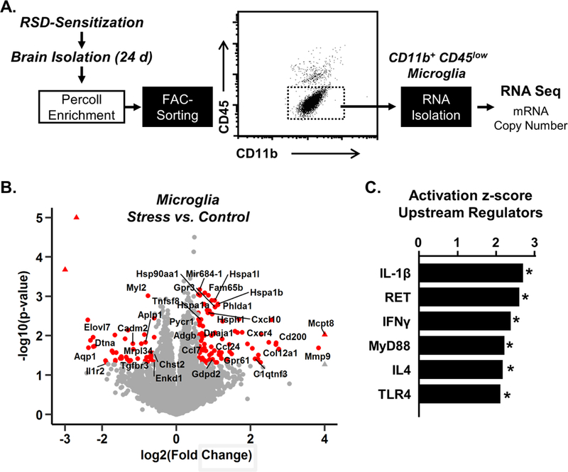
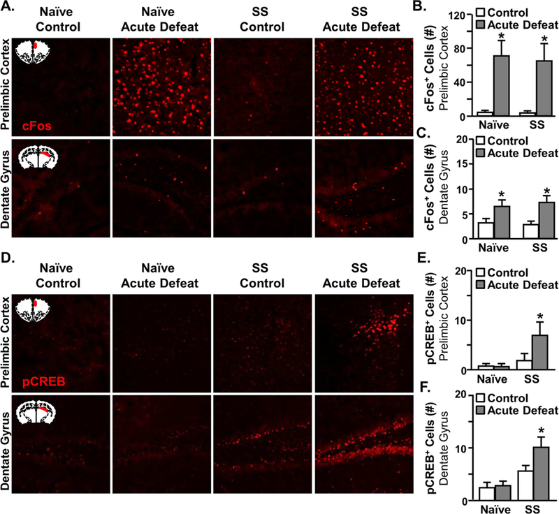
Background: Stress is associated with an increased prevalence of anxiety and depression. Repeated social defeat (RSD) stress in mice increases the release of monocytes from the bone marrow that are recruited to the brain by microglia. These monocytes enhance inflammatory signaling and augment anxiety. Moreover, RSD promotes stress sensitization, in which exposure to acute stress 24 days after cessation of RSD causes anxiety recurrence. The purpose of this study was to determine whether microglia were critical to stress sensitization and exhibited increased reactivity to subsequent acute stress or immune challenge.

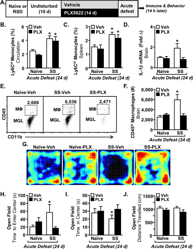
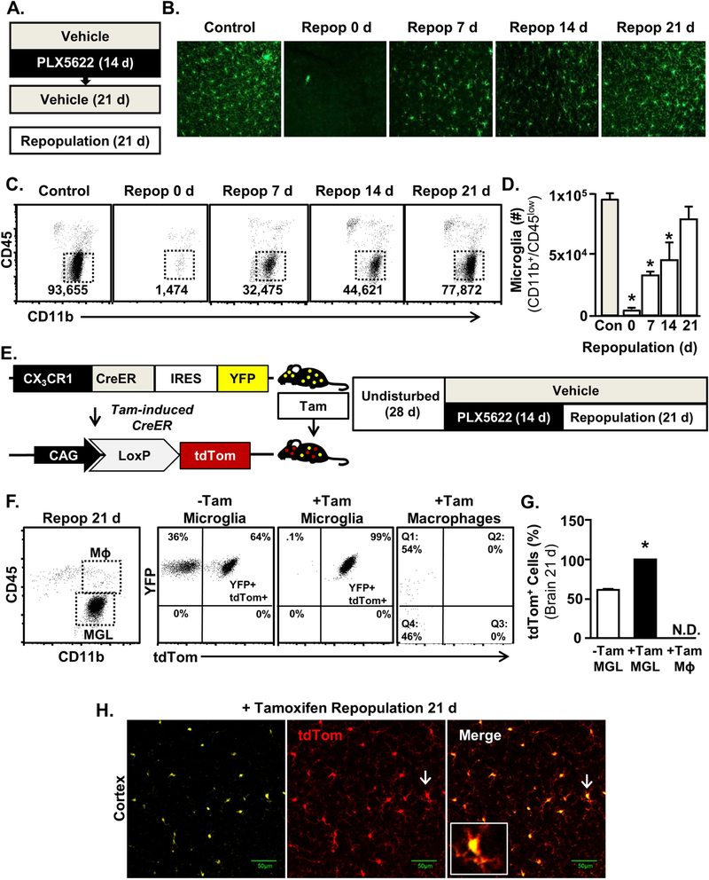
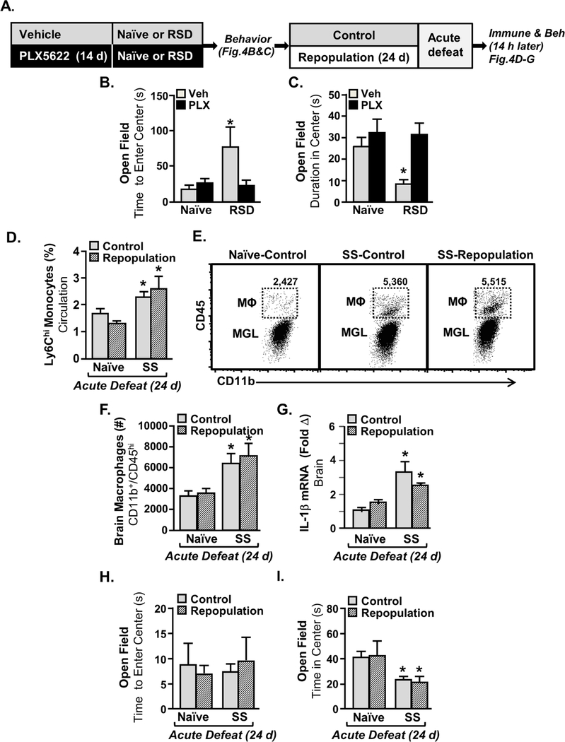
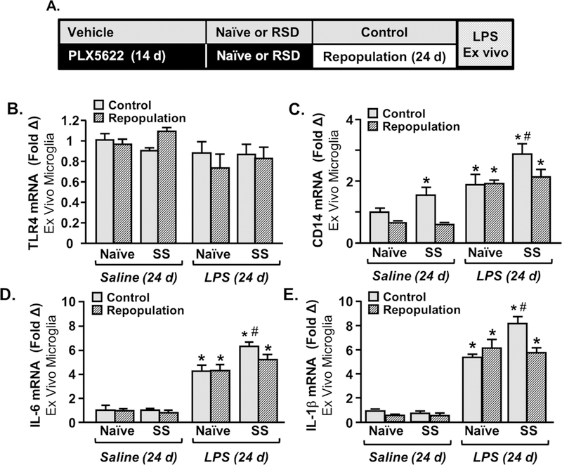
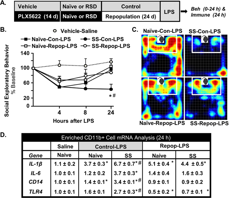
Methods: Mice were exposed to RSD, microglia were eliminated by colony-stimulating factor 1 receptor antagonism (PLX5622) and allowed to repopulate, and responses to acute stress or immune challenge (lipopolysaccharide) were determined 24 days after RSD sensitization.

Results: Microglia maintained a unique messenger RNA signature 24 days after RSD. Moreover, elimination of RSD-sensitized microglia prevented monocyte accumulation in the brain and blocked anxiety recurrence following acute stress (24 days). When microglia were eliminated prior to RSD and repopulated and mice were subjected to acute stress, there was monocyte accumulation in the brain and anxiety in RSD-sensitized mice. These responses were unaffected by microglial elimination/repopulation. This may be related to neuronal sensitization that persisted 24 days after RSD. Following immune challenge, there was robust microglial reactivity in RSD-sensitized mice associated with prolonged sickness behavior. Here, microglial elimination/repopulation prevented the amplified immune reactivity ex vivo and in vivo in RSD-sensitized mice.

Conclusions: Microglia and neurons remain sensitized weeks after RSD, and only the immune reactivity component of RSD-sensitized microglia was prevented by elimination/repopulation.

Keywords: Anxiety; CSF1R antagonist; Microglia; Monocytes; Repeated social defeat; Stress.


Conflict of interest statement

The authors report no biomedical financial interests or potential conflicts of interest.

Figures

Figure 1:
Figure 1:. Evidence for Primed Microglial profile 24 days after RSD.
A) Male C57BL/6 mice were stress-sensitized (SS) by RSD or left undisturbed as controls (Naϊve). At 24 d after stress, microglia were Percoll-enriched, FAC-sorted, and RNA was collected for RNASeq (n=6). B) Volcano plots of RSD vs control of differentially expressed genes. Red points indicate differentially expressed genes (p<0.05, absolute fold change>1.5). C) Ingenuity Pathway Analysis of significantly altered upstream regulators in microglia. Graphs with (*) are significantly different from controls.
Figure 2:
Figure 2:. Monocyte Recruitment to the Brain and Recurrence of Anxiety in Stress-Sensitized Mice was Abrogated by the Elimination of Microglia.
A) Male C57BL/6 mice were stress-sensitized (SS) by RSD or left undisturbed as controls (Naϊve). Ten days later, mice were provided diets formulated with vehicle (Veh) or a CSF1R antagonist (PLX5622). 24 days after stress-sensitization (SS), all mice were subjected to one cycle of social defeat (Acute Defeat) and blood, spleen, and brain samples were collected 14 h later (n=9–10). The percentage of monocytes (CD11b+/Ly6Chi) in the B) blood (main effect of SS, F1,39=21.45, p<0.0001) and spleen (main effect of SS, F1,39=28.63, p<0.002) were determined 14 h after acute defeat. D) IL-1β mRNA expression in a coronal brain section was determined after acute defeat (SS x intervention, F1,38=4.35, p<0.04). E) Representative bivariate dot plots of CD11b and CD45 labeling of Percoll-enriched microglia (CD11b+/CD45low) and macrophages (CD11b+/CD45high) in the brain after acute defeat. F) Number of CD45+ macrophages in the brain (SS x intervention, F1,36=10.68, p<0.003). G) Representative heat maps of activity during open-field testing. H) Time to enter center of the open field (SS x intervention, F1,40=5.20, p<0.02). I) Time spent in center in the open field after acute defeat (SS x intervention, F1,40=7.03, p<0.01). J) Total distance traveled in the open field (not significant). Bars represent the mean ± SEM. Means with (*) are significantly different from Naïve-Vehicle controls.
Figure 3:
Figure 3:. Microglia Repopulated from Non-Progenitor CX3CR1+ cells after CSF1R Antagonist-Mediated Elimination.
A) Male C57BL/6 mice were provided diets formulated with vehicle or CSF1R antagonist (PLX5622) for 14 days. Next, the CSF1R antagonist diet was removed and all mice were provided vehicle diets for 1, 7, 14 or 21 days to allow for repopulation of microglia. B) Representative images of Iba-1 labeling in the cortex 1, 7, 14 or 21 days after the cessation of the CSF1R antagonist. C) Representative bivariate dot plots of CD11b/CD45 labeling of Percoll-enriched cells at each time point. D) Number of microglia (CD11b+/CD45low) in the after brain 1, 7, 14, or 21 days of repopulation (main effect of time, F4,16= 8.57, p<0.001). E) Schematic representation of the experimental design using CX3CR1CreER/+/R26tdTOM/+ mice, which were administered 4 daily injections of control or tamoxifen (20 mg/kg, i.p.) at 3 weeks of age. Mice were left undisturbed for 28 d, provided diets formulated with a CSF1R antagonist (PLX5622) for 14 d and then provided vehicle diets for an additional 21 days to allow for repopulation of microglia. F) Representative bivariate dot plots of YFP and tdTom expression in microglia (CD11b+/CD45low) and macrophages (CD11b+/CD45high) isolated from +/− tamoxifen-injected CX3CR1CreER/+/R26tdTOM/+ mice Subjected to microglial elimination/repopulation. G) Percentage of tdTom+ microglia (CD11b+/CD45low) and macrophages (CD11b+/CD45high) in the brain isolated from +/− tamoxifen-injected CX3CR1CreER/+/R26tdTOM/+ mice subjected to microglial elimination/repopulation. H) Representative images of YFP and tdTom expression in tamoxifen-injected CX3CR1CreER/+/R26tdTOM/+ mice subjected to microglial elimination/repopulation. Inset shows YFP+/tdTom+ microglia identified by white arrows. Bars represent the mean ± SEM. Means with (*) are significantly different from control (p<0.05).
Figure 4:
Figure 4:. Microglia Repopulation in Stress-Sensitized Mice Re-established Monocyte Trafficking and Anxiety-Like Behavior Induced by Acute Defeat.
A) Male C57BL/6 mice were provided diets formulated with vehicle or CSF1R antagonist (PLX5622) for 14 days. Next, mice were stress-sensitized (SS) by RSD or left undisturbed as controls (Naϊve). Anxiety-like behavior was determined 14 h after the last cycle of RSD in the open field (n=9–10) by B) time to enter the center (Intervention x SS interaction; F1,38=10.36, p<0.003) and C) time spent in the center (Intervention x SS interaction; F1,38=3.675, p=0.06). After RSD, all mice were provided vehicle diets for an additional 24 d to allow for repopulation of microglia. After 24 days of repopulation, all mice were exposed to one cycle of social defeat (acute defeat). D) Percentage of monocytes (CD11b+/CD45high) in circulation 14 h after acute defeat (main effect of SS, F1,37=16.46, p<0.0002). E) Representative bivariate dot plots of CD11b and CD45 labeling on enriched microglia and macrophages. F) Number of brain macrophages (CD11b+/CD45high) 14 h after acute defeat (main effect of SS, F1,37=16.46, p<0.0002). G) mRNA levels of IL-1β were determined in a coronal brain section (n=4) collected 14 h after acute defeat (main effect of SS; F1,14=20.1, p<0.001). Anxiety-like behavior (n=10) was determined by H) Time to enter (not significant) and I) time spent in the center of the open field 0.5 d after acute defeat (main effect of SS; F1,39=14.85, p<0.001). Bars represent the mean ± SEM. Means with (*) are significantly different from Control-Naïve (p<0.05).
Figure 5:
Figure 5:. Evidence of Neuronal Sensitization with RSD.
Male C57BL/6 mice were stress-sensitized (SS) by RSD or left undisturbed as controls (Naϊve). At 24 d after stress, all mice were exposed to one cycle of social defeat (acute defeat). Immediately after acute defeat, brains were perfused, fixed, sectioned, and labeled for c-Fos or pCREB (n=6). A) Representative images of c-Fos expression in the prelimbic cortex (top panel) and dentate gyrus (bottom panel). The number of c-Fos+ cells in the B) prelimbic cortex (F1,23=27.6,p<0.001) and C) dentate gyrus of the hippocampus in control and SS mice 14 h after acute defeat (F1,23=75.8,p<0.001). D) Representative images of pCREB expression in the prelimbic cortex (top panel) and dentate gyrus (bottom panel). Number of pCREB+ cells in the E) prelimbic cortex (SS, F1,24=4.7 p<0.04), SS x acute stress interaction (F1,24=2.9, p=0.1) and F) dentate gyrus of control and SS mice 0.5 d after acute defeat (SS, F1,24=17.7, p<0.001). Bars represent the mean ± SEM. Means with (*) are significantly different from Control-Naïve (p<0.05).
Figure 6:
Figure 6:. Microglial Hyperactivity to ex vivo LPS Stimulation in Stress-Sensitized Mice was Attenuated by Microglial Elimination and Repopulation.
A) Male C57BL/6 mice were provided diets formulated with vehicle or CSF1R antagonist (PLX5622) for 14 days. Next, mice were stress-sensitized (SS) by RSD or left undisturbed as controls (Naϊve). After RSD-sensitization, all mice were provided vehicle diets for an additional 24 days to allow for repopulation of microglia. After 24 days of repopulation, microglia were collected by Percoll-enrichment and were cultured ex vivo with saline or LPS (100 ng/ml). mRNA levels of B) TLR4, CD14, D) IL-6, and E) IL-1β were determined in ex vivo microglia 4 h after LPS stimulation. Means with (*) are significantly different from Control-Naïve (p<0.05) and means with (#) are significantly different from SS-Naïve (p<0.05).
Figure 7:
Figure 7:. Microglial Hyperactivity to in vivo LPS Challenge in Stress-Sensitized Mice was Attenuated by Microglial Elimination and Repopulation.
A) Male C57BL/6 mice were provided diets formulated with vehicle or CSF1R antagonist (PLX5622) for 14 days. Next, mice were stress-sensitized (SS) by RSD or left undisturbed as controls (Naϊve). After RSD-sensitization, all mice were provided vehicle diets for an additional 24 days to allow for repopulation of microglia. After 24 days of repopulation, mice were injected with LPS (0.5 mg/kg; i.p.) and B) social exploratory behavior (percent of baseline) was determined at baseline and 4, 8, and 24 h after LPS challenge (main effect of SS: F1, 83=18.59, p<0.03, SS x Time F3,83=3.42, p=.066). C) Representative heat maps of social exploratory behavior 24 h after LPS.D) mRNA levels of IL-1β (SS x Repop interaction; F1,18=14.6, p<0.001), IL-6 (SS x Repop interaction; F1,18=14.2, p<0.001, CD14 (SS x Repop interaction; F1,18=10.1, p<0.005), and TLR4 SS x Repop interaction; F1,18=5.715, p<0.03) were determined in Percoll-enriched microglia collected 24 h after LPS challenge. Bars represent the mean ± SEM. Means with (*) are significantly different from Control-Naive (p<0.05) and means with (#) are significantly different from saline control (p<0.05).

Comment in

References

    1. Kessler RC (1997): The effects of stressful life events on depression. Annual review of psychology 48:191–214. - PubMed
    1. Gilman SE, Trinh NH, Smoller JW, Fava M, Murphy JM, Breslau J (2013): Psychosocial stressors and the prognosis of major depression: a test of Axis IV. Psychol Med 43:303–316. - PMC - PubMed
    1. Post RM (1992): Transduction of Psychosocial Stress into the Neurobiology of Recurrent Affective-Disorder. Am J Psychiat 149:999–1010. - PubMed
    1. McLaughlin KA, Conron KJ, Koenen KC, Gilman SE (2010): Childhood adversity, adult stressful life events, and risk of past-year psychiatric disorder: a test of the stress sensitization hypothesis in a population-based sample of adults. Psychol Med 40:1647–1658. - PMC - PubMed
    1. Setiawan E, Wilson AA, Mizrahi R, Rusjan PM, Miler L, Rajkowska G, et al. (2015): Role of Translocator Protein Density, a Marker of Neuroinflammation, in the Brain During Major Depressive Episodes. Jama Psychiat 72:268–275. - PMC - PubMed

Publication types

MeSH terms