. 2019 Jan 16;101(2):307-320.e6.
doi: 10.1016/j.neuron.2018.11.021. Epub 2018 Dec 4.

Fractionating Blunted Reward Processing Characteristic of Anhedonia by Over-Activating Primate Subgenual Anterior Cingulate Cortex

Affiliations

Fractionating Blunted Reward Processing Characteristic of Anhedonia by Over-Activating Primate Subgenual Anterior Cingulate Cortex

Laith Alexander et al. Neuron. .

Abstract

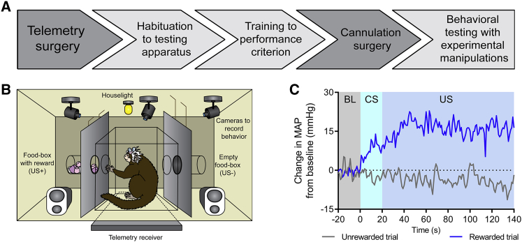
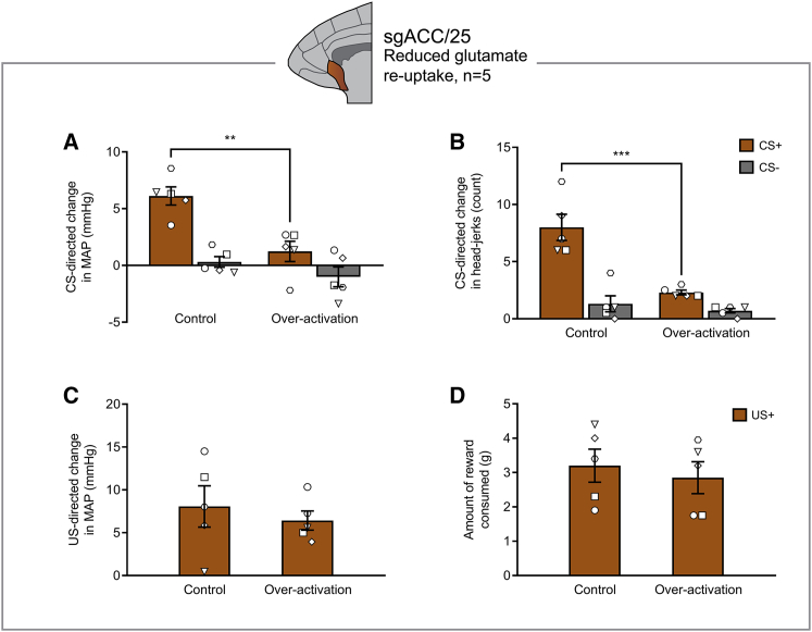
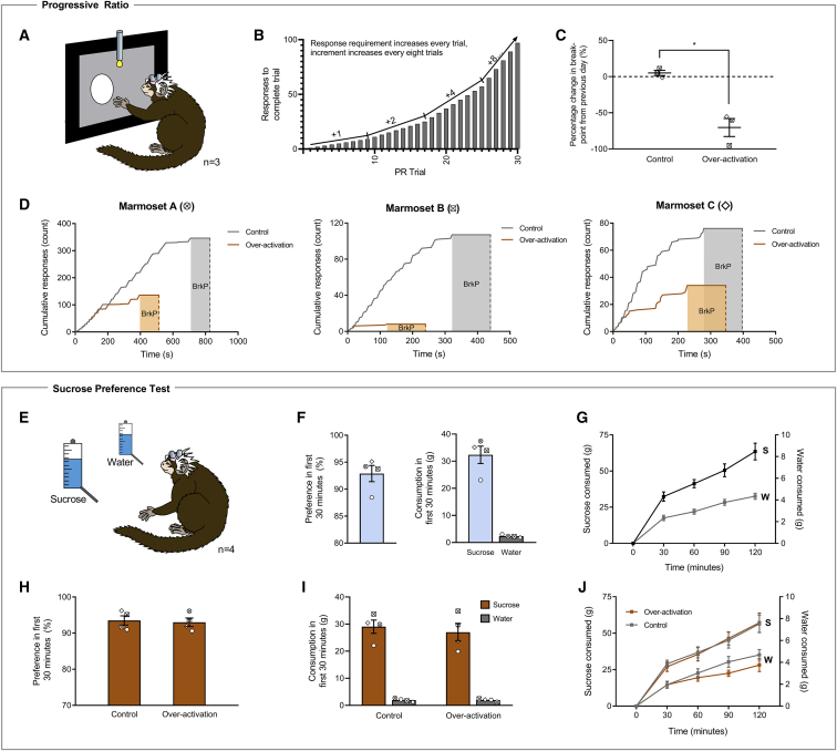
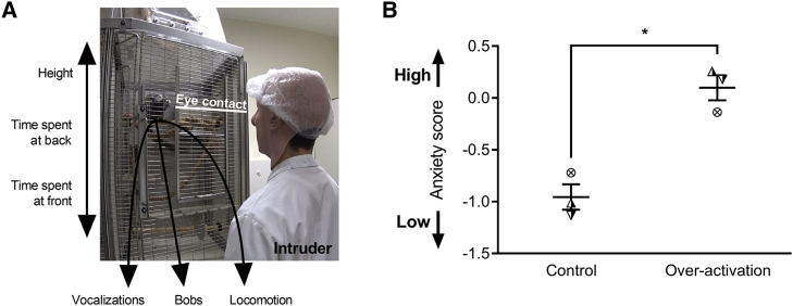
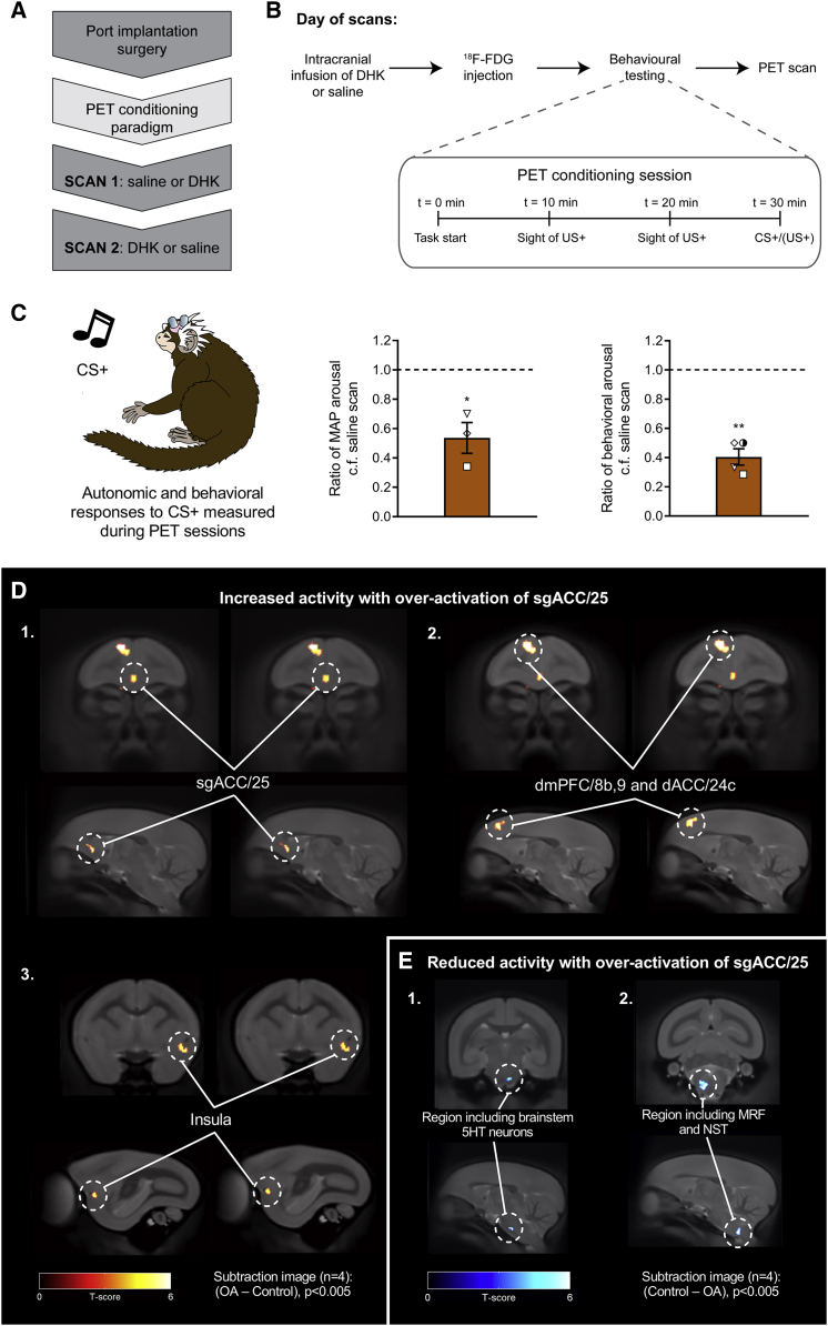
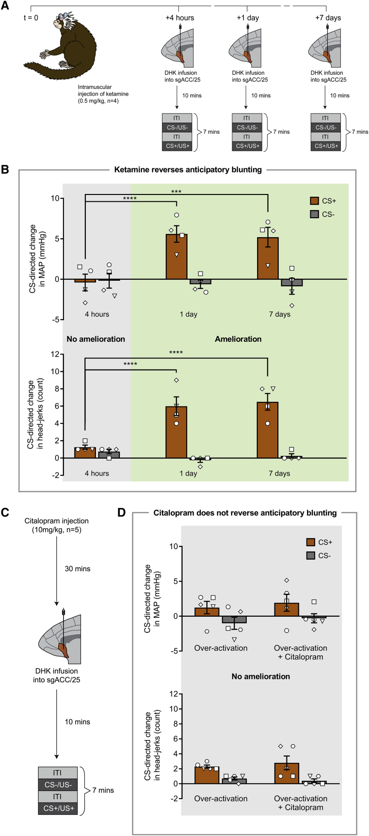
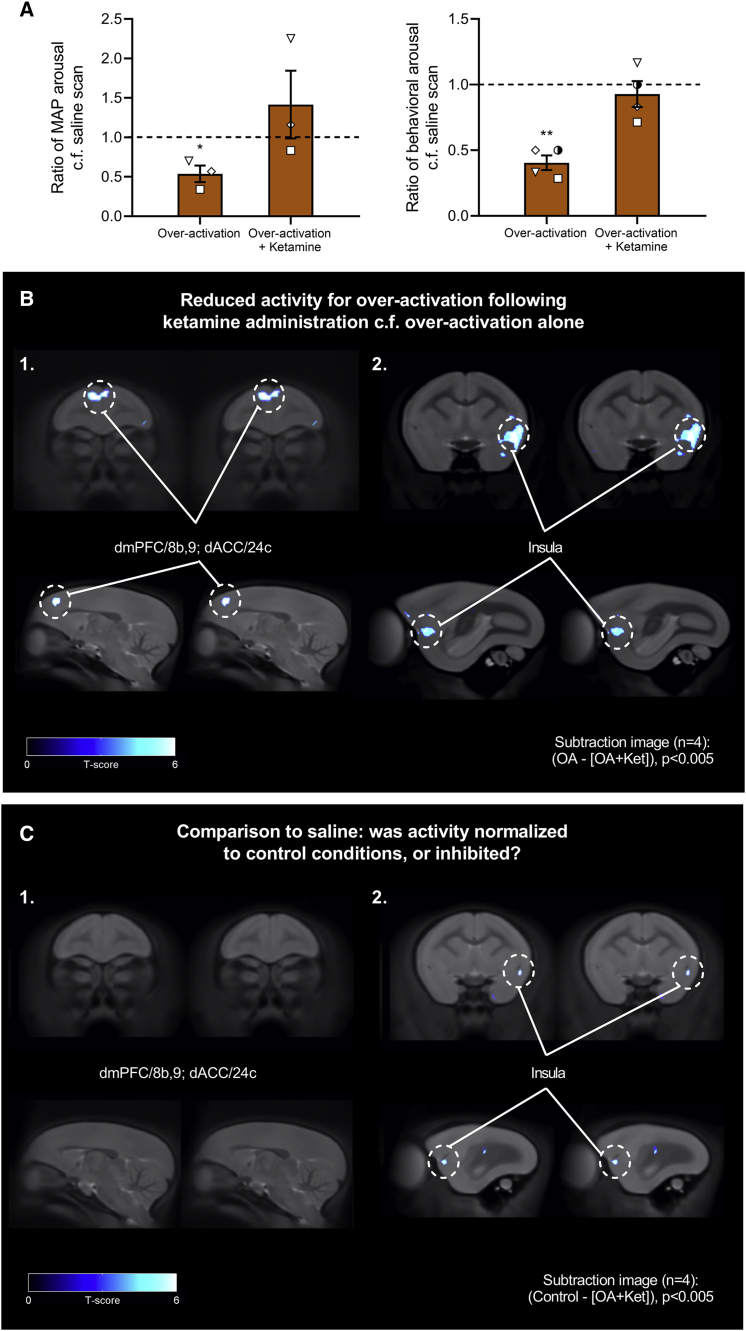
Anhedonia is a core symptom of depression, but the underlying neurobiological mechanisms are unknown. Correlative neuroimaging studies implicate dysfunction within ventromedial prefrontal cortex, but the causal roles of specific subregions remain unidentified. We addressed these issues by combining intracerebral microinfusions with cardiovascular and behavioral monitoring in marmoset monkeys to show that over-activation of primate subgenual anterior cingulate cortex (sgACC, area 25) blunts appetitive anticipatory, but not consummatory, arousal, whereas manipulations of adjacent perigenual ACC (pgACC, area 32) have no effect. sgACC/25 over-activation also reduces the willingness to work for reward. 18F-FDG PET imaging reveals over-activation induced metabolic changes in circuits involved in reward processing and interoception. Ketamine treatment ameliorates the blunted anticipatory arousal and reverses associated metabolic changes. These results demonstrate a causal role for primate sgACC/25 over-activity in selective aspects of impaired reward processing translationally relevant to anhedonia, and ketamine's modulation of an affective network to exert its action.

Keywords: PET imaging; anhedonia; anterior cingulate; area 25; depression; ketamine; marmoset; motivation; reward; subgenual.


Figures

Figure 1
Figure 1
Experimental Outline and Conditioned Discrimination Mean arterial pressure (MAP) values are recorded in mmHg. (A) Experimental overview. Following telemetry surgery, marmosets were habituated to the testing apparatus for 5–10 sessions, trained on the appetitive discrimination task until criterion was reached (significant MAP discrimination over three CS+/CS− sessions, two-tailed paired t test), and then cannulated to target sgACC/25 alone or both sgACC/25 and pgACC/32. Following re-attainment of criterion post-surgery, experimental manipulations took place. (B) Diagram of conditioning apparatus. During discrimination sessions, two auditory cues predicted either the presence (CS+/US+) or absence (CS−/US−) of a high-incentive food reward (marshmallow). A telemetry receiver placed underneath the apparatus recorded cardiovascular measurements, which were sent to a computer in an adjacent room. (C) Example MAP trace during baseline (BL, 20 s immediately prior to CS), CS (20 s), and US (120 s) periods for a rewarded and non-rewarded trial within a conditioning session. Values are calculated as a difference from the mean MAP during baseline. Animals showed an anticipatory MAP rise during the CS+ and a further consummatory rise during the US+.
Figure 2
Figure 2
sgACC/25 Over-Activation by Reducing Glutamate Reuptake Blunts Appetitive Anticipatory Arousal, but Not Consummatory Arousal Relevant graphs show mean ± SEM (n = 5). (A) sgACC/25 over-activation by reducing glutamate reuptake (DHK) blunted anticipatory cardiovascular arousal in a CS-dependent manner (manipulation × CS, F1,4 = 10.63, p = 0.031), decreasing responding to the CS+, but not the CS− (effect of manipulation: CS+, p = 0.006; CS−, p = 0.301). (B) The same manipulation also blunted anticipatory behavioral arousal in a CS-dependent manner (manipulation × CS, F1,4 = 72.25, p = 0.001), decreasing responding to the CS+, but not the CS− (effect of manipulation: CS+, p < 0.001; CS−, p = 0.407). (C) There was no significant effect on consummatory cardiovascular arousal during the US+ (two-tailed paired t test, p = 0.451). (D) There was no significant effect on reward consumption during the US+ (two-tailed paired t test, p = 0.241).
Figure 3
Figure 3
sgACC/25 Over-Activation Impairs Reward Motivation on a Progressive-Ratio Schedule of Reinforcement but Has No Effect on Sucrose Preference or Sucrose Consumption Relevant graphs show mean ± SEM (n = 3 for progressive ratio; n = 4 for sucrose preference). S, sucrose; W, water. (A) Marmosets were trained to press a circular stimulus on a touchscreen to earn milkshake reward under increasing response demands until breakpoint was reached (2 min with no response). (B) Task design. The response increment from trial n to n+1 starts at +1 and doubles every eight trials until a maximum increment of +8 (trials 1–8, responses 1–8; trials 9–16, responses 10–24, etc.). (C) sgACC/25 over-activation by reducing glutamate reuptake (DHK) decreased the number of responses marmosets made before breakpoint was reached (two-tailed paired t test, p = 0.042). (D) Response profiles in control and over-activation sessions for each animal. The 2-min timeout period signifying the breakpoint (BrkP) is shaded. (E) In the sucrose preference test, marmosets were presented with two identical bottles in their home cage: one containing sucrose, and one containing water. A single session lasted 2 hr with measurements taken every 30 min. The first 30-min time point was of a priori interest owing to the rapid actions of the intracranial infusions. (F) Prior to experimental manipulations, marmosets showed a high preference for sucrose during the first 30 min of the session (92.9% ± 1.5%), consuming 32.3 ± 3.2 g sucrose and 2.3 ± 0.2 g water (mean ± SEM). (G) Cumulative consumption profile in the session prior to experimental manipulations. Marmosets consumed significantly more sucrose at every time point measured (solution [water, sucrose] × time point [four, 30-min time bins], F3,9 = 26.97, p < 0.0001; effect of solution, p < 0.0001 at every time point). (H) Compared to a control infusion of saline, over-activation of sgACC/25 by reducing glutamate reuptake had no effect on sucrose preference in the first 30 min of the session (two-tailed paired t test, p = 0.800). (I) Over-activation of sgACC/25 had no effect on sucrose or water consumption in the first 30 min of the session (solution × manipulation, F1,3 = 1.05, p = 0.381; main effect of manipulation, F1,3 = 1.70, p = 0.283). (J) Across the 2-hr session, over-activation of sgACC/25 had no effect on cumulative sucrose or water consumption (solution × manipulation, F < 1, NS).
Figure 4
Figure 4
sgACC/25 Over-Activation Does Not Cause a General Blunting in Emotional Arousal Relevant graphs show mean ± SEM (n = 3). (A) In the human intruder test, marmosets are divided into a quadrant of their home cage and are confronted with a human intruder who maintains eye contact for 2 min. Marmosets display a range of behaviors in response to the intruder, including vocalizations (tsik, tse, tsik-egg, tse-egg, and egg calls), bobbing (rapid side-to-side movements), and locomotion (translational movements). These behaviors—together with average height, the time spent at the back of the cage, and the time spent at the front of the cage—are measured and loaded onto a single EFA-extracted factor representing anxiety (see Table S5 and STAR Methods). (B) Anxiety (factor) score following control infusions and over-activation of sgACC/25. Over-activation increased marmosets’ anxiety toward the human intruder reflected by an increased anxiety score (two-tailed paired t test, p = 0.047).
Figure 5
Figure 5
18F-FDG PET Imaging Revealed Metabolic Changes in a Network of Brain Regions Associated with Reward Processing and Interoception Following sgACC/25 Over-Activation Relevant graphs show mean ± SEM (n = 3 for cardiovascular arousal; n = 4 for behavioral arousal). n = 4 for all PET images; clusters discussed are significant at the level of p < 0.005 with an extent threshold adjusted for search volume of 26 voxels. (A) Following implantation of a subcutaneous port into the internal jugular vein, marmosets were trained on a modified version of the appetitive Pavlovian conditioning paradigm (see B) in preparation for scanning. Saline control and DHK scans were counterbalanced. (B) On the day of a scan, animals received an infusion of DHK or saline immediately followed by 18F-FDG injection through the port. The PET conditioning session (inset) lasted 30 min (to increase the sensitivity of 18F-FDG uptake to perturbation by the behavioral paradigm), consisting of two 20 s periods of the sight of reward without access, and a final 20 s CS+ period. During training, the CS+ was followed by a 120 s US+. On scan days, the animals were immediately removed from the apparatus when the CS+ period terminated, anesthetized, and then scanned. (C) Cardiovascular and behavioral responses were measured during the CS+ period in the PET conditioning sessions immediately prior to scanning. Compared to saline scans, over-activation of sgACC/25 significantly blunted cardiovascular (ratio of MAP response to saline scans; one-sample t test to 1.0, p = 0.048) and behavioral (ratio of head-jerk response to saline scans; one sample t test to 1.0, p < 0.001) arousal. (D) Subtraction images calculated from standardized uptake value ratio (SUVR) maps for over-activation (OA) scans—saline control scans, showing brain regions with increased activity following sgACC/25 over-activation. Increased metabolic activity was observed in sgACC/25 (1), together with a region of dorsomedial prefrontal cortex spanning dmPFC/8b,9 and dACC/24c (surviving p < 0.001; 2). More caudally, increased metabolic activity was observed in the left ventral insula (3). (E) Subtraction images calculated from SUVR maps for saline control scans—over-activation scans, showing brain regions with reduced activity following sgACC/25 over-activation. Reduced metabolic activity was observed in a region encompassing brainstem 5HT neurons (1) and, more caudally, brainstem autonomic control centers including the medullary reticular formation (MRF) and the nucleus of the solitary tract (NST; 2).
Figure 6
Figure 6
A Single Intramuscular Injection of Ketamine Ameliorates the Cardiovascular and Behavioral Anticipatory Impairment Induced by Over-Activating sgACC/25 in a Time-Dependent Manner, whereas Acute Citalopram Has No Effect Relevant graphs show mean ± SEM (n = 4 for ketamine study; n = 5 for citalopram study). (A) Timeline of ketamine study. Marmosets received a single intramuscular injection of ketamine (t = 0) followed by over-activation of sgACC/25 (DHK) 4 hr, 1 day, and 7 days later. (B) Ketamine had a time-dependent effect to reverse the cardiovascular (time point × CS, F2,12 = 14.71, p < 0.001) and behavioral (time point × CS, F2,12 = 19.59, p < 0.001) aspects of the anticipatory blunting induced by sgACC/25 over-activation (DHK infusions). Compared to control infusions of saline vehicle (not shown), sgACC/25 over-activation 4 hr after ketamine administration still resulted in significant blunting of cardiovascular (manipulation × CS, F1,3 = 60.46, p = 0.004; effect of manipulation on CS+, p = 0.003) and behavioral (manipulation × CS, F1,3 = 25.59, p = 0.015; effect of manipulation on CS+, p = 0.012) arousal. Over-activation 1 day following ketamine administration evidenced amelioration of the cardiovascular (4 hr versus 1 day: CS+, p < 0.0001; CS−, p = 0.863) and behavioral (4 hr versus 1 day: CS+, p < 0.0001; CS−, p = 0.371) impairments compared to 4 hr. Similarly, over-activation 7 days following ketamine administration evidenced amelioration of the cardiovascular (4 hr versus 7 days: CS+, p < 0.001; CS−, p = 0.704) and behavioral (4 hr versus 7 days: CS+, p < 0.0001; CS−, p = 0.767) impairments compared to 4 hr. (C) Timeline of acute citalopram study. Marmosets received a single intramuscular injection of citalopram followed by over-activation of sgACC/25 (DHK) 30 min later. (D) Compared to sgACC/25 over-activation alone, acute citalopram had no effect on the cardiovascular (manipulation × CS, F < 1, NS) or behavioral (manipulation × CS, F1,4 = 1.19, p = 0.338) components of the anticipatory blunting. Compared to control infusions of saline vehicle (not shown), sgACC/25 over-activation with acute citalopram still resulted in significant blunting of cardiovascular (manipulation × CS, F1,4 = 8.74, p = 0.042; effect of manipulation on CS+, p = 0.016) and behavioral (manipulation × CS, F1,4 = 462, p < 0.0001; effect of manipulation on CS+, p < 0.0001) arousal.
Figure 7
Figure 7
Reversal of Blunted Anticipatory Arousal by Ketamine Is Associated with Metabolic Changes within dmPFC, dACC, Insula, and sgACC/25 Relevant graphs show mean ± SEM (n = 3 for cardiovascular arousal; n = 4 for behavioral arousal). n = 4 for all PET images; clusters discussed are significant at the level of p < 0.005 with an extent threshold adjusted for search volume of 26 voxels. (A) Ketamine administration 1 day earlier ameliorated the blunted cardiovascular arousal associated with over-activation alone, returning cardiovascular arousal to levels no different from the saline scan (one-sample t test on ratio values compared to 1.0, over-activation alone, p = 0.048; over-activation + ketamine, p = 0.435). Ketamine administration also ameliorated the blunted behavioral arousal associated with over-activation alone, returning behavioral arousal to levels no different from the saline scan (one-sample t test on ratio values compared to 1.0, over-activation alone, p = 0.007; over-activation + ketamine, p = 0.435). (B) Subtraction images calculated from SUVR maps for over-activation (OA) scans—[over-activation + ketamine (Ket)] scans, showing brain regions with reduced activation following ketamine administration 1 day earlier. Reversal of blunted anticipatory arousal was associated with reduced activity in dmPFC/8b,9, dACC/24c (1) and left ventral insula (2). (C) Subtraction images calculated from SUVR maps for control scans—[over-activation + ketamine] scans revealed that activity in the dmPFC/8b,9 and dACC/24c region was no different from control scans, indicating that ketamine had normalized over-activity in these regions to control levels (1). However, activity in the left insula was reduced even compared to control conditions, suggesting that ketamine administration caused deactivation of the insula (2).

Similar articles

Cited by

References

    1. Abdallah C.G., Sanacora G., Duman R.S., Krystal J.H. Ketamine and rapid-acting antidepressants: a window into a new neurobiology for mood disorder therapeutics. Annu. Rev. Med. 2015;66:509–523. - PMC - PubMed
    1. Allen T.A., Narayanan N.S., Kholodar-Smith D.B., Zhao Y., Laubach M., Brown T.H. Imaging the spread of reversible brain inactivations using fluorescent muscimol. J. Neurosci. Methods. 2008;171:30–38. - PMC - PubMed
    1. Alvarez R.P., Chen G., Bodurka J., Kaplan R., Grillon C. Phasic and sustained fear in humans elicits distinct patterns of brain activity. Neuroimage. 2011;55:389–400. - PMC - PubMed
    1. Amat J., Baratta M.V., Paul E., Bland S.T., Watkins L.R., Maier S.F. Medial prefrontal cortex determines how stressor controllability affects behavior and dorsal raphe nucleus. Nat. Neurosci. 2005;8:365–371. - PubMed
    1. Amemori K., Amemori S., Graybiel A.M. Motivation and affective judgments differentially recruit neurons in the primate dorsolateral prefrontal and anterior cingulate cortex. J. Neurosci. 2015;35:1939–1953. - PMC - PubMed

Publication types

MeSH terms

LinkOut - more resources