. 2019 Apr;17(2):310-317.
doi: 10.1016/j.jtos.2018.12.001. Epub 2018 Dec 4.

Hypoxia: A breath of fresh air for the meibomian gland

Affiliations

Hypoxia: A breath of fresh air for the meibomian gland

Yang Liu et al. Ocul Surf. 2019 Apr.

Abstract

Purpose: Optimal meibomian gland (MG) function is critically important for the health and wellbeing of the ocular surface. We hypothesize that low oxygen (O2) conditions promote the function of human MG epithelial cells (HMGECs) and that human MGs exist in a relatively hypoxic environment. The purpose of this study was to test our hypotheses.

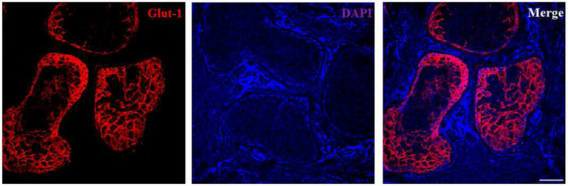
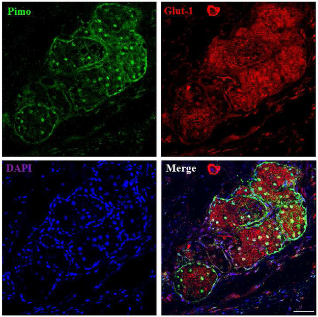
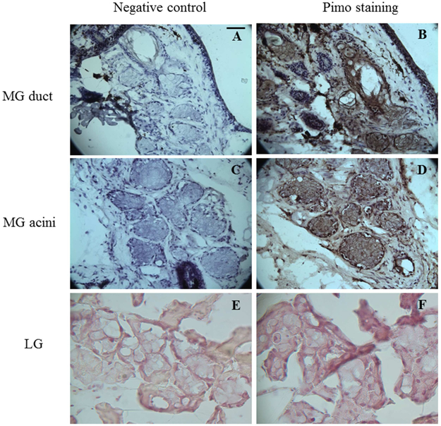
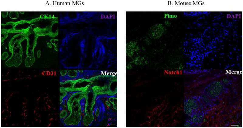
Methods: We used human and mouse eyelid segments, and immortalized human MG epithelial cells (IHMGECs) in our studies. To evaluate oxygen (O2) levels in the mouse MG and vicinity, we injected pimonidazole (pimo), a hypoxia marker, before sacrifice. Human eyelid samples were stained with the hypoxia marker glucose transporter 1 (Glut-1). To determine the effect of low O2 levels on IHMGECs, we cultured cells under proliferating and differentiating conditions in both normoxic (21% O2) and hypoxic (3% O2) conditions for 5-15 days. IHMGECs were evaluated for cell number, neutral lipid content, lysosome accumulation, expression of biomarker proteins and DNase II activity.

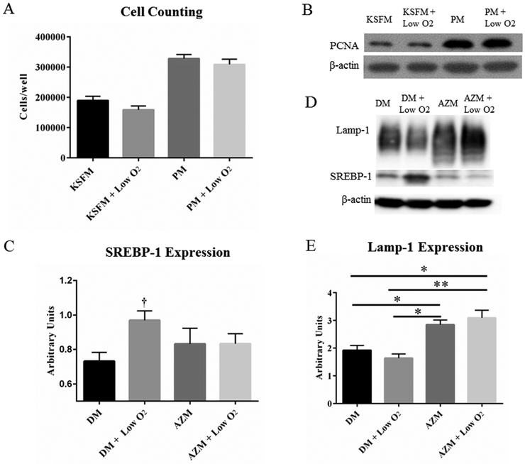
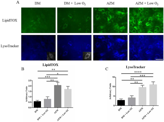
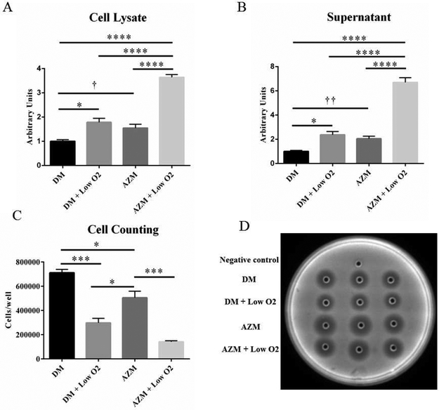
Results: Our results demonstrate that human and mouse MGs, but not the surrounding connective tissue, exist in a relatively hypoxic environment in vivo. In addition, our findings show that hypoxia does not influence IHMGEC numbers in basal or proliferating culture conditions, but does stimulate the expression of SREBP-1 in differentiating IHMGECs. Hypoxia also significantly increased DNase II activity, and apparently IHMGEC terminal differentiation.

Conclusions: Our Results support our hypotheses, and indicate that relative hypoxia promotes MG function.

Keywords: DNase II; Glucose transporter 1; Hypoxia; Meibomian gland; Pimonidazole.


Figures

Figure 1.
Figure 1.
Glut-1 staining inhuman MGs. High intensity Glut-1 staining is found in human MG acini, but not in the peripheral connective tissues. Data from one experiment are shown as a representative of three independent repeats. Scale bar = 50 μM
Figure 2.
Figure 2.
The correlation of pimo and Glut-1 staining in mouse MGs. High intensity staining of pimo and Glut-1 is found in mouse MG acini. Pimo staining is found both in the nucleus and cytoplasm, and Glut-1 staining shows a membranous pattern. Data from one experiment are shown as a representative of three independent studies. Scale bar = 50 μM.
Figure 3.
Figure 3.
Pimo staining of mouse MGs and LGs. Both the MG duct (arrows) and acini (notched arrows) show high intensity of Pimo, as do the mouse SGs (* in B). LG shows no staining of pimo. Data from one experiment are shown as a representative of three studies performed under the same conditions. Scale bar = 5 μM.
Figure 4.
Figure 4.
The vasculature of human and mouse MGs. (A) CK14 (green) and vessels (red) staining in human MGs. (B) Pimo (green) and vessels (red) staining in mouse MGs. The experiment was repeated three times. Scale bar = 50 μM.
Figure 5.
Figure 5.
Effects of low O2 on IHMGEC proliferation and differentiation. Low O2 treatment did not impact the cell growth (A) or the expression of PCNA (B). SREBP-1, but not Lamp-1, expression is significantly increased in a hypoxic environment (C and D, † p < 0.05, n = 3 experiments). AZM with or without low O2 significantly increased the levels of Lamp-1 protein. (E, * p < 0.05, ** p< 0.01, n=3 experiments). DM = Differentiation Medium (DMEM/F12 + 10% FBS). PM = Proliferation Medium (KSFM + EGF/BPE).
Figure 6.
Figure 6.
Influence of low O2 on lipid and lysosome accumulation in IHMGECs. IHMGECs were treated were cultured in normoxic (21% O2) and hypoxic (3% O2) conditions environments in the presence or absence of AZM and then stained for neutral lipids (LipidTox Neutral Green) and lysosomes (LysoTracker blue). (* p < 0.05, ** p < 0.01, *** p< 0.001, **** p < 0.0001, one-way ANOVA, n = 3 experiments). Inserts show a section of the LysoTracker staining in black and white. Scale bar = 50 μM.
Figure 7.
Figure 7.
Impact of low O2 on DNase II activity and cell number in IHMGECs under differentiating conditions. DNase II activity is significantly increased by low O2 treatment in both cell lysate (A) and supernatant (B). Low O2 also significantly decreased the cell number under differentiating condition (C). DM = Differentiation Medium (DMEM/F12 + 10% FBS). (* p < 0.05, *** p< 0.001, **** p < 0.0001, one-way ANOVA. f p < 0.05, †† p < 0.01, two-tailed t-test. n = 3 experiments)

References

    1. Green-Church KB, Butovich I, Willcox M, Borchman D, Paulsen F, Barabino S, et al. The international workshop on meibomian gland dysfunction: report of the subcommittee on tear film lipids and lipid-protein interactions in health and disease. Investigative ophthalmology & visual science. 2011;52:1979–93. - PMC - PubMed
    1. Knop E, Knop N, Millar T, Obata H, Sullivan DA. The international workshop on meibomian gland dysfunction: report of the subcommittee on anatomy, physiology, and pathophysiology of the meibomian gland. Investigative ophthalmology & visual science. 2011;52:1938–78. - PMC - PubMed
    1. Bron AJ, Tiffany JM. The contribution of meibomian disease to dry eye. The ocular surface. 2004;2:149–65. - PubMed
    1. Bron AJ, Tiffany JM, Gouveia SM, Yokoi N, Voon LW. Functional aspects of the tear film lipid layer. Experimental eye research. 2004;78:347–60. - PubMed
    1. McCulley JP, Shine WE. Meibomian gland function and the tear lipid layer. The ocular surface. 2003;1:97–106. - PubMed

Publication types