

Emerging imaging technologies in dermatology: Part I: Basic principles
- PMID: 30528311
- PMCID: PMC7469876
- DOI: 10.1016/j.jaad.2018.11.042
Emerging imaging technologies in dermatology: Part I: Basic principles
Abstract
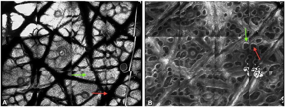
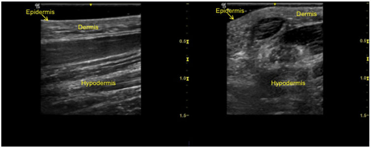
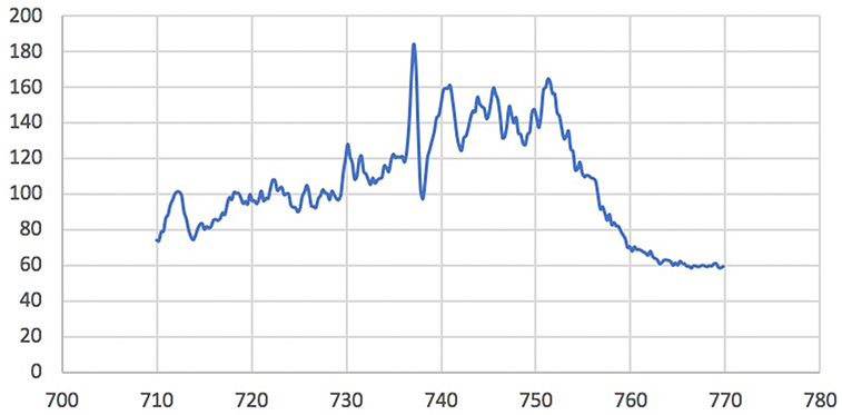
Dermatologists rely primarily on clinical examination in combination with histopathology to diagnose conditions; however, clinical examination alone might not be sufficient for accurate diagnosis and skin biopsies have associated morbidity. With continued technological advancement, there are emerging ancillary imaging technologies available to dermatologists to aid in diagnosis and management. This 2-part review article will discuss these emerging technologies including: digital photographic imaging, confocal microscopy, optical coherence tomography, and high-frequency ultrasound, as well as several additional modalities in development. In this first installment, the authors describe the breadth of technologies available and the science behind them. Then, in the second article, the authors discuss the applications and limitations of these technologies and future directions.
Keywords: Raman spectroscopy; confocal microscopy; dermatology; dermoscopy; digital photographic imaging; fluorescence imaging; high-frequency ultrasound; machine-based learning; multispectral optoacoustic tomography; optical coherence tomography.
Copyright © 2019. Published by Elsevier Inc.
Conflict of interest statement
Conflicts of interest: Dr Schneider has no relevant conflicts to disclose. Dr Kohli has served as a subinvestigator for Estee Lauder, Unigen, Ferndale laboratories, Allergan, Chromaderm, Pfizer, Johnson & Johnson, and Bayer. Dr Hamzavi has served as research investigator for Estee Lauder, Unigen, Ferndale laboratories, Allergan, Bayer, Johnson & Johnson, and Incyte Corporation. Dr Council has served as consultant for MD Outlook and Medline Industries. Dr Rossi has served as consultant for Canfield Scientific Inc. Dr Ozog has served as investigator for MiRagen and Biofrontera, on the advisory board for Allergan, and was on the past medical board for DermOne.
Figures
References
-
- Bae EJ, Seo SH, Kye YC, Ahn HH. A quantitative assessment of the human skin surface using polarized light digital photography and its dermatologic significance. Skin Res Technol. 2010;16(3):270–274. - PubMed
