. 2019 Jan:39:145-158.
doi: 10.1016/j.ebiom.2018.11.039. Epub 2018 Dec 6.

Installation of a cancer promoting WNT/SIX1 signaling axis by the oncofusion protein MLL-AF9

Affiliations

Installation of a cancer promoting WNT/SIX1 signaling axis by the oncofusion protein MLL-AF9

Li-Shu Zhang et al. EBioMedicine. 2019 Jan.

Abstract

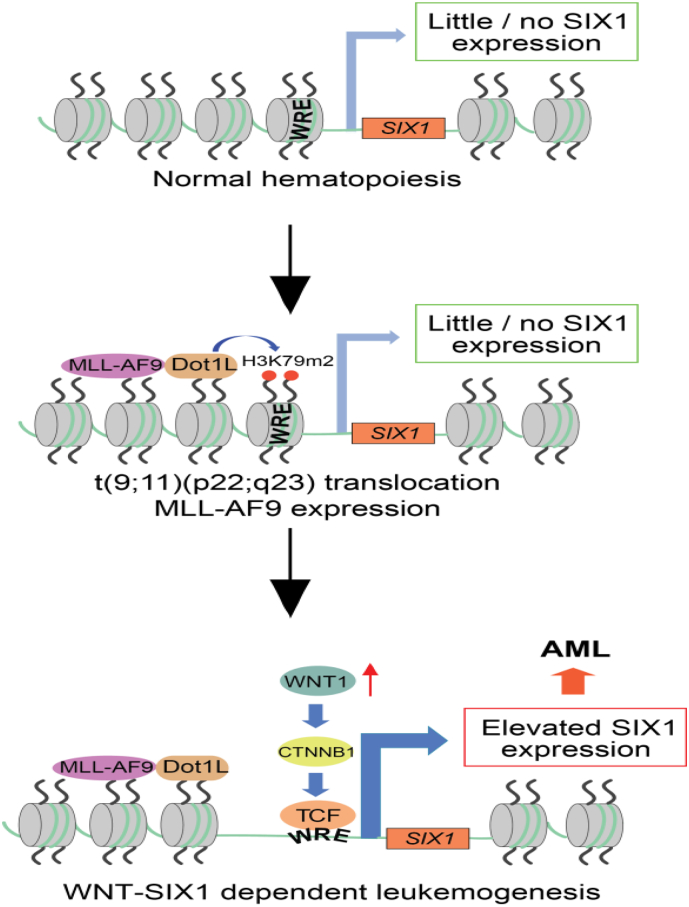
Background: Chromosomal translocation-induced expression of the chromatin modifying oncofusion protein MLL-AF9 promotes acute myelocytic leukemia (AML). Whereas WNT/β-catenin signaling has previously been shown to support MLL-AF9-driven leukemogenesis, the mechanism underlying this relationship remains unclear.

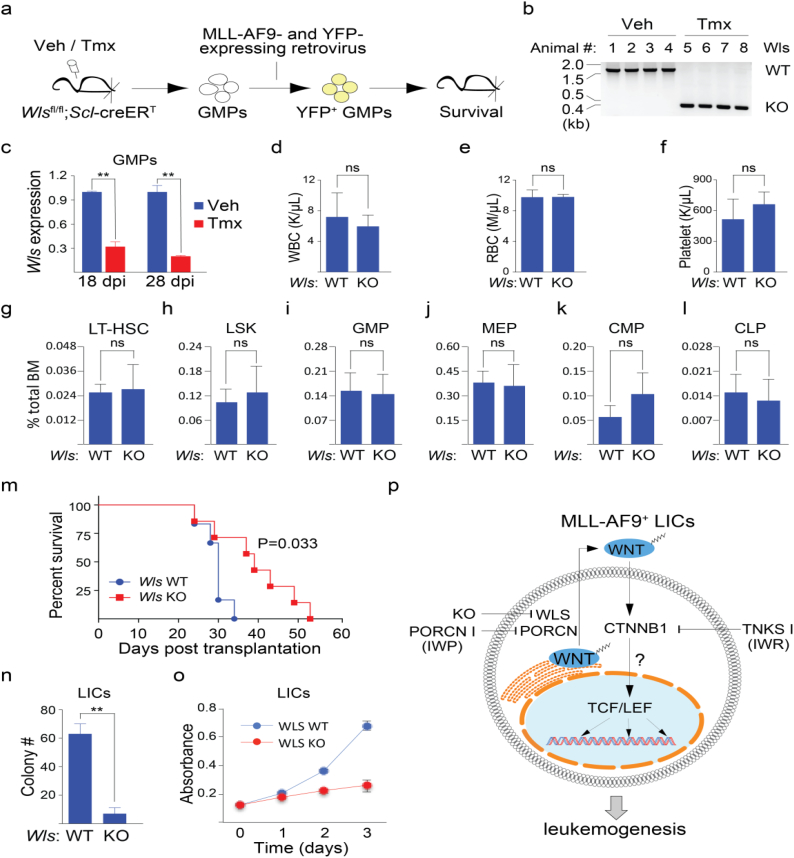
Methods: We used two novel small molecules targeting WNT signaling as well as a genetically modified mouse model that allow targeted deletion of the WNT protein chaperone Wntless (WLS) to evaluate the role of WNT signaling in AML progression. ATAC-seq and transcriptome profiling were deployed to understand the cellular consequences of disrupting a WNT signaling in leukemic initiating cells (LICs).

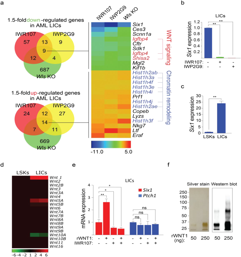
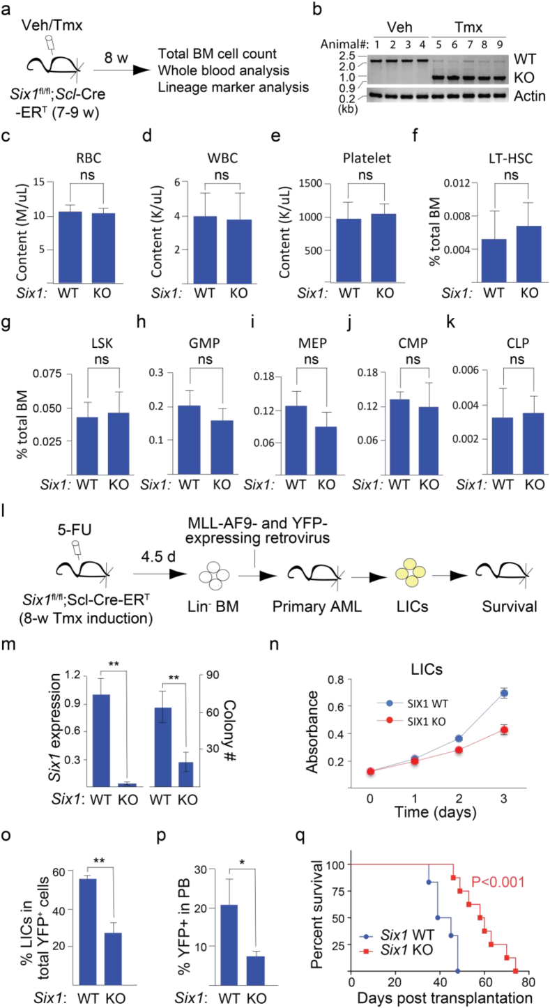
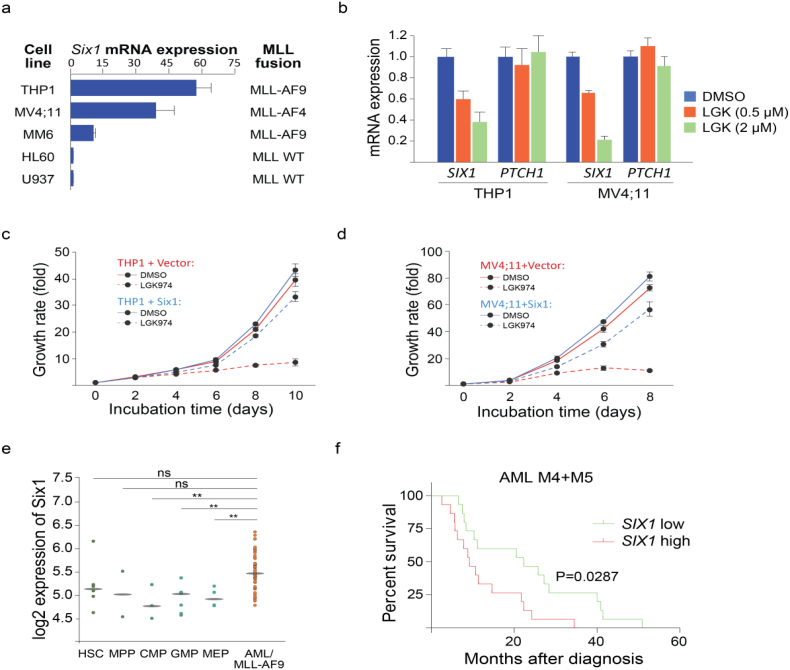
Findings: We identified Six1 to be a WNT-controlled target gene in MLL-AF9-transformed leukemic initiating cells (LICs). MLL-AF9 alters the accessibility of Six1 DNA to the transcriptional effector TCF7L2, a transducer of WNT/β-catenin gene expression changes. Disruption of WNT/SIX1 signaling using inhibitors of the Wnt signaling delays the development of AML.

Interpretation: By rendering TCF/LEF-binding elements controlling Six1 accessible to TCF7L2, MLL-AF9 promotes WNT/β-catenin-dependent growth of LICs. Small molecules disrupting WNT/β-catenin signaling block Six1 expression thereby disrupting leukemia driven by MLL fusion proteins.


Figures

Fig. 1
Fig. 1
WNT/β-catenin signaling is a chemical vulnerability in MLL-AF9-driven AML. (a) Next generation WNT pathway inhibitors based on the IWR1 and IWP2 scaffolds [20]. (b) IWR107 and IWP2G9 exhibit WNT pathway inhibitory activity in murine bone marrow cells and leukemic cells (BM-derived Lin cells with MLL-AF9 expression identified by sorting for co-expressed YFP signal). Both cell populations were exposed for 24 h to the inhibitors (10 μM) in vitro prior to Western blot analysis. (c) Evaluating the effects of WNT pathway inhibition in a mouse model of AML. Wnt inhibitors were orally administered in AML mice for 4 wks and leukemic initiating cells (LICs, YFP+ Mac1+ cKit+) from treated mice transplanted into healthy mice for survival analysis. (d) Abundance of circulating YFP+ leukemic cells is decreased in mice transplanted with LICs exposed to IWR107 (n = 9) or IWP2G9 (n = 8) compared to the Vehicle control (n = 9). PB = peripheral blood. Data is expressed as mean ± S.D. Error bars represent indicated numbers of biological replicates. **P < .01 [Student's t-test]. (e) Animals transplanted with LICs exposed to WNT inhibitors IWR107 (n = 9) and IWP2G9 (n = 8) exhibit extended survival compared to the Vehicle control (n = 9). Kaplan-Meier curves were plotted and the Log-Rank test used for the statistical analysis. (f) Liver tissues in animals transplanted with LICs previously exposed to systemically delivered WNT inhibitors exhibit less leukemic cell infiltration compared to the Vehicle control. Scale bar: 500 μm.
Fig. 2
Fig. 2
Cell-autonomous WNT signaling contributes to MLL-AF9-induced AML. (a) An experimental approach for evaluating the contribution of cell-autonomous WNT signaling in MLL-AF9-driven AML. GMPs: granulocyte-monocyte progenitors (Lin Sca1 c-Kit+ FcγR+ CD34+). (b) Tamoxifen (Tmx) induction results in excision of the Wls locus. Genomic DNA was extracted from whole blood of mice induced with Tmx or Vehicle (Veh, corn oil) for 8 wks. (c) Decreased mRNA expression of Wls in bone marrow GMPs derived from Scl-creERT; Wls-flox mice compared with those from WT animals. Dpi = days post Tmx induction. n = 3 per group. (d-l) Loss of Wls does not affect normal adult hematopoiesis as measured using contents of white blood cells (WBC, d), red blood cells (RBC, e) and platelets (f), and the percentage of long-term hematopoietic stem cells (LT-HSC, Lin Sca1+ cKit+ Flt3 CD34, g), hematopoietic stem/progenitor cells (LSK, Lin Sca1+ cKit+, h), GMP (i), megakaryocyte and erythroid progenitors (MEP, Lin Sca1 c-Kit+ CD34 FcγR, j), common myeloid progenitors (CMP, Lin Sca1 c-Kit+ CD34+ FcγR; k), and common lymphoid progenitors (CLP, Lin Sca1low c-Kitlow FLT3+ IL7R+; l) in the total BM cell population at week 8 after Tmx induction. n = 4 per group. (m) Animals transplanted with Wls null leukemic cells (n = 7) exhibit extended survival compared with its WT counterparts (n = 6). Kaplan-Meier curves were generated and the Log-Rank test used for statistical analysis. (n) Wls null LICs form significantly less colony formation units (CFU) compared to WT LICs. n = 3 per group. (o) Wls null LICs proliferate more slowly compared to their WT counterparts. BrdU incorporation was used for the measurement of proliferation at the indicated time points. n = 3 per group. (p) Model of cell-autonomous WNT signaling in LICs of AML driven by MLL-AF9. In c-l and n-o, data is expressed as mean ± S.D. Error bars represent indicated numbers of biological replicates. ** P < .01. ns: not significant. [Student's t-test].
Fig. 3
Fig. 3
Transcriptional profiling identifies the homeobox gene Six1 as an atypical target of WNT/β-catenin signaling in AML. (a) Heat map revealing a gene signature of LICs perturbed by IWR107 and IWP2G9 and by Wls deletion. Total RNA was extracted from LICs isolated by FACS and subjected to microarray analysis. The cutoff for differentiated gene expression is set at 1.5 folds. Red = WNT signaling regulators, blue = histone genes. (b) Six1 mRNA expression in LICs isolated from animals exposed to IWP2G9 or IWR107 is significantly reduced compared to the Vehicle control. n = 3 per group. (c) MLL-AF9 LICs exhibit elevated Six1 mRNA expression compared to normal LSKs. n = 3 per group. (d) Profiling of mRNA expression of 19 WNT ligands in normal LSKs and enriched LIC population of AML. Fold change of mRNA expression in LICs was normalized to LSKs and used to generate the heat map. (e) Recombinant human WNT1 (rWNT1) induced expression of Six1 in LICs can be countered with IWR107. Cells were incubated with either rWNT (300 ng/ml) or IWR107 (10 μM) or both for 30 h and isolated cDNA subjected to qPCR analysis. The Hedgehog pathway target gene Ptch1 serves as a control. (f) Silver stain and Western blot using a WNT1 antibody of rWNT1 samples used in e. In b, c and f, data is expressed as mean ± S.D. Error bars represent indicated numbers of biological replicates. ** P < .01. * P < .05. ns: not significant. [Student's t-test].
Fig. 4
Fig. 4
The homeobox gene Six1 is essential for the development of MLL-AF9-driven AML but not normal adult hematopoiesis. (a) Experimental approach for evaluating the role of SIX1 in normal hematopoiesis of adult mice. (b) Tmx induction results in excision of the Six1 locus. Genomic DNA was extracted from whole blood of mice induced with Tmx or Veh for 8 wks. (c-k) Loss of Six1 does not affect mouse normal adult hematopoiesis as measured using contents of RBC (c), WBC (d) and platelets (e) from whole blood; and percentage of BM cells that are LT-HSC (f), LSK (g), GMP (h), MEP (i), CMP (j) and CLP (k) at week 8 after Tmx induction. n = 4 per group. (l) Experimental approach for evaluating the role of SIX1 in MLL-AF9-dependent leukemogenesis. (m) Significant reduction of Six1 mRNA levels and CFU counts were observed in Tmx-exposed MLL-AF9-transformed Six1-fl/fl;Scl-creERT LICs compared to WT controls. n = 3 per group. (n) Six1 null LICs proliferate more slowly compared to WT control cells. BrdU incorporation was used for the measurement of proliferation at the indicated time points. n = 3 per group. (o) Six1 null AML mice exhibit decreased LICs in total YFP+ leukemic cells of BM than WT controls. n = 4 per group. (p) Abundance of circulating YFP+ leukemic cells is decreased in mice transplanted with Six1 null LICs (n = 8) compared to WT controls (n = 6). (q) Loss of Six1 in LICs (n = 8) slows disease progression in animals engrafted with AML LICs as compared with WT controls (n = 6). Log-Rank test is used for statistical analysis. In c-k and m-p, data is expressed as mean ± S.D. Error bars represent indicated numbers of biological replicates. ** P < .01. * P < .05. ns: not significant. [Student's t-test].
Fig. 5
Fig. 5
Chromatin remodeling by MLL-AF9 confers WNT-dependent regulation of Six1 expression in LICs. (a) Assay for Transposase Accessible Chromatin with high-throughput sequencing (ATAC-Seq) was used to identify shared and unique chromatin regions (peaks) accessible to transposase activity in MLL-AF9 AML LICs vs. WT LSKs. Numbers of mapped genes from identified peaks are shown in the inset. Two biological replicates were used in the assay. (b) Putative direct target genes of MLL-AF9 identified by superimposing genes associated with regions of chromatin change induced by MLL-AF9 and previously identified by ChIP-Seq analysis using MLL-AF9 pull-down [6]. Six1 and its binding partner gene Eya1 (in bold) are among them. (c) Normalized ATAC-Seq profiles at Six1 locus. Signals of AML LICs and normal LSKs are depicted in red and blue, respectively. Two elevated peaks are observed: one immediately upstream of the transcriptional start site (TSS) and another ~8 K further upstream of TSS. (d) TCF7L2 (TCF4) binding to DNA regions within each peak was determined by ChIP-qPCR. Amplicons used in the analysis and whether or not they harbor putative TCF/LEF binding sites are outlined. Data is expressed as means ± S.D. Error bars represent three biological replicates. * P < .05, ** P < .01, ns: not significant. [Student's t-test].
Fig. 6
Fig. 6
Six1 is a potential biomarker for targeting WNT signaling in some forms of human MLL rearranged AML. (a) Profiling of Six1 mRNA expression in a panel of human AML lines. Expression levels are normalized to U937 cells (lowest expression) and rank ordered. THP1 (MLL-AF9 re-arrangement) and MV4;11 (MLL-AF4 re-arrangement) cells have higher Six1 mRNA expression among 6 cell lines tested. n = 3 technical replicates. (b) The PORCN inhibitor LGK974 dose-dependently inhibits Six1 mRNA expression in THP1 and MV4;11 cells. Cells were incubated with LGK974 for 30 h at the indicated doses. PTCH1 was used as a control for evaluating the on-target effect of the compound. (c-d) The growth of THP1 (c) or MV4;11 (d) cells is inhibited by LGK974 (2 μM) in the indicated time course. Stably expressing Six1 in THP1 (c) or MV4;11 (d) cells partially rescues the growth inhibition by LGK974. (e) SIX1 expression in human normal hematopoietic lineage cells (GSE42519) vs. human AML cells [GSE13159, GSE15434, GSE61804, GSE14468, The Cancer Genome Atlas (TCGA)]. ** P < .01. ns: not significant. [Student's t-test] AML/MLL-AF9: AML with t(11q23)/MLL-AF9 rearrangement. (f) High SIX1 expression is correlated with poor survival in AML subtype M4 and M5 patients [based on the French-American-British (FAB) classification system]. The Kaplan-Meier plots were generated using gene expression data of 183 AML patient samples from TCGA [39]. High and low SIX1 expression is defined as above and below the median of expression values across all patient samples, respectively.
Fig. 7
Fig. 7
Model of WNT/SIX1 signaling promoting AML. MLL-AF9 recruits the histone methyltransferase Dot1L to TCF/LEF binding sites proximal to SIX1 thus installing a LIC-intrinsic WNT/SIX1 signaling axis that promotes leukemogenesis. WRE = WNT Responsive Element.

Similar articles

Cited by

References

    1. Hanahan D., Weinberg R.A. Hallmarks of cancer: the next generation. Cell. 2011;144(5):646–674. - PubMed
    1. Bennett R.L., Licht J.D. Targeting epigenetics in cancer. Annu Rev Pharmacol Toxicol. 2018;58:187–207. - PMC - PubMed
    1. Brien G.L., Valerio D.G., Armstrong S.A. Exploiting the epigenome to control cancer-promoting gene-expression programs. Cancer Cell. 2016;29(4):464–476. - PMC - PubMed
    1. Jones P.A., Issa J.P., Baylin S. Targeting the cancer epigenome for therapy. Nat Rev Genet. 2016;17(10):630–641. - PubMed
    1. Hu D., Shilatifard A. Epigenetics of hematopoiesis and hematological malignancies. Genes Dev. 2016;30(18):2021–2041. - PMC - PubMed

MeSH terms

Substances