. 2019 Jan 15;29(2):148-154.
doi: 10.1016/j.bmcl.2018.12.008. Epub 2018 Dec 4.

Dual inhibition of Kif15 by oxindole and quinazolinedione chemical probes

Affiliations

Dual inhibition of Kif15 by oxindole and quinazolinedione chemical probes

Megan E Dumas et al. Bioorg Med Chem Lett. .

Abstract

The mitotic spindle is a microtubule-based machine that segregates a replicated set of chromosomes during cell division. Many cancer drugs alter or disrupt the microtubules that form the mitotic spindle. Microtubule-dependent molecular motors that function during mitosis are logical alternative mitotic targets for drug development. Eg5 (Kinesin-5) and Kif15 (Kinesin-12), in particular, are an attractive pair of motor proteins, as they work in concert to drive centrosome separation and promote spindle bipolarity. Furthermore, we hypothesize that the clinical failure of Eg5 inhibitors may be (in part) due to compensation by Kif15. In order to test this idea, we screened a small library of kinase inhibitors and identified GW108X, an oxindole that inhibits Kif15 in vitro. We show that GW108X has a distinct mechanism of action compared with a commercially available Kif15 inhibitor, Kif15-IN-1 and may serve as a lead with which to further develop Kif15 inhibitors as clinically relevant agents.

Keywords: Eg5; Kif15; Kinesin; Mitosis; Oxindole.


Figures

Figure 1.
Figure 1.
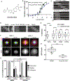
GW406108X (GW108X) inhibits and displays preference for Kif15. (A) Structure of GW108X. (B) Concentration response curves (CRC) generated from the ATPase (blue line) and MT gliding (green line) assay. CRC from the ATPase assay was developed from 10 concentrations from 30 to 0.001 μM. Each concentration was repeated in triplicate. CRC from the MT gliding assay was developed from 12 concentrations from 10 to 0.2 μM. Error bars show SEM. (C) Representative montage of fluorescent MT in gliding assay with Kif15-N700 with DMSO (top) or 10 μM of GW108X. Numbers indicate time in seconds after initial frame. Scale bar, 5 μm. (D) Representative montage of fluorescent MTs from the double wash out experiment. Numbers indicate time in seconds after initial frame, which are not the same MT for each condition. Scale bar, 5 μm. (E) Percent inhibition of indicated mitotic motors treated with 30 μM of GW108X. n >20 for all conditions, **** p < 0.0001, ** p = 0.0074. (F) Max intensity z-projections of TP53−/− RPE-1 (left) and KIRC-1 (right) cells treated with DMSO or 25 μM GW108X for 24 hours and stained with antibodies targeting Kif15 (grayscale and red), tubulin (green) and DNA (blue). Lookup tables (LUTs) for grayscale, red, and green channel are scaled identically. Scale bar, 10 μm. (G) Quantitation of % of pre-anaphase structures in TP53−/− RPE-1 and KIRC-1 cells treated with DMSO or 25 μM GW108X for 24 hours. Each condition was tested in triplicate and n ≥ 100 cells per replicate were counted. Errors bars show SD. (H) Quantitation of Kif15 on metaphase MTs in TP53−/− RPE-1 cells treated with DMSO or 25 μM GW108X. Shown are ratios of Kif15 fluorescence intensities to tubulin intensities. Box-and-whisker plots describe the median value as well as the 10th, 25th, 75th and 90th percentiles. ****, p < 0.0001. n ≥ 25 cells from triplicate experiments.
Figure 2.
Figure 2.
Scheme 1. Reagents and conditions: a. AlCl3, DCE. 0 °C, 65% b. RCHO, piperidine, EtOH, 80 °C, 40–70 % c. ArB(OH)2, Pd(PH3)4, Na2CO3, 2:1 dioxane/water,110 °C, 20–79 % d. amine, HATU, DIPEA, DMF, 57–64 %.
Figure 3.
Figure 3.
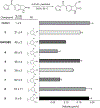
Structure-activity relationship (SAR) analysis of GW108X derivatives. Representative compounds from SAR library of phenol derivatives tested at 750 nM. Data is the average microtubule gliding velocity of Kif15-N700 with each compound. Every compound was tested in triplicate with n ≥ 30 measurements for each replicate, except 8 and 5, which were performed in duplicate. Error bars show SEM. See Table S1 for complete list of SAR compounds.
Figure 4.
Figure 4.
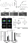
Kif15-IN-1 is a sub-micromolar inhibitor of Kif15. (A) Structure of Kif15-IN-1. (B) Concentration response curve using the MT-gliding assay, developed from 5 concentrations of Kif15-IN-1, ranging from 30 to 0.1 μM. Error bars show SD. (C) Representative montage of fluorescent MTs from the double wash out experiment. Each vertical frame represents 10 seconds. Scale bar, 5 μm. (D) Representative single optical sections of RPE-1 cells treated with DMSO or 25 μM Kif15-IN-1 for 24 hours and stained for antibodies targeting Kif15 (grayscale and red), tubulin (green) and DNA (blue). LUTs for grayscale, red and green channels are scaled identically. Scale bar, 5 μM. (E) Quantitation of RPE-1 cells treated with DMSO or 25 μM Kif15-IN-1 for 24 hours. Shown are ratios of Kif15 fluorescence intensities to tubulin intensities. Box-and-whisker plots describe the median value as well as the 10th, 25th, 75th and 90th percentiles. n = 30 cells from triplicate experiments. (F) Quantitation of the % pre-anaphase structures in RPE-1 cells treated with DMSO or 25 μM Kif15-IN-1 for 24 hours. Each condition was tested in triplicate and graph displays average percent from each triplicate. Errors bars show SD.
Figure 5.
Figure 5.
Kif15-IN-1 is a competitive ATP inhibitor and GW108X is a non-competitive ATP inhibitor. (A) Double reciprocal Lineweaver-Burk plot of ATP titration at 0.5, 1 and 2 μM GW108X. (B) Double reciprocal Lineweaver-Burk plot of MT titration at 0.5 and 2 μM GW108X. (C) Double reciprocal Lineweaver-Burk plot of ATP titration using 5 μM Kif15-IN-1. Each reaction in A-C was performed in triplicate. All error bars show SEM.

References

    1. van Vuuren RJ; Visagie MH; Theron AE; Joubert AM, Antimitotic drugs in the treatment of cancer. Cancer Chemother Pharmacol 2015, 76 (6), 1101–12. - PMC - PubMed
    1. Huang TC; Campbell TC, Comparison of weekly versus every 3 weeks paclitaxel in the treatment of advanced solid tumors: a meta-analysis. Cancer Treat Rev 2012, 38 (6), 613–7. - PubMed
    1. Ferrier J; Pereira V; Busserolles J; Authier N; Balayssac D, Emerging trends in understanding chemotherapy-induced peripheral neuropathy. Curr Pain Headache Rep 2013, 17 (10), 364. - PubMed
    1. Dominguez-Brauer C; Thu KL; Mason JM; Blaser H; Bray MR; Mak TW, Targeting Mitosis in Cancer: Emerging Strategies. Mol Cell 2015, 60 (4), 524–36. - PubMed
    1. Mayer TU; Kapoor TM; Haggarty SJ; King RW; Schreiber SL; Mitchison TJ, Small molecule inhibitor of mitotic spindle bipolarity identified in a phenotype-based screen. Science 1999, 286 (5441), 971–4. - PubMed

Publication types

MeSH terms

LinkOut - more resources