. 2019 Jun;139(6):1349-1361.
doi: 10.1016/j.jid.2018.11.024. Epub 2018 Dec 6.

Identification of a Robust Methylation Classifier for Cutaneous Melanoma Diagnosis

Affiliations

Identification of a Robust Methylation Classifier for Cutaneous Melanoma Diagnosis

Kathleen Conway et al. J Invest Dermatol. 2019 Jun.

Abstract

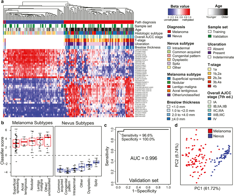
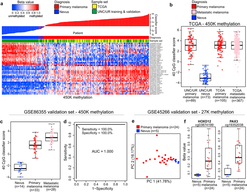
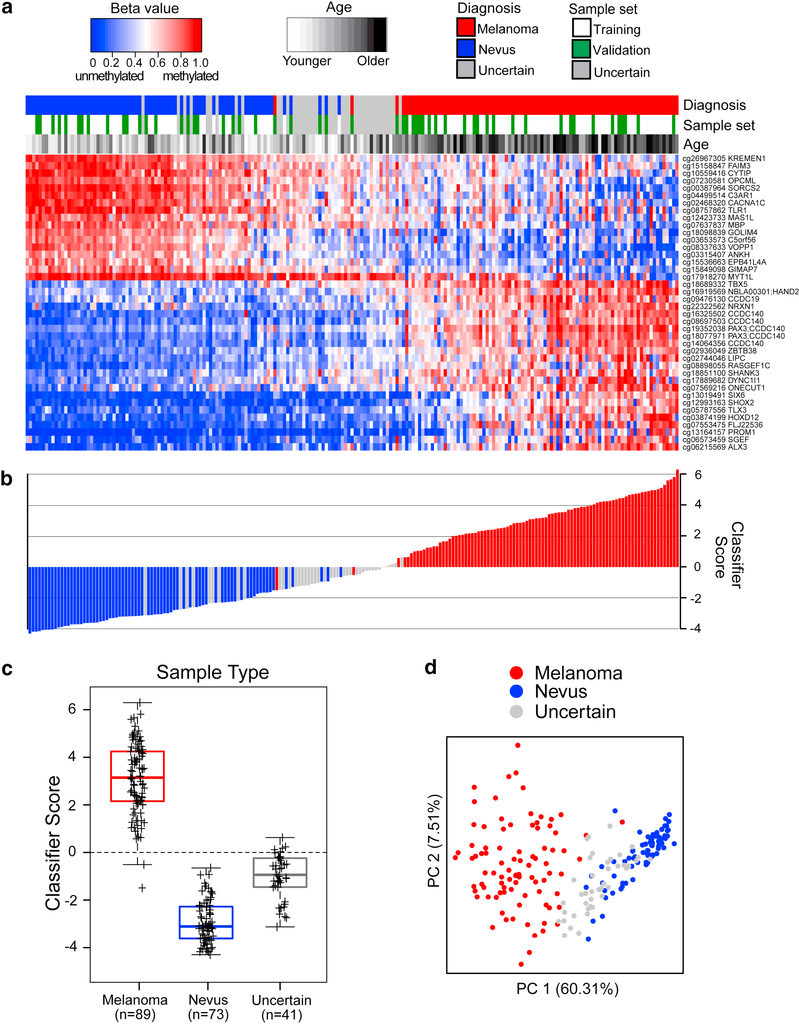
Early diagnosis improves melanoma survival, yet the histopathological diagnosis of cutaneous primary melanoma can be challenging, even for expert dermatopathologists. Analysis of epigenetic alterations, such as DNA methylation, that occur in melanoma can aid in its early diagnosis. Using a genome-wide methylation screening, we assessed CpG methylation in a diverse set of 89 primary invasive melanomas, 73 nevi, and 41 melanocytic proliferations of uncertain malignant potential, classified based on interobserver review by dermatopathologists. Melanomas and nevi were split into training and validation sets. Predictive modeling in the training set using ElasticNet identified a 40-CpG classifier distinguishing 60 melanomas from 48 nevi. High diagnostic accuracy (area under the receiver operator characteristic curve = 0.996, sensitivity = 96.6%, and specificity = 100.0%) was independently confirmed in the validation set (29 melanomas, 25 nevi) and other published sample sets. The 40-CpG melanoma classifier included homeobox transcription factors and genes with roles in stem cell pluripotency or the nervous system. Application of the 40-CpG melanoma classifier to the diagnostically uncertain samples assigned melanoma or nevus status, potentially offering a diagnostic tool to assist dermatopathologists. In summary, the robust, accurate 40-CpG melanoma classifier offers a promising assay for improving primary melanoma diagnosis.


Conflict of interest statement

Disclosure of Potential Conflicts of Interest: No potential conflicts of interest were disclosed

Figures

Figure 1.
Figure 1.. Performance of the 40-CpG melanoma classifier in training and/or validation sets.
Specimens in the training (60 melanomas and 48 nevi) and validation (29 melanomas and 25 nevi) sets had diagnostic consensus on interobserver review. The 40 diagnostic probes were identified from the model that analyzed annotated probes with IQR > 0.2 β between melanomas and nevi. (a) Heatmap showing methylation at 40 classifier probes in melanomas (red) and nevi (blue) from the combined training (white) and validation sets (green). Red represents highly methylated and blue represents unmethylated. (b) Boxplots of classifier scores for histological subtypes of nevi and melanomas. (c) ROC plot showing diagnostic accuracy in the validation set. (d) PCA showing the segregation of melanoma and nevus samples based on the 40 CpG classifier.
Figure 2.
Figure 2.. Independent validation of differential methylation at classifier CpG loci.
Validation of the diagnostic classifier was conducted in three public datasets. (a) 40-CpG methylation heatmap and waterfall plot of classifier scores in 105 primary melanomas from TCGA (TCGA, 2015) (yellow) compared with 89 melanomas and 73 nevi from UNC/UR (green). (b) Boxplots showing classifier scores for TCGA primary or metastatic melanomas and UNC/UR primary melanomas and nevi. (c) Boxplots showing classifier scores for 33 primary and 28 metastatic melanomas, and 14 nevi, and (d) ROC plot showing the diagnostic accuracy of the 40 CpG classifier comparing nevi to primary melanomas in the GSE86355 450K methylation dataset. In the GSE45266 27K methylation dataset, (e) PCA of methylation at 44 CpGs associated with diagnostic classifier genes illustrates segregation of 24 primary melanomas from 5 nevi, and (f) boxplots showing methylation differences at the 2 CpG loci (cg3874199 and cg19352038) directly matching 450K probes in the diagnostic classifier.
Figure 3.
Figure 3.. Diagnostic 40-CpG melanoma classifier calls on melanomas, nevi, and diagnostically uncertain samples.
Interobserver dermatopathologic review identified 89 melanomas, 73 nevi, and 41 uncertain samples. (a) Supervised heatmap, ordered left to right from lowest to highest diagnostic classifier score, showing methylation levels at the 40 diagnostic CpGs in melanomas (red) or nevi (blue) from the training (white) or validation sets (green), or uncertain samples (gray). (b) Waterfall plot of classifier scores, ordered as in the heatmap, and color-coded for diagnosis. (c) Boxplots of classifier scores for each diagnostic category, with median and interquartile range encompassed by each box. The broken lines indicate the classifier score threshold for distinguishing melanomas from nevi. (d) PCA plot shows sample segregation based on the 40 CpG classifier.

References

    1. Alexandrescu DT, Kauffman CL, Jatkoe TA, Hartmann DP, Vener T, Wang H, et al. Melanoma-specific marker expression in skin biopsy tissues as a tool to facilitate melanoma diagnosis. J Invest Dermatol 2010;130(7):1887–92. - PubMed
    1. Andtbacka RH, Kaufman HL, Collichio F, Amatruda T, Senzer N, Chesney J, et al. Talimogene Laherparepvec Improves Durable Response Rate in Patients With Advanced Melanoma. J Clin Oncol 2015;33(25):2780–8. - PubMed
    1. Arai E, Kanai Y. DNA methylation profiles in precancerous tissue and cancers: carcinogenetic risk estimation and prognostication based on DNA methylation status. Epigenomics 2010;2(3):467–81. - PubMed
    1. Balch CM, Gershenwald JE, Soong SJ, Thompson JF, Atkins MB, Byrd DR, et al. Final version of 2009 AJCC melanoma staging and classification. J Clin Oncol 2009;27(36):6199–206. - PMC - PubMed
    1. Baras A, Yu Y, Filtz M, Kim B, Moskaluk CA. Combined genomic and gene expression microarray profiling identifies ECOP as an upregulated gene in squamous cell carcinomas independent of DNA amplification. Oncogene 2009;28(32):2919–24. - PubMed

Publication types

MeSH terms

Substances