Review
. 2019 Feb:140:18-25.
doi: 10.1016/j.prostaglandins.2018.12.001. Epub 2018 Dec 4.

VIP modulates the ALX/FPR2 receptor axis toward inflammation resolution in a mouse model of bacterial keratitis

Affiliations
Review

VIP modulates the ALX/FPR2 receptor axis toward inflammation resolution in a mouse model of bacterial keratitis

Thomas W Carion et al. Prostaglandins Other Lipid Mediat. 2019 Feb.

Abstract

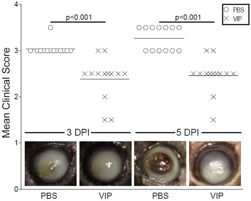
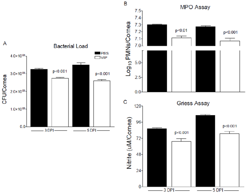
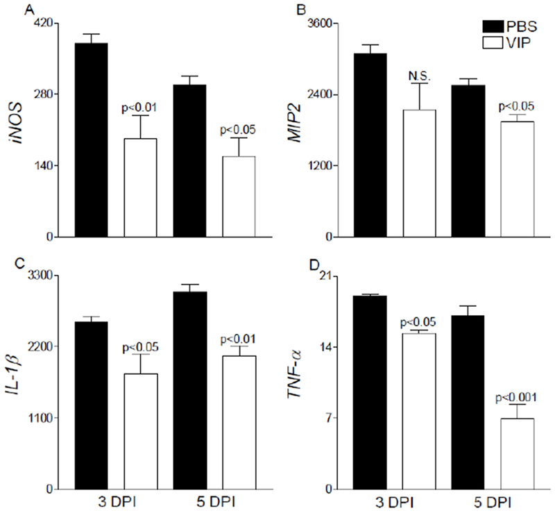
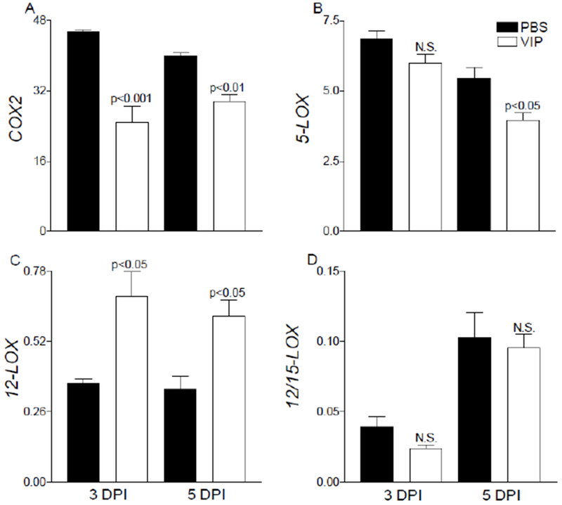
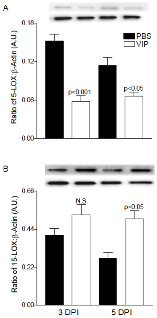
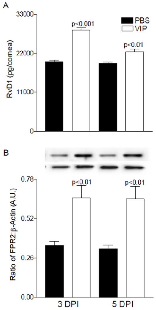
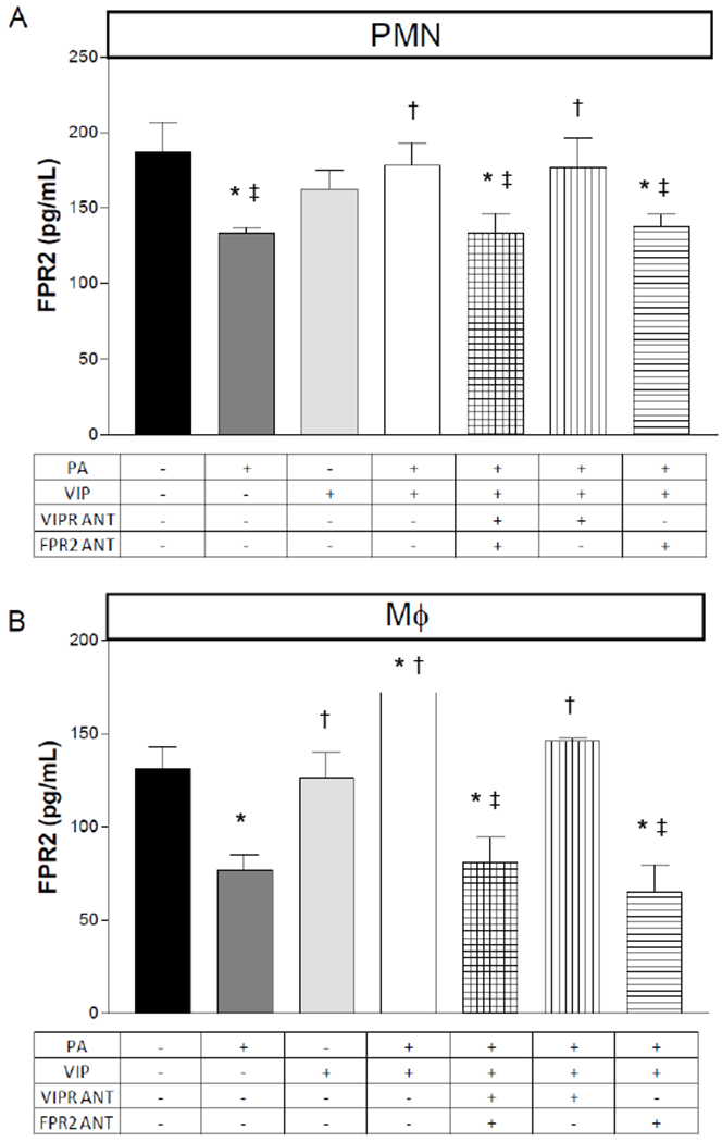
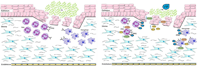
Vasoactive intestinal peptide (VIP) has been shown to regulate corneal inflammation. Formyl peptide receptor 2 (FPR2) is a transmembrane protein belonging to the GPCR family. Ligands include pro-resolving lipids, lipoxin A4 (LXA4) and resolvin D1 (RvD1). The current study focuses on the effect of VIP regarding the FPR2 receptor axis in improving disease outcome in a mouse model of bacterial keratitis. Infection was induced in C57BL/6 (B6) mice using P. aeruginosa (PA) ATCC 19660. Mice received topical treatment (VIP or PBS) 3× daily after infection. Mean clinical scores, bacterial plate counts, Griess and myeloperoxidase (MPO) assays indicate that topical VIP effectively abrogates the disease response. Findings also reveal that VIP influences FPR2 pathway activation independent of archetypal VIP receptors. Exploring the immunoresolving role of FPR2, its ligand RvD1 and related enzymes (5-LOX, 12/15-LOX), our results suggest a mechanism by which VIP treatment influences the disease response in bacterial keratitis, which could offer a therapeutic point of intervention for enhancing this pro-resolving circuit.

Keywords: Inflammation; Lipid mediators; Mouse; Ocular infection; Resolution.


Figures

Figure 1.
Figure 1.
Ocular disease response of P. aeruginosa-infected B6 mice after topical treatment with PBS or VIP. Results are mean clinical scores ± SEM (n = 13 mice/group). Significant differences between the two treatment groups were observed at both 3 and 5 days p.i. Representative eyes from PBS- and VIP-treated mice were photographed at the same time points and illustrate the disease response visually. Magnification = ×35.
Figure 2.
Figure 2.
A, Viable bacteria detected by direct plate counts from corneas of PBS- and VIP-treated B6 mice at 3 and 5 days p.i. Results are reported as CFU/cornea ± SEM (n = 6 corneas/group). B, Estimation of PMN as calculated from MPO levels detected from corneas of PBS- and VIP-treated B6 mice after infection. Results are reported as log10/cornea + SEM (n = 5 corneas/group). C, Nitrite levels from infected corneas after PBS and VIP treatment. Data are reported as μM/cornea ± SEM (n = 5 corneas/group).
Figure 3.
Figure 3.
Select pro-inflammatory cytokine/chemokine transcript levels were assessed after VIP treatment in infected corneas of B6 mice. Significant decreases were observed in iNOS (A), MIP-2 (B), IL-1β (C), and TNF-α (D) mRNA levels in VIP- versus PBS-treated mice. Results are presented as relative fold change for the gene of interest normalized to β-actin ± SEM (n = 5 corneas/group).
Figure 4.
Figure 4.
mRNA expression of lipid mediator biosynthetic enzymes, COX-2 (A), 5-LOX (B), 12-LOX (C), and 12/15-LOX (D) are provided from infected corneas of PBS- and VIP-treated B6 mice. Results are reported as relative fold change of the gene of interest normalized to β-actin ± SEM (n = 5 corneas/group).
Figure 5.
Figure 5.
Protein levels of 5-LOX (A) and 15-LOX (B) enzymes as detected by Western blot in infected corneas of PBS- and VIP-treated mice. Data shown are representative of three independent experiments in duplicate and expressed as mean ± SEM (n = 6 corneas/group).
Figure 6.
Figure 6.
Levels of RvD1 (A) and FPR2 (B) as detected by ELISA and Western blot (respectively) in infected corneas of PBS- and VIP-treated B6 mice. Results are reported as mean (pg/cornea) ± SEM for ELISA analysis. For Western blot, a representative image is provided from three independent experiments in duplicate and expressed as mean ± SEM (n = 6 corneas/group).
Figure 7.
Figure 7.
Protein levels of FPR2 as detected by ELISA in PMN and macrophages after an in vitro stimulation assay. Peritoneal-derived PMN (A) and macrophages (B) from B6 mice were exposed to P. aeruginosa (PA) +/− the presence of VIP, VIP receptor antagonist and FPR2 antagonist. Data are reported as pg/mL ± SD (n = 6). *p<0.05 vs. CTRL; †p<0.05 vs. PA; ‡p<0.05 vs. PA+VIP expressed as mean ± SEM (n = 6 corneas/group).
Figure 8.
Figure 8.
Schematic representation summarizing the influence of VIP on the cornea after P. aeruginosa-induced infection.

References

    1. Lichtinger A, et al., Shifting trends in bacterial keratitis in Toronto: an 11-year review. Ophthalmology, 2012. 119(9): p. 1785–90. - PubMed
    1. Keay L, Stapleton F, and Schein O, Epidemiology of contact lens-related inflammation and microbial keratitis: a 20-year perspective. Eye Contact Lens, 2007. 33(6 Pt 2): p. 346–53, discussion 362–3. - PubMed
    1. Stapleton F, et al., The incidence of contact lens-related microbial keratitis in Australia. Ophthalmology, 2008. 115(10): p. 1655–62. - PubMed
    1. Yildiz EH, et al., Trends in contact lens-related corneal ulcers at a tertiary referral center. Cornea, 2012. 31(10): p. 1097–102. - PubMed
    1. Lotti R and Dart JK, Cataract as a complication of severe microbial keratitis. Eye (Lond), 1992. 6 (Pt 4): p. 400–3. - PubMed

Publication types