Review
. 2019 Feb;7(2):173-186.
doi: 10.1016/S2213-2600(18)30345-X. Epub 2018 Dec 7.

Acute respiratory failure in immunocompromised adults

Affiliations
Review

Acute respiratory failure in immunocompromised adults

Elie Azoulay et al. Lancet Respir Med. 2019 Feb.

Abstract

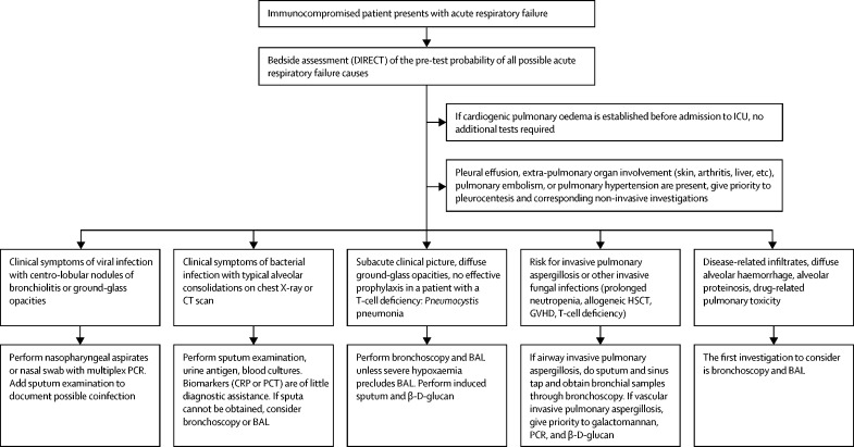
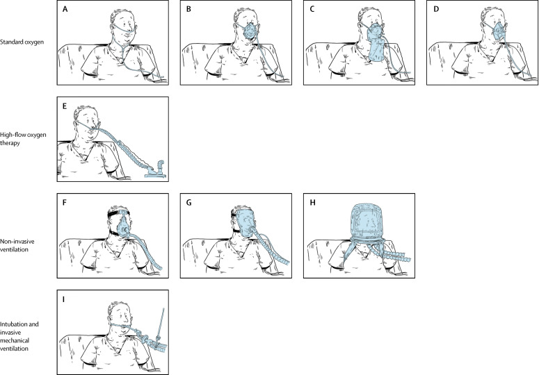
Acute respiratory failure occurs in up to half of patients with haematological malignancies and 15% of those with solid tumours or solid organ transplantation. Mortality remains high. Factors associated with mortality include a need for invasive mechanical ventilation, organ dysfunction, older age, frailty or poor performance status, delayed intensive care unit admission, and acute respiratory failure due to an invasive fungal infection or unknown cause. In addition to appropriate antibacterial therapy, initial clinical management aims to restore oxygenation and predict the most probable cause based on variables related to the underlying disease, acute respiratory failure characteristics, and radiographic findings. The cause of acute respiratory failure must then be confirmed using the most efficient, least invasive, and safest diagnostic tests. In patients with acute respiratory failure of undetermined cause, a standardised diagnostic investigation should be done immediately at admission before deciding whether to perform more invasive diagnostic procedures or to start empirical treatments. Collaborative and multidisciplinary clinical and research networks are crucial to improve our understanding of disease pathogenesis and causation and to develop less invasive diagnostic strategies and more targeted treatment options.


Figures

Figure 1
Figure 1
Causes of acute respiratory failure by time since diagnosis of haematological malignancy CT scans with red text refer to myeloid malignancies and those with blue text to lymphoid malignancies. The x-axis indicates time since diagnosis of the malignancy. For each scenario, the three most common diagnoses (in decreasing order) are reported at each timepoint and for each condition.
Figure 2
Figure 2
Risk for specific pathogens according to the type of haematological malignancy or treatment This figure illustrates the most frequently encountered types of infection according to the main disease-related or treatment-related immunological deficiency. It focuses mainly on secondary immunosuppression in adults, as data for primary immune deficiencies are scarce.
Figure 3
Figure 3
Five clinical vignettes for which the application of the DIRECT mnemonic leads to different diagnostic probabilities Diagnoses such as bacterial infection, pulmonary oedema, and alveolar haemorrhage need to ruled out or treated in every case. BAL=bronchoalveolar lavage.
Figure 4
Figure 4
Causes of pulmonary infiltrates in immunocompromised patients with acute respiratory failure Mean percentage is reported in the first column in red for each cause, with 95% confidence intervals reported underneath. The study by Gruson and colleagues included only bone marrow transplant recipients and the studies by Wohlfarth and colleagues and Schmidt and colleagues included only patients with severe acute respiratory distress syndrome treated with extra-corporeal membrane oxygenation.
Figure 5
Figure 5
First-line diagnostic strategy according to clinical and radiographic presentation ICU=intensive care unit. HSCT=haemopoietic stem cell therapy. GVHD=graft versus host disease. CRP=C-reactive protein. PCT=procalcitonin. BAL=bronchoalveolar lavage.
Figure 6
Figure 6
Nine oxygenation and ventilation methods Standard oxygen can be administered through a wide array of devices. Low-flow oxygen systems comprise a nasal cannula (or nasal catheters; A) providing supplemental oxygen at flows below the total minute ventilation, leading to oxygen dilution with ambient air and lowering the inspired oxygen concentration. A standard nasal cannula delivers an inspiratory FiO2 of 0·24–0·44 at supply flows 1–8 L/min, depending on respiratory rate and tidal volume. A humidification device is recommended for flows >4 L/min. Reservoir masks (B) can deliver FiO2 values of 0·40–0·60 at 5–10 L/min. The reservoir system (∼100–300 cm3) stores oxygen. A flow rate >5 L/min must be set to ensure the washout of exhaled gas and avoid CO2 retention. The non-rebreathing facemask (C) may deliver up to 0·90 FiO2 at flow settings >10 L/min. It should be used only for short periods, as humidification is difficult and there is also a risk of CO2 retention if the mask's reservoir bag is allowed to collapse on inspiration. Lastly, the Venturi system (D) mixes oxygen with room air (humidification is not necessary) and provides an accurate and constant FiO2 despite variations in respiratory rate and tidal volume. It is employed when concern arises about CO2 retention or when the respiratory drive is inconsistent. High-flow oxygen therapy through nasal prongs or cannulas (high-flow nasal oxygen therapy; E) supplies an exact FiO2 (up to 1) at a flow equal to or greater than the patient's inspiratory flow demand. Nasal oxygen is administered at a flow rate of up to 60 L/min. It is warmed to body temperature and saturated to full humidity by molecular humidification. Non-invasive positive-pressure ventilation provides ventilator support without an endotracheal tube, using an oronasal (F) or total face (G) mask or a helmet (H). The main non-invasive ventilation mode used in hypoxaemic acute respiratory failure is pressure support; continuous positive airway pressure and bi-level positive airway pressure are used less often. Finally, invasive mechanical ventilation (I) uses a tracheal tube inserted into the trachea under general anaesthesia and a neuromuscular-blocking drug. The tube is then secured to the face or neck and connected to a ventilator. In patients with hypoxaemic acute respiratory failure, intubation and invasive mechanical ventilation are used after failure of standard oxygen and, in some cases, of the aforementioned non-invasive options. FiO2= fraction of inspired oxygen.

References

    1. Harpaz R, Dahl RM, Dooling KL. Prevalence of immunosuppression among US adults, 2013. JAMA. 2016;316:2547–2548. - PubMed
    1. Siegel RL, Miller KD, Jemal A. Cancer statistics, 2018. CA Cancer J Clin. 2018;68:7–30. - PubMed
    1. Israni AK, Zaun D, Rosendale JD, Schaffhausen C, Snyder JJ, Kasiske BL. OPTN/SRTR 2016 annual data report: deceased organ donation. Am J Transplant. 2018;18(suppl 1):434–463. - PubMed
    1. Weyand CM, Goronzy JJ. Clinical practice. Giant-cell arteritis and polymyalgia rheumatica. N Engl J Med. 2014;371:50–57. - PMC - PubMed
    1. Azoulay E, Thiéry G, Chevret S. The prognosis of acute respiratory failure in critically ill cancer patients. Medicine (Baltimore) 2004;83:360–370. - PubMed

Uncited Reference

    1. Rañó A, Agustí C, Jimenez P. Pulmonary infiltrates in non-HIV immunocompromised patients: a diagnostic approach using non-invasive and bronchoscopic procedures. Thorax. 2001;56:379–387. - PMC - PubMed

MeSH terms