. 2019 Feb 15;294(7):2232-2246.
doi: 10.1074/jbc.RA118.006691. Epub 2018 Dec 10.

cAMP-dependent activation of the Rac guanine exchange factor P-REX1 by type I protein kinase A (PKA) regulatory subunits

Affiliations

cAMP-dependent activation of the Rac guanine exchange factor P-REX1 by type I protein kinase A (PKA) regulatory subunits

Sendi Rafael Adame-García et al. J Biol Chem. .

Abstract

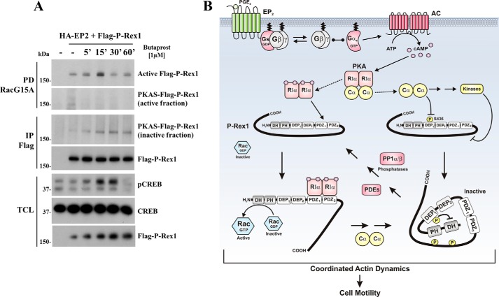
Regulatory subunits of protein kinase A (PKA) inhibit its kinase subunits. Intriguingly, their potential as cAMP-dependent signal transducers remains uncharacterized. We recently reported that type I PKA regulatory subunits (RIα) interact with phosphatidylinositol 3,4,5-trisphosphate-dependent Rac exchange factor 1 (P-REX1), a chemotactic Rac guanine exchange factor (RacGEF). Because P-REX1 is known to be phosphorylated and inhibited by PKA, its interaction with RIα suggests that PKA regulatory and catalytic subunits may fine-tune P-REX1 activity or those of its target pools. Here, we tested whether RIα acts as a cAMP-dependent factor promoting P-REX1-mediated Rac activation and cell migration. We observed that Gs-coupled EP2 receptors indeed promote endothelial cell migration via RIα-activated P-REX1. Expression of the P-REX1-PDZ1 domain prevented RIα/P-REX1 interaction, P-REX1 activation, and EP2-dependent cell migration, and P-REX1 silencing abrogated RIα-dependent Rac activation. RIα-specific cAMP analogs activated P-REX1, but lost this activity in RIα-knockdown cells, and cAMP pulldown assays revealed that P-REX1 preferentially interacts with free RIα. Moreover, purified RIα directly activated P-REX1 in vitro We also found that the RIα CNB-B domain is critical for the interaction with P-REX1, which was increased in RIα mutants, such as the acrodysostosis-associated mutant, that activate P-REX1 at basal cAMP levels. RIα and Cα PKA subunits targeted distinct P-REX1 molecules, indicated by an absence of phosphorylation in the active fraction of P-REX1. This was in contrast to the inactive fraction in which phosphorylated P-REX1 was present, suggesting co-existence of dual stimulatory and inhibitory effects. We conclude that PKA's regulatory subunits are cAMP-dependent signal transducers.

Keywords: EP2 receptors; G protein–coupled receptor (GPCR); P-REX1; Rac (Rac GTPase); Rho guanine nucleotide exchange factor (RhoGEF); cell migration; cell signaling; chemotaxis; cyclic AMP (cAMP); endothelial cell; guanine nucleotide exchange factor (GEF); heterotrimeric G protein; protein kinase A (PKA); regulatory subunit alpha (RIα); wound healing.


Conflict of interest statement

The authors declare that they have no conflicts of interest with the contents of this article

Figures

Figure 1.
Figure 1.
EP2 prostaglandin receptors promote endothelial cell migration, P-REX1/Rac activation, and interaction of endogenous PKA–RIα with active P-REX1. A, working hypothesis postulating that P-REX1 activity is fine-tuned by independent actions of type I PKA regulatory and catalytic subunits. Accordingly, Gs-coupled EP2 receptor activates P-REX1 via cAMP-dependent direct interaction with regulatory subunits and eventually, as we previously described (7), inhibited by phosphorylation leading to intramolecular inhibitory interactions. B, PGE2 (1 μm) and butaprost (1 μm) (a specific EP2 agonist) promote migration of PAE cells in wound-healing assays. The pictures represent the wound closure of PAE cells after 16 h of stimulation, and S1P (1 μm) and 10% FBS were used as positive controls. Three independent experiments were performed (n = 3). C, PGE2 (1 μm) and butaprost (1 μm) promote chemotactic migration of PAE cells in Boyden Chamber Chemotaxis assays; S1P (1 μm) and HGF (10 ng/ml) were used as positive controls. Graph shows the densitometric analysis of four independent chemotaxis assays (n = 4). Data were analyzed by one-way ANOVA followed by Tukey's multiple comparison test, and p value is indicated. D, effect of EP2 activation on actin cytoskeleton reorganization in PAE cells was assessed with phalloidin staining. PAE cells, starved for 16 h, were stimulated with PGE2 (1 μm) and S1P (1 μm) for 15 min, and DAPI was used to show the nuclei. Images are representative of 10 different fields. E and F, PGE2 (500 nm) and butaprost (500 nm) promote phosphorylation of PKA substrate CREB at serine 133. Serum-starved PAE cells were stimulated with PGE2 or butaprost at the indicated times and then lysed and processed for immunoblot against pCREB and total CREB. Graphs show the densitometric analysis of pCREB from three independent experiments (n = 3). One-way ANOVA followed by Tukey's multiple comparison test was performed; significant p values are indicated. G, EP2 stimulation induces Rac activation in endothelial cells. Serum-starved PAE cells were stimulated with butaprost (500 nm) at the indicated times and lysed and processed for pulldown using GST-PAKN beads and analyzed by immunoblot with anti-Rac, pCREB, and total CREB antibodies. H, EP2 stimulation promotes P-REX1 activation in endothelial cells. Serum-starved PAE cells were stimulated with butaprost (1 μm) at the indicated times and processed for pulldown assays to capture active P-REX1 using GST–RacG15A beads. Western blottings of anti-P-REX1, PKA–RIα, and Cα were done in pulldowns, and total cell lysates, as well as pCREB and total CREB, were used as control. PKA-Cα was not detected associated with active P-REX1 (not shown). Graph shows the densitometric analysis of active P-REX1 from three independent experiments (n = 3). One-way ANOVA followed by Tukey's multiple comparison test was performed, and significant p values are shown in the graph.
Figure 2.
Figure 2.
Direct stimulation of type I PKA promotes P-REX1 activation. A, hypothetical effect of direct stimulation of type I PKA on P-REX1. B, direct stimulation of type I PKA promotes Rac activation in endothelial cells. Serum-starved PAE cells were stimulated at the indicated times with 6-Bnz–cAMP (10 μm) and 8-AHA–cAMP (10 μm) cAMP analogs, and active Rac was isolated with GST–PAKN beads. Total and pCREB were detected by Western blotting in total cell lysates. Graph shows the densitometric analysis of four independent experiments of Rac activation (n = 4). One-way ANOVA followed by Tukey's multiple comparison test was performed, and significant p values are shown in the graph. C, direct stimulation of type I PKA promotes P-REX1 activation and association of RIα with the active fraction of P-REX1 in endothelial cells. PAE cells were stimulated as indicated in B, and active P-REX1 was isolated with GST–RacG15A and detected by immunoblot; RIα was revealed in the pulldown of active P-REX1 and in total cell lysates. pCREB and total CREB revealed in total cell lysates were used as controls. Graph shows the densitometric analysis of active P-REX1 from three independent experiments (n = 3). One-way ANOVA followed by Tukey's multiple comparison test was performed, and significant p values are shown in the graph. D, P-REX1 lacking the C-terminal region is activated in response to direct stimulation of type I PKA. HEK293T cells expressing FLAG–P-REX1–DH–PDZ2 construct were serum-starved and stimulated with cAMP analogs for type I PKA at the indicated times. Active P-REX1 was isolated with GST–RacG15A beads. Samples were processed for immunoblot against FLAG, RIα, pCREB, and total CREB. Graph shows the time course of FLAG–P-REX1 activation from three independent experiments (n = 3). One-way ANOVA followed by Tukey's multiple comparison test were performed, and the significant p value is shown in the graph.
Figure 3.
Figure 3.
P-REX1 activation by type I PKA is linked to its interaction with RIα. A, stimulation of the cAMP pathway promotes interaction between endogenous P-REX1 and RIα in endothelial cells. Serum-starved PAE cells were stimulated with forskolin (10 μm) at the indicated times. Then endogenous RIα was immunoprecipitated, and bound P-REX1 was detected by Western blotting. Total P-REX1, RIα, pCREB (stimulation control), and total CREB were revealed in total cell lysates. B, stimulation of the cAMP pathway promotes interaction between endogenous RIα and the DH–PDZ2 region of P-REX1. HEK293T cells expressing GST–P-REX1–DH–PDZ2 construct were serum-starved and stimulated with forskolin (10 μm) as indicated. P-REX1–DH–PDZ2 was isolated using GSH-Sepharose beads. Immunoblot was performed against RIα, GST, pCREB (stimulation control), and total CREB. Graph shows the time course of forskolin-induced RIα interaction with P-REX1 from three independent experiments (n = 3). One-way ANOVA followed by Tukey's multiple comparison test was performed, and significant p value is shown in the graph. C, direct stimulation of type I PKA promotes interaction of RIα with P-REX1. HEK293T cells expressing GST–P-REX1–DH–PDZ2 construct were serum-starved and then stimulated with 6Bnz-cAMP (10 μm) and 8AHA-cAMP (10 μm) as indicated. P-REX1 was isolated by GST pulldown, and samples were analyzed by Western blotting against RIα, GST, pCREB (stimulation control), and total CREB. Graph shows the time course of directly activated RIα interacting with P-REX1, from three independent experiments (n = 3). One-way ANOVA followed by Kruskal-Wallis test was performed, and significant p value is indicated. D, stimulation of the cAMP pathway promotes interaction between Z6 construct (RIα CNB-B domain) and the PDZ1–PDZ2 region of P-REX1. HEK293T cells co-expressing EGFP–Z6 and GST–P-REX1–PDZ1–PDZ2 constructs were serum-starved and stimulated with forskolin (10 μm) as indicated. GST–P-REX1–PDZ1–PDZ2 was isolated with GSH-Sepharose beads. Immunoblot was performed against GFP, GST, pCREB (stimulation control), and total CREB. Three independent experiments were performed (n = 3). E, schematic representation of WT and mutant RIα constructs R335K and acrodysostosis(1–365) and Z6 (CNB-B). F, analysis of interaction between WT RIα and mutants at CNB-B domain and P-REX1 PDZ–PDZ module. HEK293T cells co-expressing GST–P-REX1–PDZ1–PDZ2 domains and FLAG–RIα WT or mutants R335K or ACRO were serum-starved, lysed, and prepared for GST pulldown interaction assays as described previously. Immunoblots were performed against FLAG and GST. Three independent experiments were performed (n = 3). G, RIα ACRO mutant increases P-REX1 activity. MCF7 cells expressing empty vector or RI WT or R335K or ACRO mutants were serum-starved and lysed, and active P-REX1 was isolated with GST–RacG15A beads. Immunoblots were performed for P-REX1 and FLAG. Graph shows the densitometric analysis of three independent experiments (n = 3). The p value obtained by one-tail Student's t test comparing control versus ACRO is indicated.
Figure 4.
Figure 4.
P-REX1 activation by type I PKA depends on regulatory, but not catalytic, subunit expression. A and B, Rac activation in response to direct stimulation of type I PKA requires P-REX1. MCF7 cells transfected with esiRNAs (A) or individual siRNAs (B) for P-REX1 or GFP (as negative control) were serum-starved and stimulated at the indicated times with type I PKA-specific cAMP analogs. Active Rac was isolated by pulldown with GST–PAKN and detected by immunoblot. Western blottings in total cell lysates were done for Rac, P-REX1, pCREB, and total CREB. Graph in A represents the densitometric analysis of three independent experiments (n = 3). Two-way ANOVA followed by Tukey test was performed, and significant p value is indicated. n.s., no significant p > 0.05. C and D, P-REX1 activation in response to direct stimulation of type I PKA requires the RIα subunit but not catalytic Cα subunit. MCF7 cells transfected with esiRNAs for PKA–RIα or -Cα subunits (C), or siRNAs for PKA–RIα or βGal (as negative control) (D), were serum-starved and stimulated with type I PKA-specific cAMP analogs at the indicated times. Active P-REX1 was isolated with GST–RacG15A and detected by immunoblot. Phosphorylation of CREB as well as expression of P-REX1, RIα, Cα, and total CREB were assessed by Western blotting in total cell lysates. Graph in C represents the densitometric analysis of active P-REX1 from three independent experiments (n = 3). One-tail Student's t test was performed for the comparisons, and significant p values are indicated.
Figure 5.
Figure 5.
Endogenous P-REX1 preferentially interacts with cAMP-bound RIα, and in vitro they form an active RacGEF complex. A, drawing shows the experimental strategy to assess whether P-REX1 preferentially interacts with cAMP-bound RIα with respect to type I PKA holoenzyme, isolated by pulldown with affinity matrices containing cAMP or the antagonist analog (Rp)-8AHA-cAMP. B, P-REX1 preferentially interacts with free cAMP–RIα but not with inactive PKA holoenzyme. Lysates of serum-starved MCF7 cells were used for pulldown assays with cAMP or (Rp)-cAMP-agaroses. P-REX1 and RIα in the pulldown, and total cell lysate, were detected by Western blotting. C, graph represents the densitometric analysis of three independent experiments (n = 3) like the one shown in B. Results were analyzed by one-tail Student's t test, and the p value is indicated in the graph. D, Coomassie staining of isolated full-length RIα and P-REX1 used for in vitro assays. RIα was expressed in E. coli BL21 strain and purified using cAMP-agarose followed by gel filtration. HA–HaloTag–P-REX1, expressed in HEK293T cells, was isolated by pulldown with Halolink resin. P-REX1 was released from the resin using HaloTag–tobacco etch virus protease, removing the HA–HaloTag. E, isolated P-REX1 and RIα were incubated in vitro and subjected to pull down analysis with cAMP or (Rp)-agaroses. Samples were immunoblotted against P-REX1 and RIα. The graph below represents the densitometric analysis of three independent experiments (n = 3). Results were analyzed by one-tail Student's t test, and p value is indicated. F, P-REX1 is directly activated by RIα. Purified P-REX1 was incubated in the presence or absence of RIα for 15 min, and the active fraction of P-REX1 was isolated with GST–RacG15A beads. P-REX1 and RIα were detected by immunoblot. Recombinant GST–RacG15A was stained with Ponceau red. The graph below represents the densitometric analysis of three independent experiments (n = 3). Results were analyzed by one tail Student's t test, and p value is indicated.
Figure 6.
Figure 6.
P-REX1 interaction with PKA–RIα is necessary for EP2-dependent endothelial cell migration. A, drawing shows the experimental strategy to assess whether P-REX1–PDZ1 domain interferes on the interaction of PKA RIα subunits with P-REX1, preventing RIα-dependent P-REX1 activation and cell migration stimulated by EP2 receptors. B, endogenous RIα interacts with P-REX1–PDZ1–PDZ2 domains in a cAMP-dependent manner. HEK293T cells expressing GST–P-REX1–PDZ1–PDZ2 were serum-starved, stimulated with forskolin (10 μm) at the indicated times, and lysed. GST–P-REX1–PDZ1–PDZ2 was isolated with glutathione-Sepharose beads. Immunoblot was performed against RIα, GST, pCREB (stimulation control), and total CREB in pulldowns (PD GST) or total cell lysates (TCL) as indicated. Graph shows the time course of forskolin-induced RIα interaction with P-REX1–PDZ1–PDZ2 from three independent experiments (n = 3). One-way ANOVA followed by Tukey's multiple comparison test was performed, and significant p value is shown in the graph. C, P-REX1 PDZ1 domain competes with endogenous P-REX1 for the interaction with RIα pulled down with cAMP-agarose. MCF7 cells expressing EGFP or EGFP–P-REX1–PDZ1 were serum-starved and lysed. Endogenous RIα was isolated with cAMP-agarose beads. RIα, P-REX1, and GFP–PDZ1 in the pulldowns were revealed by immunoblot. Total cell lysates (TCL, right panel) were used to confirm the expression of the indicated proteins. Three independent experiments were performed (n = 3). D, expression of P-REX1–PDZ1 domain prevents P-REX1 activation in response to type I PKA-specific stimulation. MCF7 cells expressing EGFP or EGFP–P-REX1–PDZ1 were serum-starved, stimulated with type I PKA-specific cAMP analogs at the indicated times, and lysed. Active endogenous P-REX1 was isolated using GST–RacG15A beads and revealed by immunoblot. Total cell lysates (TCL) were used to detect P-REX1, pCREB (stimulation control), CREB, and GFP. Three independent experiments were performed (n = 3). E, expression of P-REX1–PDZ1 domain prevents EP2-dependent endothelial cell migration. Confluent PAE cells expressing EGFP or EGFP–P-REX1–PDZ1 were serum-starved and subjected to wound-healing assays. Cells were stimulated with PGE2 (1 μm) or butaprost (1 μm). Pictures represent the wound closure after 16 h of stimulation; 10% fetal bovine serum (FBS) was used as control. F, graph represents the results of four independent experiments (n = 4). Statistical analysis was done by two-way ANOVA followed by Tukey; p values are indicated in the graph. n.s., no significant p > 0.05.
Figure 7.
Figure 7.
PKA regulatory and catalytic subunits target distinct P-REX1 molecules. A, COS7 cells co-expressing 3×HA-EP2 receptors and 3×FLAG–P-REX1 were serum-starved and stimulated with butaprost (1 μm) at the indicated times. Active P-REX1 was isolated with GST–RacG15A beads, and inactive P-REX1 was isolated from supernatants of RacG15A pulldowns by immunoprecipitation with anti-FLAG antibodies. Phosphorylation status of active and inactive P-REX1 was assessed by PKAS. Total cell lysates (TCL) were used to reveal pCREB (stimulation control), CREB, and FLAG–P-REX1. Three independent experiments were performed (n = 3). B, model explaining the effect of PKA regulatory and catalytic subunits on different P-REX1 molecules. Gs-coupled EP2 receptors stimulate adenylate cyclase generating cAMP that binds the RIα subunits of PKA holoenzyme promoting dissociation of cAMP-bound regulatory subunits and active kinase subunits. Based on our results, cAMP–RIα directly activates a fraction of P-REX1 molecules, stimulating GTP loading to Rac (left, P-REX1 molecule, stimulatory input). In contrast, PKA Cα subunits phosphorylate and inhibit a distinct pool of P-REX1 (right, P-REX1 molecule, inhibitory input). We previously demonstrated that inhibitory phosphorylation of P-REX1 by PKA occurs at Ser-436 on the DEP1 domain and PKA-regulated kinases that phosphorylate the P-REX1 C-terminal region, promoting intramolecular inhibitory interactions (7). Gradually, the fraction of active P-REX1 decreases, and the inactive, phosphorylated form increases, indicating that eventually the PKA Cα inhibitory activity predominates. Later, P-REX1 is taken to its basal state by the intervention of protein phosphatases and cAMP-phosphodiesterases. Overall, our results suggest that P-REX1 activity is fine-tuned by the combined effects of PKA subunits leading to organized cytoskeleton dynamics and effective cell migration.

Similar articles

Cited by

References

    1. Howe A. K. (2004) Regulation of actin-based cell migration by cAMP/PKA. Biochim. Biophys. Acta 1692, 159–174 10.1016/j.bbamcr.2004.03.005 - DOI - PubMed
    1. Taylor S. S., Zhang P., Steichen J. M., Keshwani M. M., and Kornev A. P. (2013) PKA: lessons learned after twenty years. Biochim. Biophys. Acta 1834, 1271–1278 10.1016/j.bbapap.2013.03.007 - DOI - PMC - PubMed
    1. Knighton D. R., Zheng J. H., Ten Eyck L. F., Ashford V. A., Xuong N. H., Taylor S. S., and Sowadski J. M. (1991) Crystal structure of the catalytic subunit of cyclic adenosine monophosphate-dependent protein kinase. Science 253, 407–414 10.1126/science.1862342 - DOI - PubMed
    1. Torres-Quesada O., Mayrhofer J. E., and Stefan E. (2017) The many faces of compartmentalized PKA signalosomes. Cell. Signal. 37, 1–11 10.1016/j.cellsig.2017.05.012 - DOI - PubMed
    1. Lim C. J., Han J., Yousefi N., Ma Y., Amieux P. S., McKnight G. S., Taylor S. S., and Ginsberg M. H. (2007) α4 integrins are type I cAMP-dependent protein kinase-anchoring proteins. Nat. Cell Biol. 9, 415–421 10.1038/ncb1561 - DOI - PubMed

Publication types

MeSH terms

Substances