. 2019 Feb 1;294(5):1529-1540.
doi: 10.1074/jbc.RA118.003995. Epub 2018 Dec 10.

An interdomain bridge influences RNA binding of the human La protein

Affiliations

An interdomain bridge influences RNA binding of the human La protein

Stefano A Marrella et al. J Biol Chem. .

Abstract

La proteins are RNA chaperones that perform various functions depending on distinct RNA-binding modes and their subcellular localization. In the nucleus, they help process UUU-3'OH-tailed nascent RNA polymerase III transcripts, such as pre-tRNAs, whereas in the cytoplasm they contribute to translation of poly(A)-tailed mRNAs. La accumulation in the nucleus and cytoplasm is controlled by several trafficking elements, including a canonical nuclear localization signal in the extreme C terminus and a nuclear retention element (NRE) in the RNA recognition motif 2 (RRM2) domain. Previous findings indicate that cytoplasmic export of La due to mutation of the NRE can be suppressed by mutations in RRM1, but the mechanism by which the RRM1 and RRM2 domains functionally cooperate is poorly understood. In this work, we use electromobility shift assays (EMSA) to show that mutations in the NRE and RRM1 affect binding of human La to pre-tRNAs but not UUU-3'OH or poly(A) sequences, and we present compensatory mutagenesis data supporting a direct interaction between the RRM1 and RRM2 domains. Moreover, we use collision-induced unfolding and time-resolved hydrogen-deuterium exchange MS analyses to study the conformational dynamics that occur when this interaction is intact or disrupted. Our results suggest that the intracellular distribution of La may be linked to its RNA-binding modes and provide the first evidence for a direct protein-protein interdomain interaction in La proteins.

Keywords: La protein; RNA processing; RNA-binding protein; RNA–protein interaction; SSB; Sjögren syndrome antigen B; intracellular trafficking; mass spectrometry (MS); precursor tRNA (pre-tRNA); protein domain.


Conflict of interest statement

The authors declare that they have no conflicts of interest with the contents of this article

Figures
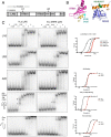
Figure 1.
Figure 1.
RNA-binding patterns of WT human La and hLa dNRE to various RNA substrates. A, domain arrangement of the human La protein. LAM, La motif; RRM1, RNA recognition motif 1; NRE, nuclear retention element; SBM, short basic motif; NLS, nuclear localization signal. B, structural representations highlighting regions of interest in the RRM1 and RRM2 domains of human La. RRM1 is in pink with RRM1-α1 in yellow, and Glu-132 and Asp-133 are in cyan. RRM2 is in violet, with RRM2-α3 in orange, and Lys-316 and Lys-317 are in green. Structures were generated from PDB codes 2VOO and 1OWX. C, EMSA showing hLa and hLa dNRE binding to U10, U20, A20, pre-tRNA AlaCGC, and a stem-loop with a 5′ single-stranded extension derived from the hepatitis C IRES (left). Binding curves for EMSA data are at right; hLa is in black and dNRE in red.
Figure 2.
Figure 2.
RNA-binding patterns of point-mutated NRE and RRM1 mutants to various RNA substrates. Left, EMSA of hLa E132K/D133K, hLa K316E/K317E, and hLa E132K/D133K/K316E/K317E to U10, U20, A20 and pre-tRNA AlaCGC. Right, binding curves for EMSA data hLa in black, hLa E132K/D133K in blue, hLa K317E/K316E in orange, and hLa E132K/D133K/K316E/K317E in purple. Supershifts of interest designated as “1” and “2”, see text.
Figure 3.
Figure 3.
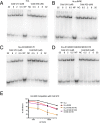
Competition experiments show that mutation of the nuclear retention element results in U10 competing better for A20 binding relative to WT hLa. A, competition experiment in which increasing amounts of cold U10 (left) or A20 (right) are tested for ability to displace radiolabeled A20 from hLa. NP, no protein added; NC, no cold competitor added. B–D, same as for A, but testing for hLa dNRE (B), hLa K316E/K317E (C), and hLa E132K/D133K/K316E/K317E (D). E, graphical representation of capacity of cold U10 to displace radiolabeled A20 from various tested mutants. Error bars, standard deviation.
Figure 4.
Figure 4.
CIU experiments on WT hLa (A), hLa dNRE (B), WT hLa (C) bound to U10, and hLa dNRE bound to U10 (D), where the unfolding and conformational changes of hLa are tracked over increasing collision energy. The left panel shows a representation of the most likely structural conformations that populate each unfolding state. The La motif, RRM1, and RRM2 are represented by the red, yellow, and green squares, respectively, in which the proposed interdomain interaction between RRM1 and RRM2 is represented by the dashed line.
Figure 5.
Figure 5.
Differences in deuterium uptake mapped onto the structures obtained from PDB (codes 1OWX and 1YTY). Changes in deuterium uptake are in the WT hLa U10 (A) and A20 complexes (B), in which C is a representation of the likely domain organization prior to RNA binding. Changes in deuterium uptake are in the hLa dNRE U10 (D) and A20 complexes (E), in which F is the proposed domain organization prior to RNA binding. The areas of the protein with decreased, increased, or no change in deuterium uptake are highlighted in blue, red, and tan, respectively. The areas colored gray mean there were no observed peptides from that area.
Figure 6.
Figure 6.
Differences in deuterium uptake mapped onto the structures obtained from PDB (codes 1OWX and 1YTY). A shows the hypothesized disrupted interdomain interaction in hLa K316E/K317E in which the key residues are highlighted in orange. Changes in deuterium uptake are in the hLa K316E/K317E U10 (B) and A20 complexes (C). D shows the re-establishment of the inter-domain interaction in hLa D132K/E133K/K316E/K317E in which the key residues are highlighted in orange. Changes in deuterium uptake are in the hLa D132K/E133K/K316E/K317E U10 (E) and A20 complexes (F). B, C, E, and F, areas of the protein with decreased, increased, or no change in deuterium uptake are highlighted in blue, red and tan, respectively. The areas colored gray means there were no observed peptides from that area.

References

    1. Wolin S. L., and Cedervall T. (2002) The La protein. Annu. Rev. Biochem. 71, 375–403 10.1146/annurev.biochem.71.090501.150003 - DOI - PubMed
    1. Kotik-Kogan O., Valentine E. R., Sanfelice D., Conte M. R., and Curry S. (2008) Structural analysis reveals conformational plasticity in the recognition of RNA 3′ ends by the human La protein. Structure 16, 852–862 10.1016/j.str.2008.02.021 - DOI - PMC - PubMed
    1. Teplova M., Yuan Y.-R., Phan A. T., Malinina L., Ilin S., Teplov A., and Patel D. J. (2006) Structural basis for recognition and sequestration of UUU(OH) 3′ termini of nascent RNA polymerase III transcripts by La, a rheumatic disease autoantigen. Mol. Cell 21, 75–85 10.1016/j.molcel.2005.10.027 - DOI - PMC - PubMed
    1. Naeeni A. R., Conte M. R., and Bayfield M. A. (2012) RNA chaperone activity of human La protein is mediated by variant RNA recognition motif. J. Biol. Chem. 287, 5472–5482 10.1074/jbc.M111.276071 - DOI - PMC - PubMed
    1. Hussain R. H., Zawawi M., and Bayfield M. A. (2013) Conservation of RNA chaperone activity of the human La-related proteins 4, 6, and 7. Nucleic Acids Res. 41, 8715–8725 10.1093/nar/gkt649 - DOI - PMC - PubMed

Publication types

Grants and funding

LinkOut - more resources