. 2019 Feb 21;87(3):e00846-18.
doi: 10.1128/IAI.00846-18. Print 2019 Mar.

A Novel, Multiple-Antigen Pneumococcal Vaccine Protects against Lethal Streptococcus pneumoniae Challenge

Affiliations

A Novel, Multiple-Antigen Pneumococcal Vaccine Protects against Lethal Streptococcus pneumoniae Challenge

Win-Yan Chan et al. Infect Immun. .

Erratum in

Corrected and republished in

Abstract

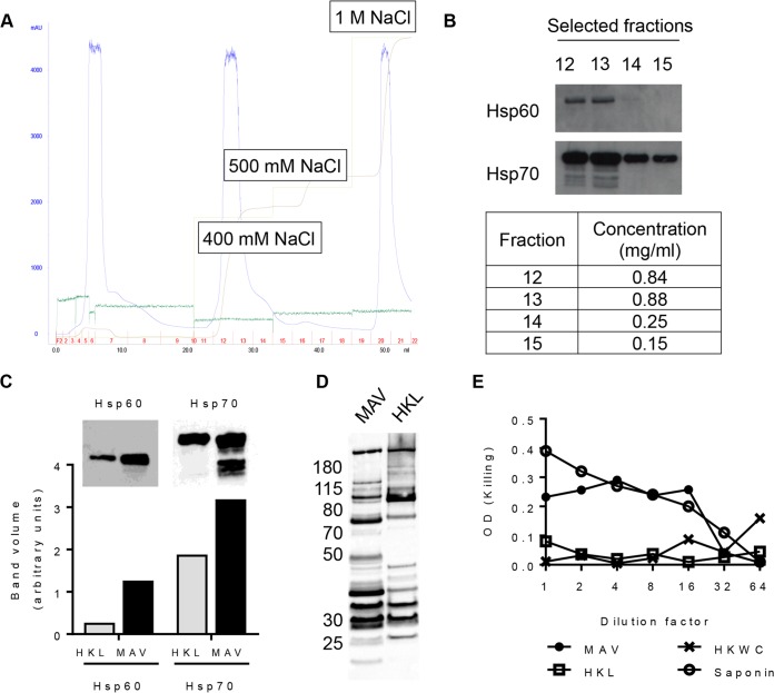
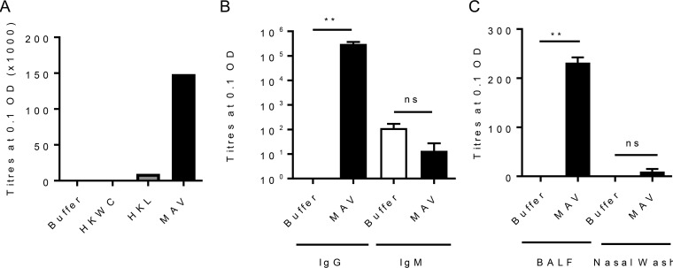
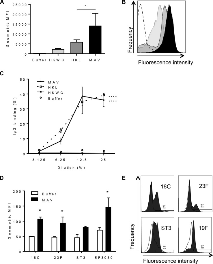
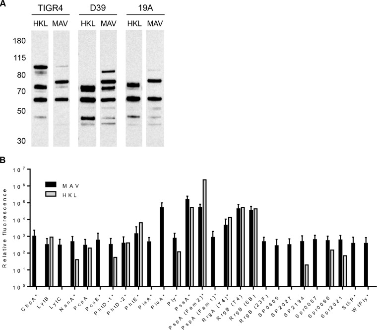
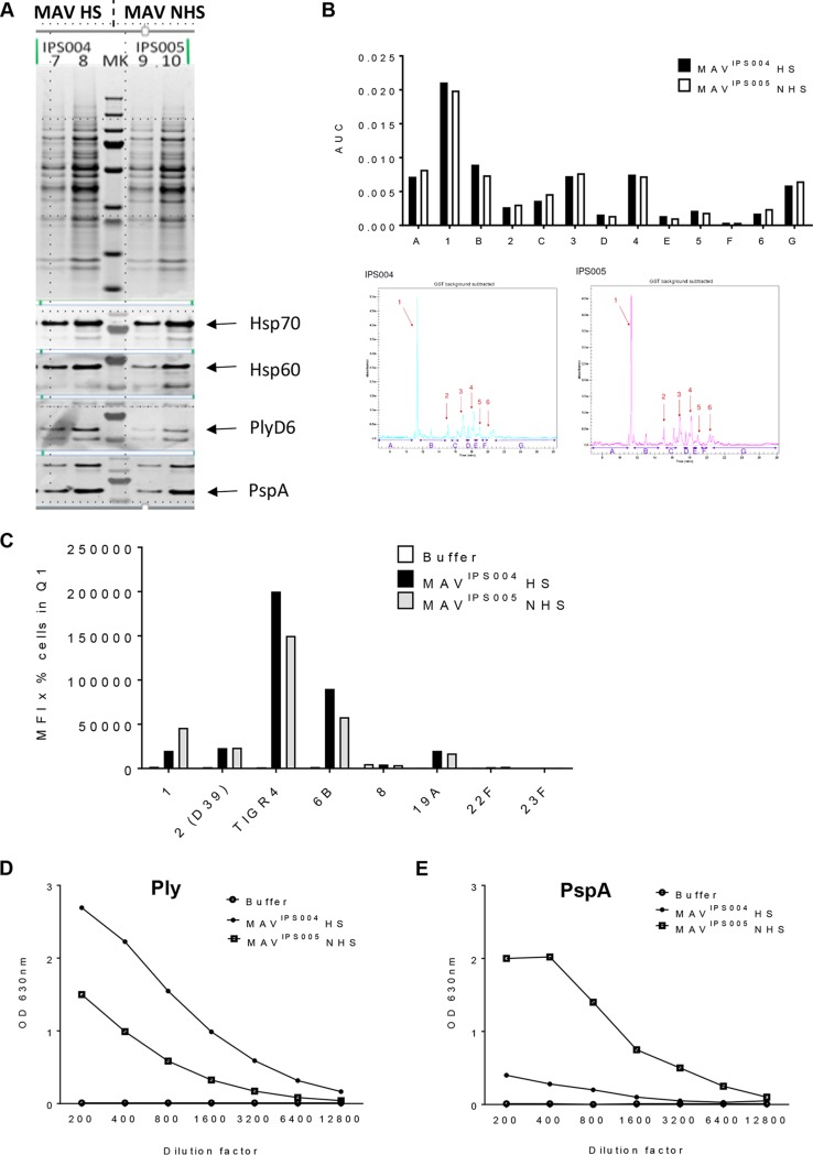
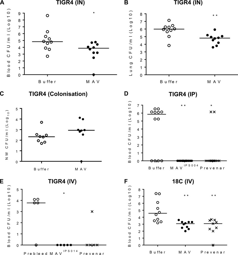
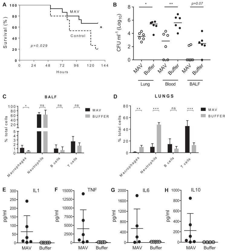
Current vaccination against Streptococcus pneumoniae uses vaccines based on capsular polysaccharides from selected serotypes and has led to nonvaccine serotype replacement disease. We have investigated an alternative serotype-independent approach, using multiple-antigen vaccines (MAV) prepared from S. pneumoniae TIGR4 lysates enriched for surface proteins by a chromatography step after culture under conditions that induce expression of heat shock proteins (Hsp; thought to be immune adjuvants). Proteomics and immunoblot analyses demonstrated that, compared to standard bacterial lysates, MAV was enriched with Hsps and contained several recognized protective protein antigens, including pneumococcal surface protein A (PspA) and pneumolysin (Ply). Vaccination of rodents with MAV induced robust antibody responses to multiple serotypes, including nonpneumococcal conjugate vaccine serotypes. Homologous and heterologous strains of S. pneumoniae were opsonized after incubation in sera from vaccinated rodents. In mouse models, active vaccination with MAV significantly protected against pneumonia, while passive transfer of rabbit serum from MAV-vaccinated rabbits significantly protected against sepsis caused by both homologous and heterologous S. pneumoniae strains. Direct comparison of MAV preparations made with or without the heat shock step showed no clear differences in protein antigen content and antigenicity, suggesting that the chromatography step rather than Hsp induction improved MAV antigenicity. Overall, these data suggest that the MAV approach may provide serotype-independent protection against S. pneumoniae.

Keywords: Streptococcus pneumoniae; multiple-antigen vaccine; protein antigen; vaccines.


Figures

FIG 1
FIG 1
Formulation of a multiple-antigen S. pneumoniae TIGR4-derived vaccine preparation (MAV). (A) Ion-exchange (IEX) chromatogram showing the purification of the MAV. Light green line, NaCl elution concentration; brown line, the resulting conductivity in the system; blue line, UV trace showing the concentrations of the eluted proteins (milli-absorbance units); dark green line, pressure in the system. The fractions collected are numbered in red; the total volume (in milliliters) is recorded on the x axis. (B) Detection of Hsp60 and Hsp70 by Western blotting in selected ion-exchange chromatography fractions; the BCA assay protein concentrations for these fractions are shown in the table. (C) A comparison of the heat shock protein content (Hsp60 and Hsp70) measured by immunoblotting of heat-killed lysate (HKL) and MAV. The bar chart shows the pixel intensity quantification (ImageQuant TL; GE Lifesciences) for Hsp60 and Hsp70 bands. (D) Immunoblots of 5 μg of total protein of either MAV or HKL probed with pooled human IgG at 1:20,000 (Pentaglobin; Paviour Pharmaceuticals, New Delhi, India). (E) Comparison of the hemolytic activity against horse red blood cells in serial 2-fold dilutions of MAV from neat to 1:64 and HKL and HKWC preparations with a saponin positive control.
FIG 2
FIG 2
MAV is immunogenic in a mouse model of subcutaneous vaccination. CD1 mice were vaccinated subcutaneously with 75 μg on day 0 and day 21 and culled at 28 days to obtain serum. (A) Results of a whole-cell IgG ELISA against S. pneumoniae TIGR4 for pooled sera harvested from tail vein bleeds (10 μl per mouse, n = 6). (B and C) Results of whole-cell IgG and IgM ELISAs against S. pneumoniae TIGR4 for pooled sera from MAV-vaccinated mice (n = 5) (B) and against S. pneumoniae TIGR4 for pooled BALF and nasal wash specimens from MAV-vaccinated mice (C). Data are presented as the mean and 95% confidence interval. P values were calculated using the Mann-Whitney t test. **, P < 0.01; ns, not significant.
FIG 3
FIG 3
Binding of immune mouse sera to the surface of S. pneumoniae strains. (A) The results of IgG surface binding assays of S. pneumoniae TIGR4 incubated in sera from vaccinated mice are shown as the geometric mean fluorescence index (MFI). Error bars represent the SD from technical replicates. Significance is calculated with the Holm-Sidak test. *, P < 0.05. (B) Representative flow cytometry histograms showing IgG-positive S. pneumoniae TIGR4 populations. White histogram, buffer negative-control serum; black histogram, serum from MAV-vaccinated mice; dark gray histogram, serum from HKL-vaccinated mice; light gray histogram, serum from HKWC-vaccinated mice. (C) IgG binding to TIGR4 in immune serum diluted to 25, 12.5, 6.25, and 3.125%. Data points are means from technical replicates; error bars represent standard deviations. Significance values between each dilution curve were calculated by using a two-way ANOVA and comparison to the buffer negative control. ****, P < 0.001. (D) Mean fluorescent IgG surface binding to S. pneumoniae serotype 18C, 23F, ST3, and 19F strains incubated in sera from MAV- or buffer-vaccinated mice. Error bars represent standard deviations from technical replicates. Significance was calculated with the Holm-Sidak test. *, P < 0.05. (E) Representative histograms showing a shift in populations positive for IgG against different strains of S. pneumoniae. White histogram, IgG binding in buffer-vaccinated mouse serum; shaded histogram, binding in MAV-vaccinated mouse serum.
FIG 4
FIG 4
Identification of protein antigens recognized by sera from vaccinated mice. (A) Immunoblots of S. pneumoniae TIGR4, D39, or 19A strain whole-cell lysates probed with serum diluted 1:1,000 from mice vaccinated with either HKL or MAV. The numbers on the left are molecular masses (in kilodaltons). (B) Identification of protein antigens recognized by sera from vaccinated mice using the MSD assay. Values are normalized to those for the negative control consisting of buffer-vaccinated mouse serum. Mean values are shown, with error bars representing standard deviations for sera from mice vaccinated with MAV (n = 3, black columns) and a mouse vaccinated with HKL (n = 1, gray columns).
FIG 5
FIG 5
Comparison of heat-shocked MAV versus non-heat-shocked MAV. (A) MAV preparations were made with (MAVIPS004) and without (MAVIPS005) the Hsp induction step. Protein bands were compared using a Coomassie gel (top). Three micrograms (lanes 7 and 9) and 5 μg (lanes 8 and 10) of each MAV was loaded; immunoblots of the MAV preparations were also probed for the presence of the key S. pneumoniae protein antigens (PlyD6, PspA) and Hsps (Hsp70, Hsp60). (B) Capillary gel electrophoresis (CGE) analysis was conducted to determine the protein constituents of each preparation. Each peak is denoted by a number, and interpeak regions are marked by a letter. Quantification of peaks is shown in the bar chart on top. CGE traces are shown below. (C) Both MAVIPS004 and MAVIPS005 were used to generate antisera using vaccination experiments in mice. Sera recovered from mice vaccinated with either preparation were analyzed using flow cytometry assays of IgG binding to live S. pneumoniae bacteria (serotypes 1, 2 [D39], 4 [TIGR4], 6B, 8, 19A, 22F, and 23F), and results are represented as the mean fluorescence intensity (MFI) in the appropriate gate. Q1, quadrant 1. (D and E) ELISAs detecting anti-Ply (D) and anti-PspA (E) responses were conducted in duplicate. Sera from the experiments described above were diluted, as shown on the x axis, and the OD450 was measured for each MAV and a buffer control. Abbreviations: MAV, multiantigen vaccine; HS, heat shocked; NHS, non-heat shocked; MK, molecular weight marker; AUC, area under the curve.
FIG 6
FIG 6
Vaccination with MAV preparations protects mice against S. pneumoniae challenge. (A and B) Number of CFU in the lung (A) and blood (B) 24 h after challenge by intranasal (IN) inoculation with 1 × 107 CFU of the S. pneumoniae TIGR4 strain in mice vaccinated twice subcutaneously with 75 μg of MAV or a negative-control buffer (n = 10 per group). (C) Number of CFU in nasal wash (NW) specimens 2 weeks after nasopharyngeal colonization with 5 × 106 CFU of S. pneumoniae TIGR4 of mice vaccinated twice subcutaneously with 75 μg of MAV or a negative control buffer (n = 8 per group). (D) Number of CFU in blood 6 h after challenge by intraperitoneal (IP) inoculation of 1 × 104 CFU of the S. pneumoniae TIGR4 strain into mice passively vaccinated with 200 μl of sera from rabbits vaccinated three times with 375 μg of MAVIPS004 or twice with 0.2 ml of the Prevenar vaccine or a negative-control buffer (n = 12 per group). (E and F) Number of CFU in the blood of mice 4 h after challenge by intravenous (IV) inoculation with 5 × 105 CFU of the S. pneumoniae TIGR4 (E) or ATCC BAA-1662 (18C) (F) strain that was incubated preinoculation in sera obtained from rabbits vaccinated with MAVIPS014, the Prevenar vaccine, or a negative-control buffer (n = 5 to 10). For all panels, each symbol represents data from a single mouse, and horizontal bars represent median values. Statistical significances were calculated using a Mann-Whitney t test (A to D) or Dunnett’s multiple-comparison test (E and F). Significance abbreviations: *, P < 0.05; **, P < 0.01.
FIG 7
FIG 7
Vaccination with MAV preparations increases the survival of mice and alters the inflammatory response after S. pneumoniae TIGR4 pneumonia challenge. (A) Percent survival of mice over 6 days after challenge by intranasal inoculation with 1 × 107 CFU of the S. pneumoniae TIGR4 strain of mice that had been vaccinated three times (days 1, 10, and 22) intraperitoneally with 75 μg of MAVIPS014 or a negative-control buffer (n = 15 per group). Significance was calculated using the log-rank (Mantel-Cox) test. (B to D) Numbers of CFU in target organs (B) and inflammatory cell populations in BALF (C) and lung (D) 24 h after challenge with 1 × 107 CFU of the S. pneumoniae TIGR4 strain in MAVIPS014-vaccinated and control mice. Inflammatory cell data are shown as a percentage of the total cells recovered from the lungs of MAV- and buffer-vaccinated mice; CFU data show the number of CFU in lung, blood, or BALF recovered 24 h after challenge, with each symbol representing data from a single mouse and horizontal bars representing median values. (E to H) Lung homogenate cytokine levels 24 h after challenge with 1 × 107 CFU of the S. pneumoniae TIGR4 strain in MAVIPS014-vaccinated and control mice. For panels B to H, statistical significances were calculated using a Mann-Whitney t test. Significance abbreviations: *, P < 0.05; **, P < 0.01; ***, P < 0.001; ns, not significant.

References

    1. Said MA, Johnson HL, Nonyane BAS, Deloria-Knoll M, O’Brien KL. 2013. Estimating the burden of pneumococcal pneumonia among adults: a systematic review and meta-analysis of diagnostic techniques. PLoS One 8:e60273. doi:10.1371/journal.pone.0060273. - DOI - PMC - PubMed
    1. Monasta L, Ronfani L, Marchetti F, Montico M, Brumatti L, Bavcar A, Grasso D, Barbiero C, Tamburlini G. 2012. Burden of disease caused by otitis media: systematic review and global estimates. PLoS One 7:e36226. doi:10.1371/journal.pone.0036226. - DOI - PMC - PubMed
    1. Sørensen UB, Henrichsen J, Chen HC, Szu SC, Sorensen UBS, Henrichsen J, Chen HC, Szu SC. 1990. Covalent linkage between the capsular polysaccharide and the cell wall peptidoglycan of Streptococcus pneumoniae revealed by immunochemical methods. Microb Pathog 8:325–334. doi:10.1016/0882-4010(90)90091-4. - DOI - PubMed
    1. Maruyama T, Taguchi O, Niederman MS, Morser J, Kobayashi H, Kobayashi T, D'Alessandro-Gabazza C, Nakayama S, Nishikubo K, Noguchi T, Takei Y, Gabazza EC. 2010. Efficacy of 23-valent pneumococcal vaccine in preventing pneumonia and improving survival in nursing home residents: double blind, randomised and placebo controlled trial. BMJ 340:c1004. doi:10.1136/bmj.c1004. - DOI - PMC - PubMed
    1. Webster J, Theodoratou E, Nair H, Seong AC, Zgaga L, Huda T, Johnson HL, Madhi S, Rubens C, Zhang JSF, El Arifeen S, Krause R, Jacobs TA, Brooks AW, Campbell H, Rudan I. 2011. An evaluation of emerging vaccines for childhood pneumococcal pneumonia. BMC Public Health 11:S26. doi:10.1186/1471-2458-11-S3-S26. - DOI - PMC - PubMed

Publication types

MeSH terms