Review
. 2019 Jan;20(1):e46864.
doi: 10.15252/embr.201846864. Epub 2018 Dec 10.

Peroxisome biogenesis, membrane contact sites, and quality control

Affiliations
Review

Peroxisome biogenesis, membrane contact sites, and quality control

Jean-Claude Farré et al. EMBO Rep. 2019 Jan.

Abstract

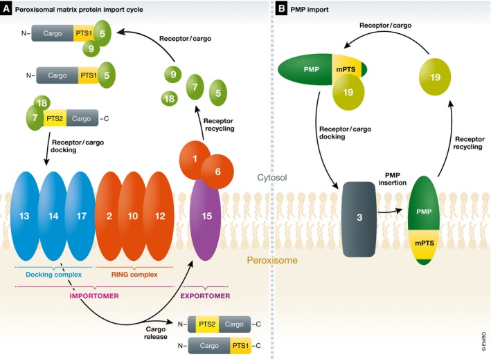
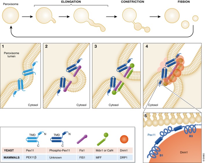
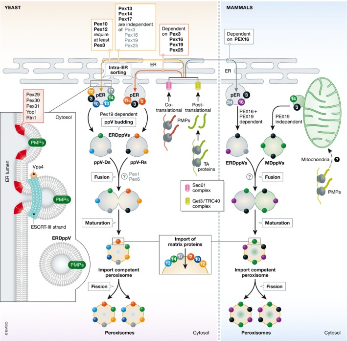
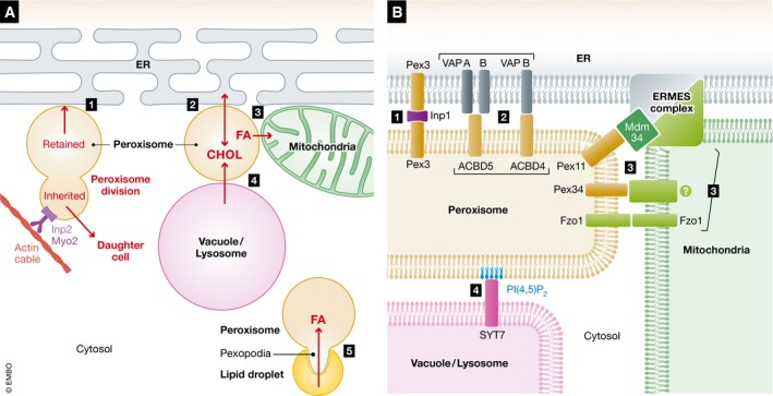
Peroxisomes are conserved organelles of eukaryotic cells with important roles in cellular metabolism, human health, redox homeostasis, as well as intracellular metabolite transfer and signaling. We review here the current status of the different co-existing modes of biogenesis of peroxisomal membrane proteins demonstrating the fascinating adaptability in their targeting and sorting pathways. While earlier studies focused on peroxisomes as autonomous organelles, the necessity of the ER and potentially even mitochondria as sources of peroxisomal membrane proteins and lipids has come to light in recent years. Additionally, the intimate physical juxtaposition of peroxisomes with other organelles has transitioned from being viewed as random encounters to a growing appreciation of the expanding roles of such inter-organellar membrane contact sites in metabolic and regulatory functions. Peroxisomal quality control mechanisms have also come of age with a variety of mechanisms operating both during biogenesis and in the cellular response to environmental cues.

Keywords: de novo peroxisome biogenesis; peroxisomal membrane contact sites; peroxisomal membrane protein biogenesis; peroxisome growth and division; peroxisome quality control.


Conflict of interest statement

The authors declare that they have no conflict of interest.

Figures

Figure 1
Figure 1. Peroxisomal matrix and membrane protein import in yeast (an overview)
Most proteins destined for import into the peroxisome matrix possess either a C‐terminal PTS1 or an N‐terminal PTS2. (A) The peroxisomal matrix protein import cycle. These cargos synthesized in the cytosol are recognized by PTS receptors, Pex5 for PTS1 and Pex7 for PTS2, respectively. Pex7 generally works with a co‐receptor (Pex18/21 in S. cerevisiae or Pex20 in P. pastoris only Pex18 is shown). Pex9 is a Pex5‐related protein found in S. cerevisiae that acts on limited PTS1 cargos as described in the text 10, 11. The PTS receptor/cargo complex, along with the co‐receptor, where applicable, docks at the peroxisome membrane with the docking complex, comprised of Pex13, Pex14, and Pex17. PTS cargos are translocated into the peroxisome matrix across translocons in the peroxisome membrane. The minimal translocon in yeast involves Pex14 and Pex5 for PTS1 import 12, and likely Pex14/Pex17 and Pex18 for PTS2 import 13. Associated with the docking complex is the RING subcomplex comprised of Pex2, Pex10, and Pex12 that have E3 ligase activities involved in ubiquitin‐dependent, PTS‐receptor recycling and QC steps (sections Brief overview of peroxisomal matrix protein import and QC during peroxisomal matrix protein import). Together, the docking and RING subcomplexes form the importomer complex 14, 15. Following PTS cargo release in the peroxisome lumen, the PTS receptors, and co‐receptors where applicable, recycle from the peroxisomes back to the cytosol for another round of import, using components collectively called the exportomer, whose components are described in the text 16. (B) The PMP import cycle for the direct import of proteins into the peroxisome membrane (section The direct import of PMPs to peroxisomes). Each PMP has at least one mPTS that is bound to, and the PMP is chaperoned by, Pex19, which docks at the peroxisomes via interactions with Pex3. The PMP is inserted into the membrane and Pex19 recycles back to the cytosol for another round of PMP import.
Figure 2
Figure 2. Peroxisome fission in the growth and division model
According to the growth and division model, peroxisome fission happens in a 3‐step process. During the first step of elongation, Pex11 (PEX11β in mammals), a transmembrane protein that imparts curvature to peroxisome membranes (panel 1), is essential for the elongation step. The topology shown here for Pex11 is based on studies in H. polymorpha 67. The second step, involving membrane constriction, is poorly understood and we do not know any proteins implicated in this step. The third step, peroxisome fission, starts in P. pastoris with the phosphorylation of Pex11(S173) that stimulates its interaction with the adaptor, Fis1 (panel 2) 78. Note that the topology of PpPex11 has not been documented, so it is unclear whether the phosphorylation is on the cytosolic or the peroxisome matrix side. Fis1 then recruits the peripheral receptors, Mdv1 and/or Caf4 (panel 3) 75. Mdv1 and/or Caf4 assemble a Dnm1 ring around the peroxisome constriction site (panel 4). Mammals do not have homologues for these proteins, and DRP1 is recruited to peroxisomes by MFF and FIS1 79. Yeast Dnm1 interacts with Fis1 and two Pex11 helices named B1 and B3 (panel 5). The hydrolysis of GTP by Dnm1, enhanced by the interaction with the B3 helix of Pex11, leads to a constriction that divides the peroxisome 82.
Figure 3
Figure 3. Schematic representation of de novo peroxisome biogenesis pathways in yeast and mammals
The first step in the de novo biogenesis is the indirect import of PMPs to the ER. Some PMPs are co‐translationally inserted into the ER membrane via the Sec61 complex 33, 108 and TA PMPs are post‐translationally incorporated into the ER membrane via the Get3 complex (yeast) 63 or ASNA1/TRC40 (mammals) 62. After PMP insertion into the ER, work in yeasts shows that an intra‐ER sorting step targets the PMPs to sub‐domains of the ER called the pER 32. Work in P. pastoris reveals that this routing of PMPs is either dependent or independent of Pex3, 16, 19, and 25 116, 117, 118. Studies from several yeasts define at least two modes of intra‐ER sorting of PMPs. One pathway is exemplified by the docking subcomplex proteins (Pex13, 14, and 17), which are independent of Pex3, 16, 19, and 25 118, 137. The other is exemplified by the RING‐domain PMPs (Pex10, 12, 2, 11c) and is dependent on Pex3, 16, 19, and 25 for intra‐ER sorting 118, 137. The exit sites for ppV budding are marked by the presence of several proteins (shown in inset on the left) including Pex29, 30, 31, which interact with Yop1 and Rtn1 and impart positive curvature in the ER 122, 124, 130. Subsequently, ESCRT‐III proteins (Vps20 and Snf7) are proposed to play a role in ppV scission 121 in an energy‐dependent manner, perhaps facilitated by Vps4 (stimulating disassembly of ESCRT‐III at the ER) 121. The ppVs bud from the pER in a Pex19‐dependent manner 115, 120. ERDppVs are of two distinct varieties—ppV‐R, containing Pex3, Pex2, and Pex11C and ppV‐D comprised of Pex13, Pex14, Pex17, Pex10, Pex12, and Pex3 116. Subsequently, these ppVs fuse heterotypically or with pre‐existing peroxisomes 40, 42, 119. In mammals, ppV formation is different in that several PMPs are sorted to the pER in a PEX16‐dependent manner 31, 146 and several other PMPs are routed to peroxisomes via mitochondria, from which MDppVs are formed in a PEX19‐independent manner 39. Subsequently, ERDppVs and MDppVs are proposed to fuse to form import‐competent peroxisomes, which subsequently import the matrix proteins and become metabolically active organelles. The question mark (?) represents uncertainty regarding either the known 42, 119, 144 or unknown proteins required for this fusion step.
Figure 4
Figure 4. Peroxisome membrane contact sites
(A) MCSs of peroxisomes with several organelles (labeled 1–5), and their suggested functions. During cell division in yeast, some peroxisomes are retained in mother cells via tethering to the ER and the new peroxisomes, produced by division, are inherited (Inh) to daughter cells moving along actin cable 163. Yeast peroxisomes are pulled by the class V myosin motor, Myo2, which is attached to the peroxisomal membrane by the Inp2 protein 164. The functions of peroxisome–mitochondria MCSs linked either by Pex11‐Mdm34, Fzo1‐Fzo1, or Pex34 with an undefined (?) mitochondrial partner are unknown (panel B), but may play a role in peroxisome fission 150, 165. The other MCSs shown with other organelles are implicated in the transfer of lipids, such as fatty acids (FA) 65, 157, 158 and cholesterol (CHOL) from or to peroxisomes 170, respectively. Arrows indicate the direction of lipid traffic or organelle movement to daughter cell. (B) Known tether components of peroxisome MCSs from panel (A) represented in a single peroxisome for simplicity. However, most likely each peroxisome may not have all of these MCSs simultaneously and these sites are also dynamic in nature. 1 and 2, ER‐peroxisome tethers: 3, mitochondria‐peroxisome tethers; 4, lysosome‐peroxisome tether; 5, peroxisome–LD tether.
Figure 5
Figure 5. Peroxisomal quality control pathways
During peroxisomal matrix protein import, after the PTS receptor protein, Pex5, has released the cargo in the peroxisomal matrix, it follows different fates. Receptor Recycling: Mono‐ubiquitination (monoUb) of Pex5 occurs at a conserved cysteine residue (C6 in yeast and C11 in mammals) catalyzed by the E2‐enzyme complex (Pex4/Pex22) in yeast or UbcH5 in mammals, and the E3 ligases Pex2, Pex10, and Pex12 21. Next, mono‐ubiquitinated Pex5 is recycled to the cytosol, mediated by the AAA‐ATPases, Pex1, and Pex6 141. Finally, ubiquitin is removed by a deubiquitinating enzyme (DUB) and Pex5 becomes available for another round of import 24. RADAR: As a quality control mechanism, poly‐ubiquitination (polyUb) of Pex5 at conserved lysine residues in yeast by the E2‐enzyme Ubc4 and E3 ligases direct Pex5 for degradation by the proteasome (RADAR) 186. In mammals, the E2‐enzyme UbcH5 has been implicated in mono‐ and poly‐ubiquitination of PEX5. Pexophagy (and its prevention): As is the case in yeast, during receptor recycling in mammals, Pex1 and Pex6 are implicated in the recycling of PEX5, but in addition their presence prevents pexophagy 142. The mechanism in mammals is through recycling PEX5 from the peroxisomal membrane, as PEX5 is also target of ubiquitination which is recognized by autophagy factors driving pexophagy 186. For example, pexophagy is induced by high levels of peroxisomal reactive oxygen species (ROS), which recruits ATM to peroxisomes. ATM phosphorylates (P) PEX5 at Ser141 (S141), which then mediates the ubiquitination of PEX5 either at Lys209 (K209) 9. Alternatively, Cys11 is ubiquitinated 7. In yeast, the pexophagy prevention by the AAA‐ATPases is most probably independent of Pex5, which is not required for pexophagy. Instead, a different factor (X) that triggers pexophagy, which could be the yeast pexophagy receptor (Atg36), might need to be removed from the peroxisome surface by Pex1 and Pex6. Other QC mechanisms: The peroxisomal Lon type AAA‐protease, Pln, degrades damaged proteins in the peroxisomal matrix 182, 187, 188, 189. This protease is absent in S. cerevisiae but is present in other yeasts. Another QC mechanism may operate to recycle back to the cytosol damaged enzymes, which could be degraded by the proteasome. Such a mechanism has been described by the release of catalase to the cytosol by a peroxisome‐localized BAK, due to the lack of mitochondrial VDAC2, which normally retains BAK at mitochondria 179.

References

    1. Tabak HF, Braakman I, Distel B (1999) Peroxisomes: simple in function but complex in maintenance. Trends Cell Biol 9: 447–453 - PubMed
    1. Walker CL, Pomatto LCD, Tripathi DN, Davies KJA (2018) Redox regulation of homeostasis and proteostasis in peroxisomes. Physiol Rev 98: 89–115 - PMC - PubMed
    1. Waterham HR, Ferdinandusse S, Wanders RJ (2016) Human disorders of peroxisome metabolism and biogenesis. Biochim Biophys Acta 1863: 922–933 - PubMed
    1. Honsho M, Yamashita S, Fujiki Y (2016) Peroxisome homeostasis: mechanisms of division and selective degradation of peroxisomes in mammals. Biochim Biophys Acta 1863: 984–991 - PubMed
    1. Till A, Lakhani R, Burnett SF, Subramani S (2012) Pexophagy: the selective degradation of peroxisomes. Int J Cell Biol 2012: 512721 - PMC - PubMed

Publication types

MeSH terms

Substances