Review
. 2019 Jan;60(1):2-6.
doi: 10.1194/jlr.S087452. Epub 2018 Dec 7.

Fat-regulating phosphatidic acid phosphatase: a review of its roles and regulation in lipid homeostasis

Affiliations
Review

Fat-regulating phosphatidic acid phosphatase: a review of its roles and regulation in lipid homeostasis

George M Carman et al. J Lipid Res. 2019 Jan.

Abstract

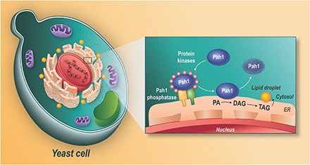
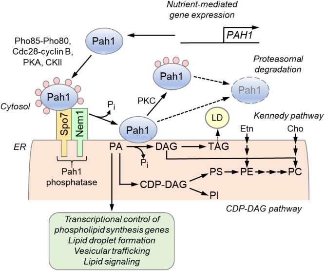
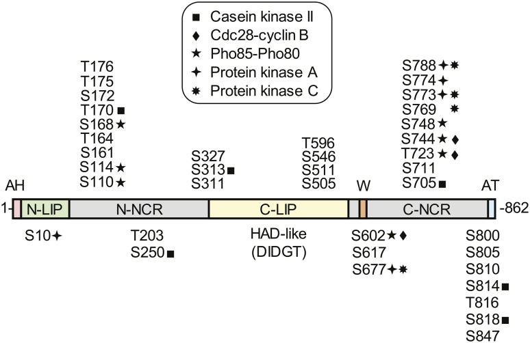
Phosphatidic acid (PA) phosphatase is an evolutionarily conserved enzyme that plays a major role in lipid homeostasis by controlling the cellular levels of its substrate, PA, and its product, diacylglycerol. These lipids are essential intermediates for the synthesis of triacylglycerol and membrane phospholipids; they also function in lipid signaling, vesicular trafficking, lipid droplet formation, and phospholipid synthesis gene expression. The importance of PA phosphatase to lipid homeostasis and cell physiology is exemplified in yeast, mice, and humans by a host of cellular defects and lipid-based diseases associated with loss or overexpression of the enzyme activity. In this review, we focus on the mode of action and regulation of PA phosphatase in the yeast Saccharomyces cerevisiae The enzyme Pah1 translocates from the cytosol to the nuclear/endoplasmic reticulum membrane through phosphorylation and dephosphorylation. Pah1 phosphorylation is mediated in the cytosol by multiple protein kinases, whereas dephosphorylation is catalyzed on the membrane surface by an integral membrane protein phosphatase. Posttranslational modifications of Pah1 also affect its catalytic activity and susceptibility to degradation by the proteasome. Additional mechanistic understanding of Pah1 regulation should be instrumental for the identification of small-molecule inhibitors or activators that can fine-tune PA phosphatase function and thereby restore lipid homeostasis.

Keywords: Nem1-Spo7 protein phosphatase complex; diacylglycerol; lipodystrophy; obesity; triacylglycerol.


Conflict of interest statement

The authors declare that there are no conflicts of interest with the contents of this article.

Figures

None
Graphical abstract
Fig. 1.
Fig. 1.
Roles and regulation of PAH1-encoded PA phosphatase in lipid synthesis. The expression of the PAH1 gene that encodes the PA phosphatase protein Pah1 is regulated throughout growth by nutrient status. Pah1 in the cytosol is phosphorylated by multiple protein kinases during vegetative growth when the synthesis of phospholipids occurs at the expense of TAG. As cells progress into stasis, the phosphorylated Pah1 (pink circles) translocates to the ER membrane through its dephosphorylation by the Pah1 phosphatase, which is composed of Nem1 (catalytic subunit) and Spo7 (regulatory subunit). Dephosphorylated Pah1 that is associated with the ER membrane catalyzes the conversion of PA to DAG, which is then acylated to form TAG that is stored in lipid droplets (LD). Dephosphorylated Pah1 or PKC-phosphorylated Pah1 that is not phosphorylated at the seven target sites for Pho85-Pho80 protein kinase is degraded by the proteasome (dashed line arrows). PA is also utilized for the synthesis of membrane phospholipids via the CDP-DAG pathway, and it has signaling functions (green). When the CDP-DAG pathway for phospholipid synthesis is blocked, phosphatidylcholine (PC) or phosphatidylethanolamine (PE) may be synthesized from the DAG derived from the PA phosphatase reaction if cells are supplemented with choline (Cho) or ethanolamine (Etn) via the Kennedy pathway. PI, phosphatidylinositol; PS, phosphatidylserine.
Fig. 2.
Fig. 2.
Domains/regions and phosphorylation sites in Pah1. The diagram shows the positions of the amphipathic helix (AH, pink) required for ER membrane interaction, the N-LIP (green) and C-LIP (contains HAD-like domain with DIDGT catalytic motif) (yellow) domains that are required for PA phosphatase activity, the acidic tail (AT, blue) required for interaction with Spo7 of the Pah1 phosphatase, N-terminal non-conserved region (N-NCR, grey), C-terminal non-conserved region (C-NCR, grey), the serine (S) and threonine (T) residues in their approximate regions that are known phosphorylation sites, and the sites that are phosphorylated by CKII, Cdc28-cyclin B, Pho85-Pho80, PKA, and PKC, and the tryptophan (W) residue within the C-terminal conserved sequence WRDPLVDID (orange) required for Pah1 function in vivo.

References

    1. Pascual F., and Carman G. M.. 2013. Phosphatidate phosphatase, a key regulator of lipid homeostasis. Biochim. Biophys. Acta. 1831: 514–522. - PMC - PubMed
    1. Smith S. W., Weiss S. B., and Kennedy E. P.. 1957. The enzymatic dephosphorylation of phosphatidic acids. J. Biol. Chem. 228: 915–922. - PubMed
    1. Kates M. 1955. Hydrolysis of lecithin by plant plastid enzymes. Can. J. Biochem. 33: 575–589. - PubMed
    1. Lin Y-P., and Carman G. M.. 1989. Purification and characterization of phosphatidate phosphatase from Saccharomyces cerevisiae. J. Biol. Chem. 264: 8641–8645. - PubMed
    1. Han G-S., Wu W-I., and Carman G. M.. 2006. The Saccharomyces cerevisiae lipin homolog is a Mg2+-dependent phosphatidate phosphatase enzyme. J. Biol. Chem. 281: 9210–9218. - PMC - PubMed

Publication types

Substances

LinkOut - more resources