. 2018 Dec 26;115(52):E12407-E12416.
doi: 10.1073/pnas.1816177115. Epub 2018 Dec 7.

MTSS1/Src family kinase dysregulation underlies multiple inherited ataxias

Affiliations

MTSS1/Src family kinase dysregulation underlies multiple inherited ataxias

Alexander S Brown et al. Proc Natl Acad Sci U S A. .

Abstract

The genetically heterogeneous spinocerebellar ataxias (SCAs) are caused by Purkinje neuron dysfunction and degeneration, but their underlying pathological mechanisms remain elusive. The Src family of nonreceptor tyrosine kinases (SFK) are essential for nervous system homeostasis and are increasingly implicated in degenerative disease. Here we reveal that the SFK suppressor Missing-in-metastasis (MTSS1) is an ataxia locus that links multiple SCAs. MTSS1 loss results in increased SFK activity, reduced Purkinje neuron arborization, and low basal firing rates, followed by cell death. Surprisingly, mouse models for SCA1, SCA2, and SCA5 show elevated SFK activity, with SCA1 and SCA2 displaying dramatically reduced MTSS1 protein levels through reduced gene expression and protein translation, respectively. Treatment of each SCA model with a clinically approved Src inhibitor corrects Purkinje neuron basal firing and delays ataxia progression in MTSS1 mutants. Our results identify a common SCA therapeutic target and demonstrate a key role for MTSS1/SFK in Purkinje neuron survival and ataxia progression.

Keywords: BAR domain proteins; Src kinase; actin cytoskeleton; neurodegeneration; spinocerebellar ataxia.


Conflict of interest statement

The authors declare no conflict of interest.

Figures
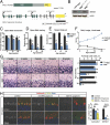
Fig. 1.
Fig. 1.
MIMEX15 mutants develop progressive spinocerebellar ataxia. (A) The structure of the Mtss1 locus with alternative promoters and the Src interacting domain deleted in MIMEX15 mutants. (B) Loss of MTSS1 protein in MIMEX15 cerebellum lysate shown with MTSS1 antibody against the N-terminal I-BAR (IMD) domain. (C and D) MIMEX15 mice show slower movement velocity (C) and less frequent rearing (D) in open field tests. (E) Impaired rotarod performance in MIMEX15 mutants is shown as reduced duration (time to fall). (F) A composite test of gait, balance, and grip strength to measure spinocerebellar ataxia symptoms. An increased score reflects reduced function with an age-dependent increase in severity in MIMEX15 mutants. (G) Age-dependent loss of Purkinje neurons in MIMEX15 mutants occurs after the onset of ataxia. (H) At 20 wk MIMLoxp/−;Pcp2-Cre and MIMLoxp/Loxp;Pcp2-Cre mutants show a dramatic reduction in Purkinje neurons that stain with MTSS1. Many Purkinje neurons persist, as there is a less dramatic reduction in calbindin+ Purkinje cell number. *P < 0.05, **P < 0.005, ***P < 5E-5, one-way ANOVA with Tukey post hoc test; ns, not significant. Error bars indicate SEM.
Fig. 2.
Fig. 2.
MIMEX15-mutant Purkinje neurons undergo autophagy. (A) MIMEX15 mutants display fused mitochondria shown by increased complex 5 ATP-synthase immunostaining and collapsed Golgi shown by reduced giantin immunostaining at 4 wk. (B) Eight-week-old MIMEX15 mutants show increased LC3-II abundance. *P < 0.005; Student’s t test. (C) MIMEX15 mutants show increased levels of mRNA for the autophagocytic marker VMP1. *P < 0.05; Student’s t test. (D) MIMEX15 mutants show increased microglial infiltration shown by Aif1 transcript. *P < 0.002; Student’s t test. (E) MIMEX15 mutants show GFAP+ glial infiltration during disease progression. (F) Western blots quantifying increased cerebellar GFAP. *P < 0.05; Student’s t test. (G) MIMEX15-mutant cerebella do not have increased TUNEL staining at 4, 8, or 16 wk of age.
Fig. 3.
Fig. 3.
Mtss1 prevents SFK-dependent firing defects and ataxia. (A) Confocal projection of an individual Purkinje cell filled with biocytin and fluorescent dye to visualize morphology. (BE) Measurement of dye-filled Purkinje neurons shows MIMEX15 mutants have reduced arbor volume (n = 3 each genotype) (B) and reduced dendritic spine density (C) but no change in dendritic spine length (D) and no change in dendritic spine width (E). MIMEX15/+, n = 3 neurons, 1,720 spines; MIMEX15, n = 3 neurons, 1,454 spines. *P < 0.05, Student’s t test. Error bars indicate SEM. (F) Western blot for active SFK-Y416 phosphorylation with actin as a loading control. Cerebellar lysate was collected from MIMEX15 mice and age-matched controls at the indicated times between postnatal day 15 (P15) and postnatal day 30 (P30). (G) Slow EPSP in wild-type (Left) and MIMEX15 (Center) cells elicited by the stimulation of parallel fibers with 10-pulse trains at 100 Hz in the presence AMPA, NMDA, and GABA receptor antagonists (control conditions). Corresponding intracellular Ca2+ signals (ΔF/F) for responses for wild-type and MIMEX15 mGluR EPSPs are illustrated. EPSPs and corresponding Ca2+ signals are blocked by mGluR1 antagonist CPCCOEt. (Right) Summary data of intracellular Ca2+ signals (ΔF/F) for responses for WT and MTSS1EX15 in control conditions and in the presence of CPCCOEt are shown. (H) Percent histograms of Purkinje neuron mean firing frequencies (Left), examples of extracellular recordings of 1-s duration of a spontaneously spiking Purkinje neuron in the respective condition (Center), and histograms of interspike intervals (ISIs) calculated for the 2-min recording periods of the same neuron (Right) are shown for the wild-type, MIMEX15, wild-type+dasatinib, and MIMEX15+dasatinib conditions. (I) Summary of data presented in H. *P = 6.1E-14; **P = 1E-13; one-way ANOVA, Tukey post hoc test. (J) Direct cerebellar administration of dasatinib maintains rotarod performance, slowing the progressive ataxia in MIMEX15 mice. q = 0.006, two-stage step-up Benjamini, Krieger, Yekutieli method. Error bars indicate SEM.
Fig. 4.
Fig. 4.
MTSS1 is an ATXN2 translation target. (A, Left) Western blot of whole-cerebellum lysate from 24-wk-old mice shows 90% reduction (arrow) in the band that corresponds MTSS1 in ATXN2Q127 mice, while calbindin (CALB1) was reduced 50%. Actin is included as a loading control. (Right) Quantitation of Western blot results. *P < 0.01, **P < 0.001; Student’s t test. (B, Left) Western blots for active SFK-Y416 phosphorylation and total Src in cerebellar lysate from 24-wk-old Atxn2Q127 mice show an eightfold increase in SFK-Y416 abundance. Tubulin was used as a loading control. (Right) Quantitation of Western blot results. (C) Percent histograms of Purkinje neuron mean firing frequencies (Left), examples of extracellular recordings of 1-s duration of a spontaneously spiking Purkinje neuron in the respective condition (Center), and histograms of interspike intervals calculated for the 2-min recording periods of the same neuron for ATXN2Q127 and ATXN2Q127+dasatinib. (D) Mean firing rates. **P = 3.77E-8; one-way ANOVA and Tukey post hoc test. (E) Western blot for Atxn2 in cerebellar lysate from 4-wk-old MIMEX15 cerebellum and age-matched controls with tubulin as a loading control. (F) RNA-immunoprecipitation in HEK-293 cells for Flag-ATXN2Q22 and Flag-ATXN2Q108 shows enrichment for MTSS1 but not GAPDH mRNA. Error bars indicate SD. (G) Polyribosome fractionation in 293T cells transfected with the MTSS1 UTR reporter and pcDNA, ATXN2Q22, ATXN2Q108, or ATXN2Q22+ATXN2Q108. The green line indicates UV 254-nm absorbance (nucleic acids) with 40S, 60S, 80S, and polyribosome peaks labeled. (H) Remaining Purkinje neurons in human SCA2 cerebellum (ATXN2Q22/Q41) show reduced MTSS1 staining compared with an age-matched control (ATXN2Q22/Q22).
Fig. 5.
Fig. 5.
SFK dysregulation occurs in multiple SCAs. (A) Western blot of whole-cerebellum lysate from 15-wk-old mice shows a 95% reduction of in the band that corresponds to MTSS1 in ATXN1Q82 mice but only a 50% reduction in calbindin. Tubulin is included as a loading control. (B) Western blot of cerebellum lysate from 4-wk-old MIMEX15 mice shows no change in phospho-serine776 ATXN1 levels. (C) RNA-sequencing from ATXN1Q82 cerebella shows reduced fragments per kilobase of transcript per million mapped reads for Mtss1 mRNA in samples from 12-wk-old and 28-wk-old mice (*q < 0.005). (D) Mean firing frequency values (in Hertz) for WT and ATXN1Q82 mice, with and without dasatinib (Das) treatment. *P = 0.0094, one-way ANOVA with Tukey post hoc test. Error bars indicate SEM. (E) Western blot of whole-cerebellum lysate from 3-wk-old mice shows no change in MTSS1 in βIII-spectrin−/− mice, but active SFK-Y416 phosphorylation is increased. Calbindin and total Src are included as loading controls. (F) Mean firing frequency values (in Hertz) for wild-type and βIII-spectrin−/− mice, with and without dasatinib treatment. *P < 0.05, one-way ANOVA, Tukey post hoc test. Error bars indicate SEM. (G) SPTNB2 abundance is not changed in 4-wk-old MIMEX15 mice. (H) βIII-spectrin levels are reduced 40% in 24-wk-old ATXN2Q127 mice. (I) A model in which pathogenic alleles of ATNX1 (ATXN1Q82) and ATXN2 (ATXN2Q42) prevent the accumulation of MTSS1 and SPTBN2 which restrain SFK activity to prevent abnormal firing patterns and neurodegeneration.

References

    1. Hansen ST, Meera P, Otis TS, Pulst SM. Changes in Purkinje cell firing and gene expression precede behavioral pathology in a mouse model of SCA2. Hum Mol Genet. 2013;22:271–283. - PMC - PubMed
    1. Inoue T, et al. Calcium dynamics and electrophysiological properties of cerebellar Purkinje cells in SCA1 transgenic mice. J Neurophysiol. 2001;85:1750–1760. - PubMed
    1. Perkins EM, et al. Loss of beta-III spectrin leads to Purkinje cell dysfunction recapitulating the behavior and neuropathology of spinocerebellar ataxia type 5 in humans. J Neurosci. 2010;30:4857–4867. - PMC - PubMed
    1. Hourez R, et al. Aminopyridines correct early dysfunction and delay neurodegeneration in a mouse model of spinocerebellar ataxia type 1. J Neurosci. 2011;31:11795–11807. - PMC - PubMed
    1. Dell’Orco JM, et al. Neuronal atrophy early in degenerative ataxia is a compensatory mechanism to regulate membrane excitability. J Neurosci. 2015;35:11292–11307. - PMC - PubMed

Publication types

MeSH terms