. 2018 Dec 26;115(52):E12228-E12234.
doi: 10.1073/pnas.1813458115. Epub 2018 Dec 10.

SREBP-1a-stimulated lipid synthesis is required for macrophage phagocytosis downstream of TLR4-directed mTORC1

Affiliations

SREBP-1a-stimulated lipid synthesis is required for macrophage phagocytosis downstream of TLR4-directed mTORC1

Jae-Ho Lee et al. Proc Natl Acad Sci U S A. .

Abstract

There is a growing appreciation for a fundamental connection between lipid metabolism and the immune response. Macrophage phagocytosis is a signature innate immune response to pathogen exposure, and cytoplasmic membrane expansion is required to engulf the phagocytic target. The sterol regulatory element binding proteins (SREBPs) are key transcriptional regulatory proteins that sense the intracellular lipid environment and modulate expression of key genes of fatty acid and cholesterol metabolism to maintain lipid homeostasis. In this study, we show that TLR4-dependent stimulation of macrophage phagocytosis requires mTORC1-directed SREBP-1a-dependent lipid synthesis. We also show that the phagocytic defect in macrophages from SREBP-1a-deficient mice results from decreased interaction between membrane lipid rafts and the actin cytoskeleton, presumably due to reduced accumulation of newly synthesized fatty acyl chains within major membrane phospholipids. We show that mTORC1-deficient macrophages also have a phagocytic block downstream from TLR4 signaling, and, interestingly, the reduced level of phagocytosis in both SREBP-1a- and mTORC1-deficient macrophages can be restored by ectopic SREBP-1a expression. Taken together, these observations indicate SREBP-1a is a major downstream effector of TLR4-mTORC1 directed interactions between membrane lipid rafts and the actin cytoskeleton that are required for pathogen-stimulated phagocytosis in macrophages.

Keywords: SREBPs; TLR4; lipid synthesis; mTORC1; macrophages.


Conflict of interest statement

The authors declare no conflict of interest.

Figures
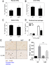
Fig. 1.
Fig. 1.
Reduced phagocytosis in SREBP-1aDF/B6 mice in vivo. (A) Plasma IC-IP6 levels in WT and SREBP-1aDF/B6 mice. Quantification of Fe3+ level in the plasma of mice i.v. injected with 10 μM IC-IP6 for 4 h. Fe concentrations in the livers (B) and spleens (C) of WT and SREBP-1aDF/B6 mice. (D) Uptake level of Fe3+ in peritoneal macrophages of WT and SREBP-1aDF/B6 mice. Data are represented as mean ± SEM; *P < 0.05. (E) Costaining of Prussian blue and F4/80 for assessment of IC-IP6 uptake by liver macrophages isolated from WT and SREBP-1aDF/B6 mice. (F) Quantification of data from E. ***P < 0.001.
Fig. 2.
Fig. 2.
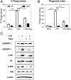
LPS-stimulated phagocytosis requires mTOR signaling and SREBP-1a. (A) Percentage of phagocytosis after LPS and rapamycin treatment in WT and SREBP-1aDF/B6 BMDMs. LPS (100 ng/mL) and rapamycin (25 μM) were treated respectively or together in BMDMs from WT and SREBP-1aDF/B6 mice for 16 h; BMDMs were incubated with sheep red blood cells for 30 min at 37 °C, and targets taken into 200 cells were counted. The percent of cells that displayed active phagocytosis was calculated. (B) Phagocytic index levels in WT and SREBP-1aDF/B6 BMDMs. The phagocytic index indicates the average number of targets per 200 cells. (C) Protein induction level for SREBP-1 and mTOR signaling by LPS and Rapamycin challenge in BMDMs. Values are expressed as mean ± SEM; **P < 0.01, ***P < 0.001.
Fig. 3.
Fig. 3.
SREBP-1a reverses phagocytosis defect in SREBP-1aDF/B6 BMDMs. Phagocytosis was repaired by SREBP-1a overexpression in SREBP-1aDF/B6 macrophages. A control (GFP) or SREBP-1a adenovirus was infected into the BMDM of SREBP-1aDF/B6 mice. After incubating adenoviruses for 48 h, BMDMs were treated with 100 ng/mL of LPS and 20 μM rapamycin for 16 h. Percentage of phagocytosis (A) and phagocytic index levels (B) were measured in WT BMDMs. Percentage of phagocytosis (C) and phagocytic index level (D) were measured in BMDMs from SREBP-1aDF/B6 mice. Data are represented as mean ± SEM; *P < 0.05, **P < 0.01, ***P < 0.001, vs. Ad-GFP.
Fig. 4.
Fig. 4.
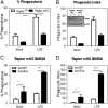
SREBP-1a restores phagocytosis defect in raptor mKO BMDMs. (A) Percentage of phagocytosis is increased by LPS in WT BMDMs, and phagocytosis is reduced in raptor mKO BMDMs. (B) Phagocytic index level by LPS in raptorfl/fl and raptor mKO BMDMs. Adenovirus of GFP or SREBP-1a (10 MOI) was infected in SREBP-1aDF/B6 BMDMs for 48 h, and BMDMs were treated with 100 ng/mL of LPS for 16 h. (C) Percentage of phagocytosis and (D) phagocytic index were measured. Data are represented as mean ± SEM; *P < 0.05; **P < 0.01, ***P < 0.001.
Fig. 5.
Fig. 5.
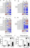
Phospholipid species with de novo synthesized fatty acyl chains are reduced in SREBP-1aDF/B6 and SCAP mKO BMDMs. (A) PC profile after LPS (100 ng/mL) treatment for 24 h, analyzed by liquid chromatography-MS in WT and SREBP-1aDF/B6 BMDMs. (B) PE profile after LPS challenge in WT and SREBP-1aDF/B6 BMDMs. (C) PC profile after LPS treatment in BMDMs from SCAPfl/fl and SCAP mKO mice. (D) PE profile after LPS treatment in BMDMs from SCAPfl/fl and SCAP mKO mice. (E) Percentage of phagocytosis with LPS plus oleic acid in WT and SREBP-1aDF/B6 BMDMs. (F) Phagocytic index level after LPS treatment plus oleic acid in WT and SREBP-1aDF/B6 BMDMs. Data are represented as mean ± SEM; **P < 0.01, ***P < 0.001.
Fig. 6.
Fig. 6.
SREBP-1a is required for actin reorganization proteins in DRM domains. (A) Flowchart illustrating the DRM isolation procedure, as detailed in Material and Methods. Fraction numbers correspond to the lane numbers in B. Each fraction was isolated after spinning down for 17 h, and we collected 1 mL from each fraction. (B) DRM proteins were confirmed by immunoblotting. Extracts isolated from BMDMs of WT and SREBP-1aDF/B6 mice were separated by ultracentrifugation for 16 h at 39,000 rpm in a Beckman SW55Ti swinging bucket rotor. (C) Effects of LPS and oleic acid (OA) on moesin association with DRMs. LPS (100 ng/mL) and OA (500 μM) were treated for 16 h and 24 h, respectively, and isolated DRMs from macrophages. (D) Effects of LPS and OA on cofilin association with DRMs.

Similar articles

Cited by

References

    1. Plüddemann A, Mukhopadhyay S, Gordon S. Innate immunity to intracellular pathogens: Macrophage receptors and responses to microbial entry. Immunol Rev. 2011;240:11–24. - PubMed
    1. Aderem A, Underhill DM. Mechanisms of phagocytosis in macrophages. Annu Rev Immunol. 1999;17:593–623. - PubMed
    1. Gagnon E, et al. Endoplasmic reticulum-mediated phagocytosis is a mechanism of entry into macrophages. Cell. 2002;110:119–131. - PubMed
    1. Castoreno AB, et al. Transcriptional regulation of phagocytosis-induced membrane biogenesis by sterol regulatory element binding proteins. Proc Natl Acad Sci USA. 2005;102:13129–13134. - PMC - PubMed
    1. Gurcel L, Abrami L, Girardin S, Tschopp J, van der Goot FG. Caspase-1 activation of lipid metabolic pathways in response to bacterial pore-forming toxins promotes cell survival. Cell. 2006;126:1135–1145. - PubMed

Publication types

MeSH terms

Substances

LinkOut - more resources