. 2019 Jan 7;8(1):bio039628.
doi: 10.1242/bio.039628.

Contribution of non-circadian neurons to the temporal organization of locomotor activity

Affiliations

Contribution of non-circadian neurons to the temporal organization of locomotor activity

Nicolás Pírez et al. Biol Open. .

Abstract

In the fruit fly, Drosophila melanogaster, the daily cycle of rest and activity is a rhythmic behavior that relies on the activity of a small number of neurons. The small ventral lateral neurons (sLNvs) are considered key in the control of locomotor rhythmicity. Previous work from our laboratory has showed that these neurons undergo structural remodeling on their axonal projections on a daily basis. Such remodeling endows sLNvs with the possibility to make synaptic contacts with different partners at different times throughout the day, as has been previously described. By using different genetic tools to alter membrane excitability of the sLNv putative postsynaptic partners, we tested their functional role in the control of locomotor activity. We also used optical imaging to test the functionality of these contacts. We found that these different neuronal groups affect the consolidation of rhythmic activity, suggesting that non-circadian cells are part of the circuit that controls locomotor activity. Our results suggest that new neuronal groups, in addition to the well-characterized clock neurons, contribute to the operations of the circadian network that controls locomotor activity in D. melanogaster.

Keywords: Connectivity; Drosophila; Locomotor rhythms; Non-circadian neurons; sLNvs.


Conflict of interest statement

Competing interestsThe authors declare no competing or financial interests.

Figures
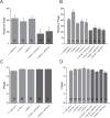
Fig. 1.
Fig. 1.
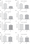
Constitutive silencing of non-circadian neurons caused a significant reduction on rhythmic power. Average rhythmic power and period under constant darkness (DD) for control lines (+>UAS-Kir2.1 and enhancer trap-GAL4>+) and experimental lines expressing the hyperpolarizing channel Kir2.1 under the expression pattern of the respective enhancer trap GAL4 lines. (A) Average rhythmic power for the experimental group one: 11-8 and 3-86 (one-way ANOVA, F12.78127, P<0.0001, genotype Tukey Comparisons, P<0.0001). (B) Average rhythmic power for the experimental group two: 7-49, 5-133, 5-43 and 4-93 (one-way ANOVA, F16.75476, P<0.0001, genotype Tukey Comparisons, P<0.05). (C) Average period for the experimental group one: 11-8 and 3-86 (one-way ANOVA, F9, P=0.0033, period Tukey Comparisons, P<0.01). (D) Average period for the experimental group two: 7-49, 5-133, 5-43 and 4-93 (one-way ANOVA, F3.2205, P=0.00187, period Tukey Comparisons, P<0.01). The data shown was calculated from 9–10 days at 25°C. The transition day between LD and DD was not used for these calculations. Data are expressed as mean±s.e.m. See text for a detailed explanation on the statistical analysis. Different letters represent statistical differences.
Fig. 2.
Fig. 2.
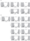
Constitutive silencing of non-circadian neurons did not cause a significant behavioral change in the daily activity profiles. Average activity plots for the first full day on LD for control lines (+>UAS-Kir2.1 and enhancer trap-GAL4>+) and experimental lines expressing the hyperpolarizing channel Kir2.1 under the expression pattern of the respective enhancer trap GAL4 lines. (A) Average activity plots for the experimental group one: 11-8 and 3-86. (B) Average activity plots for the experimental group two: 7-49, 5-133, 5-43 and 4-93. Shaded areas represent dark periods. See text for a detailed explanation on the statistical analysis.
Fig. 3.
Fig. 3.
Acute depolarization of clock neurons significantly reduced rhythmicity. (A) Representative actograms of the indicated genotypes. The different colors represent the temperature of the experiment: 22°C (gray), 28°C (pink). (B) Average rhythmic power under constant darkness (DD) at 22°C (light gray) PRE and at 28°C (dark gray) for the experimental line expressing dTrpA1 line under the control of the Clk856-GAL4. Data are expressed as mean±s.e.m. One-way ANOVA, F110.3827, P<0.0001, temperature Tukey comparisons, P<0.0001. See text for a detailed explanation on the statistical analysis.
Fig. 4.
Fig. 4.
Acute activation of non-circadian neurons triggered deconsolidation of rhythmic activity patterns. Average rhythmic power under constant darkness (DD) at 22°C (light gray) PRE and 28°C (dark gray) for the GAL4 parental control line and experimental line, expressing dTrpA1 line under the control of the different enhancer trap lines. (A) 11-8 (paired t-test, t53.76, P<0.0001), (B) 3-86 (paired t-test, t5.42, P=0.0123), (C) 4-12 (paired t-test, t5.667, P=0.0109), (D) 5-133 (paired t-test, t0.6062, P=0.5872), (E) 4-59 (paired t-test, t0.9855, P=0.397), (F) 4-93, (G) 7-49 (paired t-test, t2.619, P=0.0791) and (H) 5-43 (paired t-test, t0.2751, P=0.8091). Data are expressed as mean±s.e.m. Of note, the GRASP+ 4-93 line was analyzed in a single experiment precluding any statistical analysis.
Fig. 5.
Fig. 5.
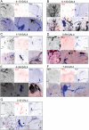
Expression pattern of novel neuronal clusters that participate in the control of locomotor activity. Confocal images that show a projection of the expression pattern of (A) 4-12-GAL4>UAS-mCD8GFP, (B) 5-133-GAL4>UAS-mCD8GFP, (C) 11-8-GAL4>UAS-mCD8GFP, (D) 3-86-GAL4>UAS-mCD8GFP, (E) 4-59-GAL4>UAS-mCD8GFP, (F) 7-49-GAL4>UAS-mCD8GFP and (G) 5-43-GAL4>UAS-mCD8GFP. All figures panels are the following: upper panels from left to right: GFP channel, PER channel and PDF channel, lower panels from left to right: merge of upper panels, zoom of PDF+ somas (merge of three channels) and sLNVs dorsal projections (merge of three channels). The magnification was 40×, except upper panels of figure C that was 20×. GFP, PDF and PER signal are shown in black, blue and red, respectively. Brains were dissected at ZT=2. lat, lateral; dor, dorsal. Scale bars: 20 µm.
Fig. 6.
Fig. 6.
Functionality of the synaptic contacts between sLNvs and putative postsynaptic targets. (A) Brains that do not express the P2X2 receptor do not show calcium changes following a stimulation with ATP (black trace). However, a high potassium stimulation does elicit a clear calcium response (gray trace). (B) Brains that express the LexAop-P2X2 but no LexA to drive it do not show calcium changes following ATP stimulation (black trace). However, a high potassium stimulation does elicit a clear calcium response (gray trace). (C) A brief 2.5 mM ATP stimulation elicits a clear calcium response measured in the PDF+ cells. Expressing the receptor and the sensor on the same cellular group controls for the delivery system and activation of the P2X2 receptor. pdf-GAL4 driver directed expression of UAS-GCaMP3 and pdf-LexA that of LexAop-P2X2. (D–E) Perfusion of ATP (activation of the sLNvs) did not elicit significant calcium responses measured by expressing the UAS-GCaMP3 under the control of 4-59 (D) or 3-86 (E) enhancer traps. (F) When sLNvs are activated by perfusion of 2.5 mM ATP, the mushroom body (MB) neuropil shows a clear calcium response, suggesting that the contacts between the sLNvs and the MB are functional. These experiments were performed within the ZT2–4 window. In all cases, the gray vertical bar represents the duration of the ATP stimulation.

Similar articles

Cited by

References

    1. Beckwith E. J. and Ceriani M. F. (2015). Experimental assessment of the network properties of the Drosophila circadian clock. J. Comp. Neurol. 523, 982-996. 10.1002/cne.23728 - DOI - PubMed
    1. Beckwith E. J., Gorostiza E. A., Berni J., Rezával C., Pérez-Santángelo A., Nadra A. D. and Ceriani M. F. (2013). Circadian period integrates network information through activation of the BMP signaling pathway. PLoS Biol. 11, e1001733 10.1371/journal.pbio.1001733 - DOI - PMC - PubMed
    1. Berni J., Beckwith E. J., Fernandez M. P. and Ceriani M. F. (2008). The axon-guidance roundabout gene alters the pace of the Drosophila circadian clock. Eur. J. Neurosci. 27, 396-407. 10.1111/j.1460-9568.2007.06010.x - DOI - PubMed
    1. Cao G., Platisa J., Pieribone V. A., Raccuglia D., Kunst M. and Nitabach M. N. (2013). Genetically targeted optical electrophysiology in intact neural circuits. Cell 154, 904-913. 10.1016/j.cell.2013.07.027 - DOI - PMC - PubMed
    1. Cavanaugh D. J., Geratowski J. D., Wooltorton J. R., Spaethling J. M., Hector C. E., Zheng X., Johnson E. C., Eberwine J. H. and Sehgal A. (2014). Identification of a circadian output circuit for rest:activity rhythms in Drosophila. Cell 157, 689-701. 10.1016/j.cell.2014.02.024 - DOI - PMC - PubMed

LinkOut - more resources