Review
. 2019 Jan;14(1):7-19.
doi: 10.4103/1673-5374.243694.

Differences in neuroplasticity after spinal cord injury in varying animal models and humans

Affiliations
Review

Differences in neuroplasticity after spinal cord injury in varying animal models and humans

Mallory E Filipp et al. Neural Regen Res. 2019 Jan.

Abstract

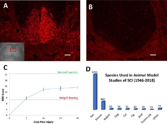
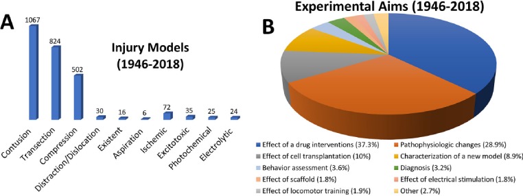
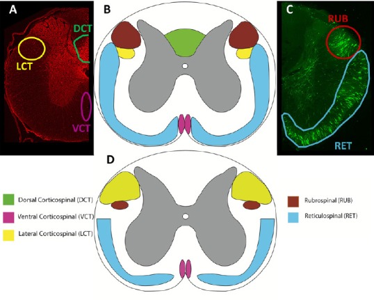
Rats have been the primary model to study the process and underlying mechanisms of recovery after spinal cord injury. Two weeks after a severe spinal cord contusion, rats can regain weight-bearing abilities without therapeutic interventions, as assessed by the Basso, Beattie and Bresnahan locomotor scale. However, many human patients suffer from permanent loss of motor function following spinal cord injury. While rats are the most understood animal model, major differences in sensorimotor pathways between quadrupeds and bipeds need to be considered. Understanding the major differences between the sensorimotor pathways of rats, non-human primates, and humans is a start to improving targets for treatments of human spinal cord injury. This review will discuss the neuroplasticity of the brain and spinal cord after spinal cord injury in rats, non-human primates, and humans. A brief overview of emerging interventions to induce plasticity in humans with spinal cord injury will also be discussed.

Keywords: animal studies; axons; functional recovery; locomotor training; plasticity; recovery; regeneration; spinal cord injury.


Conflict of interest statement

None

Figures

Figure 1
Figure 1
Animal models of SCI. After an SCI, there are major differences in the extent of recovery that occurs in different animal models. Four weeks after a spinal contusion in rats, biotinylated dextran amine, an anterograde axon tracer, was injected into the motor cortex. In a transverse spinal cord section rostral to the injury, hundreds of axons are labeled in the dorsal corticospinal tract (A), however caudal to the injury there are no labeled axons (B). Even in the absence of the dorsal corticospinal tract, the rats regain the ability to bear weight and step seven days after injury, continuing to improve for at least a month post-SCI (C). Even though this recovery is much higher than what would be expected to occur in humans, rats are still the most widely used model in SCI experiments (D). Scale bars: 100 μm (A & B). Injections performed according to Hellenbrand et al. (2013). SCI: Spinal cord injury; BBB: Basso, Beattie, and Bresnahan scale.
Figure 2
Figure 2
The goal of SCI research varies between studies. Contusion injuries are the most commonly seen injury in humans and thus the most studied animal models (A). Although contusions and compressions are thought to best represent an injury seen in humans, other models may be helpful in understanding the cellular and molecular mechanisms post-SCI and the effects of treatments on SCI. An appropriate injury model is chosen depending on the aims of the study (B). Adapted from Sharif-Alhoseini et al. (2017). SCI: Spinal cord injury.
Figure 3
Figure 3
Comparison of spinal cord tracts. Transverse sections of the rat (A-C) and human (D) demonstrating the approximate locations and sizes of the corticospinal, rubrospinal, and reticulospinal tracts. Rats were injected with an anterograde tracer, biotinylated dextran amine, into the motor cortex (A), reticular formation and red nucleus (B). Injections performed according to Hellenbrand et al. (2013). The corticospinal tract (CST) has larger lateral fibers in humans (yellow), while there is no dorsal CST in humans as compared to rats, who have a large dorsal CST (green). Both rats and humans have a ventral CST (pink). The rubrospinal tract (red) is prominent in rats, but largely reduced in humans, only passing through the upper cervical levels. All species express the reticulospinal tract (blue) prominently, with slight variations in location and size. High order non-human primates more closely resemble humans; however, tract size and exact location varies between non-human primate species. Adapted from Silva et al. (2013) and Watson et al. (2009).

References

    1. Adams MM, Hicks AL. Spasticity after spinal cord injury. Spinal Cord. 2005;43:577–586. - PubMed
    1. Alam M, He J. Lower-Limb Neuroprostheses: Restoring Walking after Spinal Cord Injury. In: Naik G, Guo Y, editors. Emerging Theory and Practice in Neuroprosthetics. Hershey, PA, USA: IGI Global; 2014. pp. 153–180.
    1. Allen AR. Surgery of experimental lesion of spinal cord equivalent to crush injury of fracture dislocation of spinal column: a preliminary report. JAMA LVLII. 1911:878–880.
    1. Angeli CA, Edgerton VR, Gerasimenko YP, Harkema SJ. Altering spinal cord excitability enables voluntary movements after chronic complete paralysis in humans. Brain. 2014;137:1394–1409. - PMC - PubMed
    1. Aoki M, Fujito Y, Satomi H, Kurosawa Y, Kasaba T. The possible role of collateral sprouting in the functional restitution of corticospinal connections after spinal hemisection. Neurosci Res. 1986;3:617–627. - PubMed