. 2019 Jan;14(1):114-123.
doi: 10.4103/1673-5374.243716.

Detecting white matter alterations in multiple sclerosis using advanced diffusion magnetic resonance imaging

Affiliations

Detecting white matter alterations in multiple sclerosis using advanced diffusion magnetic resonance imaging

Sourajit M Mustafi et al. Neural Regen Res. 2019 Jan.

Abstract

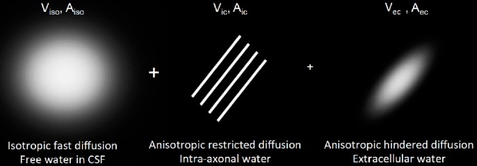
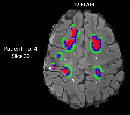
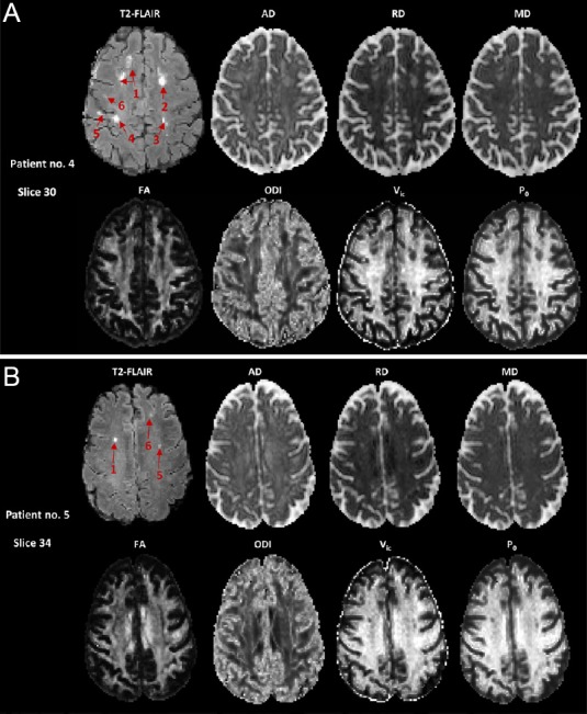
Multiple sclerosis is a neurodegenerative and inflammatory disease, a hallmark of which is demyelinating lesions in the white matter. We hypothesized that alterations in white matter microstructures can be non-invasively characterized by advanced diffusion magnetic resonance imaging. Seven diffusion metrics were extracted from hybrid diffusion imaging acquisitions via classic diffusion tensor imaging, neurite orientation dispersion and density imaging, and q-space imaging. We investigated the sensitivity of the diffusion metrics in 36 sets of regions of interest in the brain white matter of six female patients (age 52.8 ± 4.3 years) with multiple sclerosis. Each region of interest set included a conventional T2-defined lesion, a matched perilesion area, and normal-appearing white matter. Six patients with multiple sclerosis (n = 5) or clinically isolated syndrome (n = 1) at a mild to moderate disability level were recruited. The patients exhibited microstructural alterations from normal-appearing white matter transitioning to perilesion areas and lesions, consistent with decreased tissue restriction, decreased axonal density, and increased classic diffusion tensor imaging diffusivity. The findings suggest that diffusion compartment modeling and q-space analysis appeared to be sensitive for detecting subtle microstructural alterations between perilesion areas and normal-appearing white matter.

Keywords: NODDI; diffusion tensor imaging; hybrid diffusion imaging; multiple sclerosis; q-space imaging.


Conflict of interest statement

None

Figures

Figure 1
Figure 1
Visualization of diffusion compartments in the NODDI model. Viso and Vic are the volume fraction of the free isotropic diffusion and intra-axonal compartment, respectively. The extracellular volume fraction is Vec = (1 – Vic), a complement number to Vic. Aiso, Aic, and Aec are the normalized diffusion signal contributed from individual diffusion compartments. Aec: Diffusion signal from extra-axonal water; Aic: diffusion signal from intra-axonal water; Aiso: diffusion signal from isotropic fast diffusion; NODDI: neurite orientation dispersion and density imaging; Vec: volume fraction for extra-cellular water; Vic: volume fraction for intra-axonal water; Viso: volume fraction for isotropic fast diffusion; CSF: cerebrospinal fluid.
Figure 2
Figure 2
Segmentation of lesions, perilesions, and normal-appearing white matter (NAWM) on a T2-FLAIR map of patient No. 4. A set of ROIs includes a lesion (red), a matched perilesion (blue) and NAWM (green); this patient (Expanded Disability Status Scale score was 3.5) had six sets of lesions (No. 4). The set number for this patient, sizes of corresponding ROIs, and anatomic locations in the white matter are listed in Table 4. The gray scale is 0–300 AU for the T2-FLAIR map. Numbers 1–6 denote ROI sets listed in Table 3. AU: Arbitrary unit; FLAIR: fluid attenuated inversion recovery; no: number; ROIs: regions of interest.
Figure 3
Figure 3
Co-registered MRI maps of two patients with multiple sclerosis (MS). (A) Patient No. 4. Six hyperintense lesions in the T2-FLAIR image can be appreciated. Note that the two separated hyperintense regions of lesion 1 were connected in another slice (not shown). The lesion segmentation results in the same patient are shown in Figure 2. The corresponding areas in the diffusion maps appeared hyperintense in the AD, RD, and MD maps, and hypointense in the FA, Vic, and P0 maps. The gray scale is 0–300 AU for T2-FLAIR; 0–3 × 10⁻³ mm2/s for AD, RD, and MD; and 0–1 for FA, ODI, Vic, and P0. (B) Patient No. 5. Three hyperintense lesions in the T2-FLAIR image can be appreciated. AD: Axial diffusivity; AU: arbitrary unit; FA: fractional anisotropy; FLAIR: fluid attenuated inversion recovery; MD: mean diffusivity; ODI: orientation dispersion index; P0: q-space derived zero-displacement probability; RD: radial diffusivity; Vic: volume fraction for intra-axonal water.
Figure 4
Figure 4
Bar plots of T2-FLAIR and diffusion metrics in the three types of ROIs. The lesion, perilesion, and NAWM ROIs are color-coded in red, blue, and green, respectively. Error bars denote the standard deviation across 36 ROI sets. The overhead arrows denote significant differences in measurements between pairs of ROIs at a Bonferroni-corrected P threshold of 0.017 (Table 6). Note that T2-FLAIR intensity is not a quantitative measure; thus, it was not tested for statistical significance at the group level. FLAIR: Fluid attenuated inversion recovery; NAWM: normal-appearing white matter; ROIs: regions of interest.

References

    1. Assaf Y, Mayk A, Cohen Y. Displacement imaging of spinal cord using q-space diffusion-weighted MRI. Magn Reson Med. 2000;44:713–722. - PubMed
    1. Assaf Y, Chapman J, Ben-Bashat D, Hendler T, Segev Y, Korczyn AD, Graif M, Cohen Y. White matter changes in multiple sclerosis: correlation of q-space diffusion MRI and 1H MRS. Magn Reson Imaging. 2005;23:703–710. - PubMed
    1. Assaf Y, Ben-Bashat D, Chapman J, Peled S, Biton IE, Kafri M, Segev Y, Hendler T, Korczyn AD, Graif M, Cohen Y. High b-value q-space analyzed diffusion-weighted MRI: application to multiple sclerosis. Magn Reson Med. 2002;47:115–126. - PubMed
    1. Banaszek A, Bladowska J, Pokryszko-Dragan A, Podemski R, Sasiadek MJ. Evaluation of the degradation of the selected projectile, commissural and association white matter tracts within normal appearing white matter in patients with multiple sclerosis using diffusion tensor MR imaging - a preliminary study. Pol J Radiol. 2015;80:457–463. - PMC - PubMed
    1. Basser PJ, Mattiello J, LeBihan D. MR diffusion tensor spectroscopy and imaging. Biophys J. 1994;66:259–267. - PMC - PubMed