. 2018 Dec 7;8(1):17712.
doi: 10.1038/s41598-018-36277-4.

Xrp1 is a transcription factor required for cell competition-driven elimination of loser cells

Affiliations

Xrp1 is a transcription factor required for cell competition-driven elimination of loser cells

Ludovic Baillon et al. Sci Rep. .

Abstract

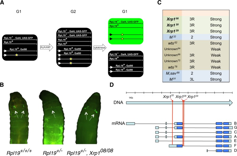
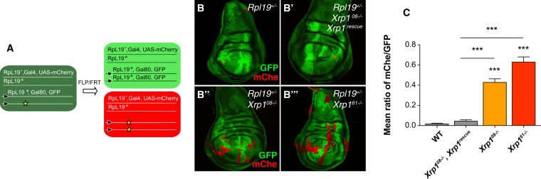
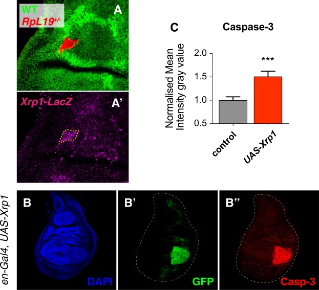
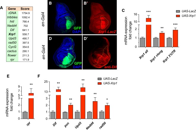
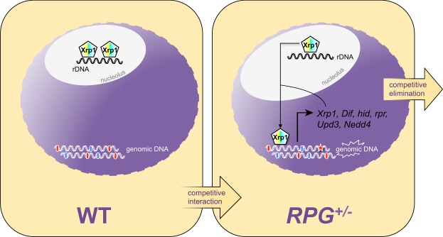
The elimination of unfit cells from a tissue is a process known in Drosophila and mammals as cell competition. In a well-studied paradigm "loser" cells that are heterozygous mutant for a haploinsufficient ribosomal protein gene are eliminated from developing tissues via apoptosis when surrounded by fitter wild-type cells, referred to as "winner" cells. However, the mechanisms underlying the induction of this phenomenon are not fully understood. Here we report that a CCAAT-Enhancer-Binding Protein (C/EBP), Xrp1, which is known to help maintaining genomic stability after genotoxic stress, is necessary for the elimination of loser clones in cell competition. In loser cells, Xrp1 is transcriptionally upregulated by an autoregulatory loop and is able to trigger apoptosis - driving cell elimination. We further show that Xrp1 acts in the nucleus to regulate the transcription of several genes that have been previously involved in cell competition. We therefore speculate that Xrp1 might play a fundamental role as a molecular caretaker of the genomic integrity of tissues.


Conflict of interest statement

The authors declare no competing interests.

Figures

Figure 1
Figure 1
Xrp1 mutations suppress cell competition driven elimination of loser cells in an EMS-based screen. Schematic of the genetics used to generate RpL19+/− loser clones in a wild-type background using the FLP/FRT system. Lines represent chromosomes, numbers at the end of each line indicate the chromosome number and triangles represent FRTs on the right arm of chromosome 3. Site-directed recombination between FRTs occurs when the expression of FLP is induced via heat shock. The yellow asterisk marks the chromosome to be tested for the presence of an EMS induced suppressor. The arrangement depicted here is a variation of the classical. MARCM technology that allows us to GFP label cells that are RpL19+/− and homozygous for a mutagenized chromosome arm 3R (A). Representative examples of living larvae displaying GFP clones in the pouch of the wing imaginal discs. SalE drives Gal4 expression in the wing pouch. (Left) Positive control for clone induction using the FRT82 RpL19+ chromosome. Recombination generates RpL19+/+/+ cells that are not eliminated. (Middle) Negative control for clone induction using the isogenized FRT82 chromosome. Recombination produces RpL19+/− cells that are efficiently eliminated. (Right) Suppressor Xrp108 rescues the elimination of RpL19+/− cells (B). List of suppressive mutations retrieved with the EMS screen. Intronic mutations Xrp108, Xrp1 and Xrp1 are strong suppressors (C). Different Xrp1 mRNA isoforms (from A to G). Blue color indicates the coding regions and light blue the untranslated regions. The red lines indicate the position of the three Xrp1 alleles retrieved from the EMS screen (Xrp108, Xrp1 and Xrp1) (D).
Figure 2
Figure 2
Xrp1 is required for the elimination of RpL19+/− loser cells. Schematic representation of the twin spot MARCM system used to generate RpL19+/− loser clones (red) and their respective twin spot clones (bright green, two copies of GFP) in a background of wild-type GFP positive cells (dark green) (A). RpL19+/− loser cells are eliminated (B). RpL19+/− cells are eliminated when Xrp1 mutations are rescued with the re-introduction of one copy of Xrp1 (B’). Loser cells elimination is rescued via either intronic Xrp108−/− mutations retrieved from the EMS screen (B”) or, even more efficiently, via Xrp161−/− mutations in the coding sequence of Xrp1 (B”’). Quantification of the mean ratio between mCherry Area and GFP2+ area (mChe/GFP). Minute loser cells, labeled with mCherry, are eliminated and the mChe/GFP ratio is close to 0. Xrp1 mutants rescue the elimination of loser cells (ratio close to 1). ***P < 0.001, *P < 0.05, Kruskal-Wallis test. Bars represent SEM. n = 52,47,45,48. Additional significance was calculated via assessing distribution normality (D’Agostino & Pearson normality test). Xrp108−/− and Xrp161−/− follow a normal distribution (P < 0.001) (C).
Figure 3
Figure 3
Xrp1 is upregulated in loser clones and functions as a driver of apoptosis. Xrp1 is upregulated in RpL19+/− loser cells as observed with a LacZ-reporter (A-A’). Overexpression of Xrp1 in the posterior compartment (en-Gal4) induces apoptosis, as observed with an anti-cleaved Caspase 3 staining (B-B”). Normalized quantification of the mean intensity gray value confirms increased apoptotic staining. Paired ratio t-test was applied. ***P < 0.001 (C).
Figure 4
Figure 4
Xrp1 regulates its own expression, the expression of pro-apoptotic genes and of genes previously linked to cell competition. ChIP-seq on Xrp1 OE wing discs reveals targets of Xrp1, including rDNA, Xrp1 itself, pro-apoptotic genes such as hid and rpr and several other genes that have been linked to either cell competition or cell proliferation (A). Using a transcriptional reporter for Xrp1 and via the overexpression of Xrp1 in the posterior compartment (en-Gal4), immunostaining reveals a positive feedback loop by which Xrp1 regulates its own transcription. Xrp1 (B-B’). The observation is confirmed by qPCR, Xrp1 is overexpressed in the wing disc 24 hours before analysis. We used primers that recognize all forms of Xrp1 including the overexpressed isoform and confirmed that the Xrp1 overexpression construct is functional (Xrp1 all). With primers that detect only the endogenous forms of Xrp1 we observe Xrp1-dependent induction of Xrp1 expression (Xrp1 vlong, Xrp1 3UTR) (C). A representative example showing upregulation of the Dif protein upon overexpression of Xrp1 in the posterior compartment (D-D’). Effects on putative Xrp1 target genes are confirmed via qPCRs. The pro-apoptotic gene rpr is upregulated in response to Xrp1 OE in wing discs. Xrp1 OE is induced 24 hours before analysis (E). Other putative target genes are also upregulated under the same conditions, respectively Dif, puc, Upd3, Nedd4 and rad50 (F). t-test was applied. *P < 0.05; **P < 0.01; ***P < 0.001.
Figure 5
Figure 5
Role of Xrp1 in cell competition-driven elimination of loser cells. Xrp1 localized in the nucleulus sitting on rDNA. In a competitive scenario, Xrp1 acts as a transcription factor in loser cells, driving the expression of Xrp1 itself, of pro-apoptotic target genes (hid, rpr), of genes involved in innate immunity (Dif), in compensatory proliferation (Upd3) and in protein degradation-dependent apoptosis and cell proliferation arrest (Nedd4). Xrp1, because of its double involvement in the elimination of loser cells and in DNA repair, putatively acts as a genomic caretaker in a p53-independent fashion.

References

    1. Morata G, Ripoll P. Minutes: mutants of drosophila autonomously affecting cell division rate. Dev Biol. 1975;42(2):211–21. doi: 10.1016/0012-1606(75)90330-9. - DOI - PubMed
    1. Clavería C, Giovinazzo G, Sierra R, Torres M. Myc-driven endogenous cell competition in the early mammalian embryo. Nature. 2013;500:39–44. doi: 10.1038/nature12389. - DOI - PubMed
    1. Marygold SJ, et al. The ribosomal protein genes and Minute loci of Drosophila melanogaster. Genome Biol. 2007;8:R216–R226. doi: 10.1186/gb-2007-8-10-r216. - DOI - PMC - PubMed
    1. Amoyel M, Bach EA. Cell competition: how to eliminate your neighbours. Development. 2014;141:988–1000. doi: 10.1242/dev.079129. - DOI - PMC - PubMed
    1. Burke R, Basler K. Dpp receptors are autonomously required for cell proliferation in the entire developing Drosophila wing. Development. 1996;122(7):2261–9. - PubMed

Publication types

MeSH terms

LinkOut - more resources