. 2019 Jul;44(8):1377-1388.
doi: 10.1038/s41386-018-0274-7. Epub 2018 Nov 15.

Impaired anandamide/palmitoylethanolamide signaling in hippocampal glutamatergic neurons alters synaptic plasticity, learning, and emotional responses

Affiliations

Impaired anandamide/palmitoylethanolamide signaling in hippocampal glutamatergic neurons alters synaptic plasticity, learning, and emotional responses

Tina Zimmermann et al. Neuropsychopharmacology. 2019 Jul.

Abstract

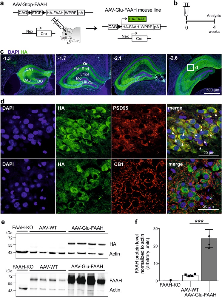
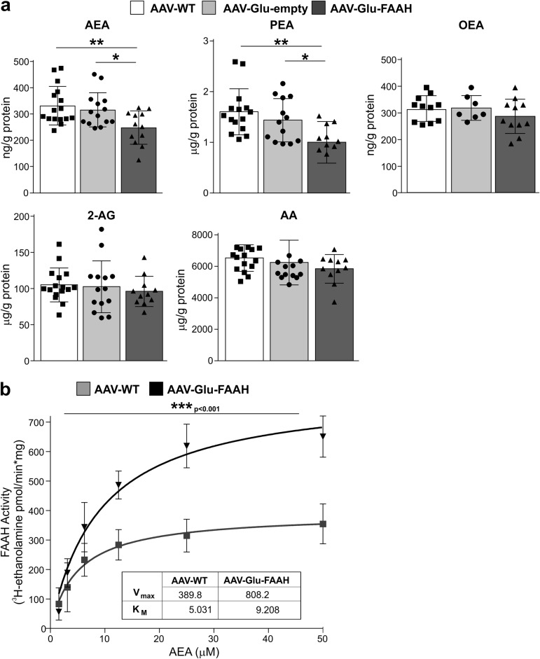
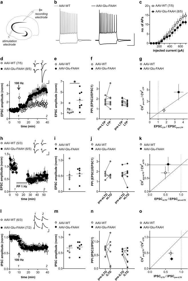
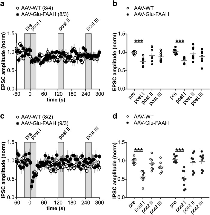
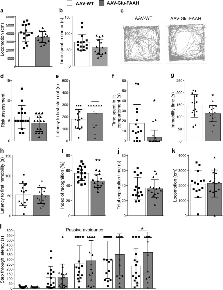
Endocannabinoid signaling via anandamide (AEA) is implicated in a variety of neuronal functions and considered a promising therapeutic target for numerous emotion-related disorders. The major AEA degrading enzyme is fatty acid amide hydrolase (FAAH). Genetic deletion and pharmacological inhibition of FAAH reduce anxiety and improve emotional responses and memory in rodents and humans. Complementarily, the mechanisms and impact of decreased AEA signaling remain to be delineated in detail. In the present study, using the Cre/loxP system combined with an adeno-associated virus (AAV)-mediated delivery system, FAAH was selectively overexpressed in hippocampal CA1-CA3 glutamatergic neurons of adult mice. This approach led to specific FAAH overexpression at the postsynaptic site of CA1-CA3 neurons, to increased FAAH enzymatic activity, and, in consequence, to decreased hippocampal levels of AEA and palmitoylethanolamide (PEA), but the levels of the second major endocannabinoid 2-arachidonoyl glycerol (2-AG) and of oleoylethanolamide (OEA) were unchanged. Electrophysiological recordings revealed an enhancement of both excitatory and inhibitory synaptic activity and of long-term potentiation (LTP). In contrast, excitatory and inhibitory long-term depression (LTD) and short-term synaptic plasticity, apparent as depolarization-induced suppression of excitation (DSE) and inhibition (DSI), remained unaltered. These changes in hippocampal synaptic activity were associated with an increase in anxiety-like behavior, and a deficit in object recognition memory and in extinction of aversive memory. This study indicates that AEA is not involved in hippocampal short-term plasticity, or eLTD and iLTD, but modulates glutamatergic transmission most likely via presynaptic sites, and that disturbances in this process impair learning and emotional responses.


Figures

Fig. 1
Fig. 1
FAAH overexpression in hippocampal CA1-CA3 pyramidal neurons. a Schematic diagram showing the experimental strategy for FAAH overexpression. AAV-FAAH contained a ubiquitous promoter (CAG), a loxP flanked (black triangles) transcriptional Stop cassette (STOP), and HA-FAAH encoding sequence. AAV-FAAH was injected into dorsal and ventral hippocampus of Nex-Cre mice. The stop cassette was excised in a Cre-dependent manner, allowing expression of HA-FAAH, termed AAV-Glu-FAAH. WPRE woodchuck hepatitis virus posttranscriptional regulatory element; pA polyadenylation signal. b Timeline of experiment. Mice were analyzed 4 weeks post AAV injection. c Coronal sections of the injected hippocampus of AAV-Glu-FAAH mice immunostained for HA, displaying the rostrocaudal extent of AAV-mediated HA-tagged FAAH expression. Numbers indicate the distance from bregma. CA1 cornu ammonis region 1, CA3 cornu ammonis region 3, DG dentate gyrus, GC granule cell layer, Hil hilar region, LMol stratum lacunosum-moleculare, Mol stratum moleculare, Or stratum oriens, Pyr pyramidal cell layer, Rad stratum radiatum. d HA and postsynaptic density marker 95 (PSD95) colocalization (upper panels; white triangles); HA and presynaptic CB1 receptor immunostaining in the CA1 region (bottom panels). e Western blot analysis of homogenized hippocampi from FAAH knockout (FAAH-KO), wild-type control (AAV-WT) and FAAH overexpressing mice (AAV-Glu-FAAH) revealed a specific FAAH signal at 63 kDa in wild-type and transgenic samples, and exclusive transgene (HA) expression in AAV-Glu-FAAH mice. f Quantification of FAAH protein levels in hippocampal homogenates showed more than seven-fold increase in transgenic mice (AAV-Glu-FAAH) as compared to wild-type controls (AAV-WT) (n = 4). ***p < 0.001, Student´s t-test. Data are represented as mean ± SEM. Statistical analysis details are reported in Supplementary Table S2.
Fig. 2
Fig. 2
FAAH overexpression in hippocampal CA1-CA3 pyramidal neurons leads to decreased anandamide (AEA) and palmitoylethanolamide (PEA) levels. a AEA and PEA levels were significantly lower in AAV-Glu-FAAH samples (n = 11-12) as compared to control AAV-WT animals (n = 15-16) and AAV-empty injected mice (n = 12-14), whereas oleoylethanolamide (OEA), arachidonic acid (AA), and 2-arachidonoylglycerol (2-AG) levels were not altered. b FAAH activity is enhanced upon FAAH overexpression in hippocampus. Along increasing amounts of substrate [3H]-AEA (0–50 μM range), FAAH activity was increased in AAV-Glu-FAAH (n = 3) as compared to AAV-WT (n = 3) mice as indicated by the increase in the maximal amount of product [3H-ethanolamine], and the statistically significant increase in Vmax (inset). No significant change in Km constant was found. Data are expressed as amount of product [3H-ethanolamine] (pmol) generated over reaction time (min) and protein amount (mg). *p < 0.05, **p < 0.01, ***p < 0.001 one-way ANOVA with Tukey’s multiple comparison test. Vmax and Km values were obtained by using a Michaelis-Menten equation-based analysis. Values are expressed as means ± SEM. Statistical analysis details are reported in Supplementary Table S3.
Fig. 3
Fig. 3
Long-term plasticity at hippocampal CA3-CA1 synapses. a Schematic drawing illustrates placement of stimulation and recording electrodes in the hippocampal slice. b Discharge pattern of CA1 neurons upon depolarizing (500 pA) and hyperpolarizing (−250 pA) current pulses. Scale bars: 50 mV, 200 ms. c Number of generated action potentials (APs) plotted against positive current injection (in pA) in CA1 pyramidal cells of AAV-WT and AAV-Glu-FAAH mice. d, e LTP in CA1 following high-frequency stimulation (HFS, 100 Hz) is significantly enhanced in AAV-Glu-FAAH mice compared to AAV-WT mice (unpaired t test, *p < 0.05). d Inset shows sample EPSC traces at indicated time points before and after HFS, scale bars: 100 pA, 25 ms. f PPI does not change after induction of LTP. g Analysis of the coefficient of variation (CV2) plotted against mean EPSC ratios indicates a presynaptic mechanism involved in LTP in AAV-Glu-FAAH mice and a postsynaptic one in AAV-WT mice. h, i Excitatory LTD (eLTD) at CA3-CA1 synapses following low-frequency stimulation (LFS, 900 paired pulses, 1 Hz) is unchanged in AAV-Glu-FAAH mice compared to AAV-WT mice. h Inset shows sample EPSC traces at indicated time points before and after LFS, scale bars: 100 pA, 25 ms. j PPI does not change after induction of eLTD. k Analysis of CV2 plotted against mean EPSC ratios point to a postynaptic mechanism involved in eLTD in both genotypes. l, m. Both AAV-WT and AAV-Glu-FAAH mice showed intact inhibitory LTD (iLTD) at CA3-CA1 synapses. In (l), inset shows sample IPSC traces at indicated time points before and after HFS, scale bars: 200 pA, 25 ms. n PPI does not change after induction of iLTD. o Analysis of CV2 plotted against mean IPSC ratios indicates a presynaptic mechanism involved in iLTD in mice of both genotypes. In e, i, m, scatter plots and corresponding mean ± SEM represent amplitudes recorded after plasticity induction (LTP and iLTD: min 36–40; eLTD: min 46–50). In c, d, h, l, numbers indicate the number of recorded cells/animals.
Fig. 4
Fig. 4
Depolarization-induced suppression of excitation (DSE) and inhibition (DSI) at hippocampal CA3-CA1 synapses. a, c Upon postsynaptic depolarization (−70 to 0 mV, 10 s duration, at time zero), both AAV-WT and AAV-Glu-FAAH mice showed comparable DSE and DSI. Numbers indicate the number of recorded cells/animals. b, d Scatter plot and corresponding mean ± SEM of amplitudes recorded immediately before (pre), immediately after (post I), 2 min (post II) and 4 min after depolarization (post III). Repeated measure-ANOVA, factor time in b: F(3, 42) = 13.22, p < 0.0001, factor time in d: F(3, 45) = 36.88, p < 0.0001, ***p < 0.001 pre vs. post I, by post hoc Bonferroni.
Fig. 5
Fig. 5
Effect of FAAH overexpression on anxiety-like behavior and memory formation. a Animals tested in the open field test showed equal locomotor activity, although (b) AAV-Glu-FAAH mice tended to spend less time in the center than AAV-WT mice. c Representative movement pattern of individual mice exposed to the open field, showing a center-avoiding behavior in AAV-Glu-FAAH mice. d In the light dark test, AAV-Glu-FAAH mice showed unaltered risk assessments, (e) no change in latency to first step out, and (f) a decrease in time spent in the lit compartment. g In the forced swim test, AAV-Glu-FAAH mice were less immobile compared to control, whereas (h) the latency to first immobility was unchanged. (i) Spatial memory was assessed by the spatial object recognition test. AAV-Glu-FAAH mice showed a decrease in the index of recognition, while (j) total exploration time and (k) locomotor activity were not affected. (l) In the fear-motivated avoidance task AAV-Glu-FAAH mice displayed equal memory acquisition (first two bars), and equal step-through latencies 1 h, 24 h and 1 week after conditioning, but a significant increase in the step-through latencies 2 weeks after conditioning (last two bars). n = 15 mice per group. *p < 0.05, **p < 0.01, Student´s t-test and two-way repeated measures ANOVA with Sidak’s multiple comparison test. Values are expressed as means ± SEM. Statistical analysis details are reported in Supplementary Tables S2 and S4.

Comment in

Similar articles

Cited by

References

    1. Duman RS, Aghajanian GK, Sanacora G, Krystal JH. Synaptic plasticity and depression: New insights from stress and rapid-acting antidepressants. Nat Med. 2016;22:238–49. doi: 10.1038/nm.4050. - DOI - PMC - PubMed
    1. Bannerman DM, Sprengel R, Sanderson DJ, McHugh SB, Rawlins JN, Monyer H, et al. Hippocampal synaptic plasticity, spatial memory and anxiety. Nat Rev Neurosci. 2014;15:181–92. doi: 10.1038/nrn3677. - DOI - PubMed
    1. Lutz B, Marsicano G, Maldonado R, Hillard CJ. The endocannabinoid system in guarding against fear, anxiety and stress. Nat Rev Neurosci. 2015;16:705–18. doi: 10.1038/nrn4036. - DOI - PMC - PubMed
    1. Maccarrone M. Metabolism of the endocannabinoid anandamide: open questions after 25 years. Front Mol Neurosci. 2017;10:166. doi: 10.3389/fnmol.2017.00166. - DOI - PMC - PubMed
    1. Basavarajappa BS, Nagre NN, Xie S, Subbanna S. Elevation of endogenous anandamide impairs LTP, learning, and memory through CB1 receptor signaling in mice. Hippocampus. 2014;24:808–18. doi: 10.1002/hipo.22272. - DOI - PMC - PubMed

Publication types

MeSH terms

LinkOut - more resources