Review
. 2019 Jan;19(1):9-31.
doi: 10.1038/s41568-018-0081-9.

The lung microenvironment: an important regulator of tumour growth and metastasis

Affiliations
Review

The lung microenvironment: an important regulator of tumour growth and metastasis

Nasser K Altorki et al. Nat Rev Cancer. 2019 Jan.

Abstract

Lung cancer is a major global health problem, as it is the leading cause of cancer-related deaths worldwide. Major advances in the identification of key mutational alterations have led to the development of molecularly targeted therapies, whose efficacy has been limited by emergence of resistance mechanisms. US Food and Drug Administration (FDA)-approved therapies targeting angiogenesis and more recently immune checkpoints have reinvigorated enthusiasm in elucidating the prognostic and pathophysiological roles of the tumour microenvironment in lung cancer. In this Review, we highlight recent advances and emerging concepts for how the tumour-reprogrammed lung microenvironment promotes both primary lung tumours and lung metastasis from extrapulmonary neoplasms by contributing to inflammation, angiogenesis, immune modulation and response to therapies. We also discuss the potential of understanding tumour microenvironmental processes to identify biomarkers of clinical utility and to develop novel targeted therapies against lung cancer.


Conflict of interest statement

Competing interests

The authors declare no competing interests.

Figures
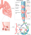
Fig. 1 |
Fig. 1 |. The heterogeneous microenvironment of the lung.
A schematic of the normal lung showing anatomic regions encompassing the proximal and distal airways is shown. The proximal airways are composed of ciliated cells, secretory club cells, undifferentiated basal cells, mucus-producing goblet cells and neuroendocrine cells; the distal airways are composed of alveolar type I and type II cells. Other cell types in the lung microenvironment include smooth muscle cells, fibroblasts, endothelial cells and immune cells, including resident alveolar macrophages and dendritic cells. Vascular capillaries line the alveolar walls to facilitate gaseous exchange and infiltration of circulating immune cells. Endothelium-derived angiocrine signalling induces and sustains regenerative lung alveolarization,. Resident alveolar macrophages maintain immunological homeostasis; however, they can also contribute to inflammation and the development of pre-malignant lung lesions in mice. Patients with chronic obstructive pulmonary disease (COPD) are at a higher risk of developing lung cancer, and their lungs show remodelling of the airway epithelium and alterations in lung inflammatory cells including neutrophils, monocytes and macrophages, which express elevated levels of pro-inflammatory mediators. A population of tissue-resident memory T cells resides in the lung airways, and they are generated by a diverse set of pathogens penetrating mucosal barriers and are believed to confer protective immunity to secondary infections. ECM, extracellular matrix.
Fig. 2 |
Fig. 2 |. The heterogeneous microenvironment of lung cancer.
The lung tumour microenvironment (TME) includes a variety of tumour-reprogrammed stromal cells. A major hallmark of immunosuppression in the TME is the inactivation of cytotoxic CD8+T cells, which is achieved through diverse pathways. Immature dendritic cells (DCs) produce transforming growth factor-β (TGFβ), which expands the population of immunosuppressive forkhead box P3 (FOXP3)+ regulatory T (Treg) cells, which in turn inhibit CD8+T cells. DCs also express programmed cell death protein 1 (PD1) ligand 1 (PDL1) to directly suppress CD8+ T cells. Myeloid-derived suppressor cells (MDSCs) express arginase 1 (ARG1) and inducible nitric oxide synthase (iNOS), which contribute to T cell inhibition. Cancer-associated fibroblasts (CAFs) can also suppress the activity of CD8+ T cells by inducing Treg cells or by expressing PDL1, PDL2 and FAS ligand (FASL). Furthermore, tumour cells can directly produce inhibitory molecules including cyclooxygenase 2 (COX2), prostaglandin E2 (PGE2), PDL1 and indoleamine 2,3-dioxygenase (IDO), which can blunt the activity of CD8+ tumour-infiltrating lymphocytes. Neutrophils are major players in the lung TME, as they impact tumour progression through secretion of cytokines (interleukin-1β (IL-1β) and IL-6). Tumours secrete soluble receptor for advanced glycosylation end product (sRAGE), which systemically activates osteoblasts in the bone marrow to produce tumour-infiltrating sialic acid-binding immunoglobulin-like lectin F (SiglecF) neutrophils, which promote tumour growth by increasing angiogenesis, myeloid cell differentiation, T cell suppression and tumour cell proliferation. Endothelial cells respond to vascular endothelial growth factor (VEGF) from various sources including tumour cells, natural killer (NK) cells and tumour-associated macrophages (TAMs) to induce angiogenesis. In addition to promoting angiogenesis, TAMs may also activate other pathways to promote invasion and metastasis through secretion of various factors including IL-6, COX2, matrix metalloproteinase 9 (MMP9) and MMP14. Besides inducing angiogenesis, NK cells also mediate immune suppression by undergoing dysfunctional phenotypes characterized by downregulation of NK receptors, loss of degranulation potential and reduced interferon-γ (IFNγ) expression. B cells in tertiary lymphoid structures (TLS) can either generate tumour-specific antibodies or undergo a dysfunctional state that induces Treg cells. BTLA, B and T lymphocyte attenuator; CSCs, cancer stem cells; CTLA4, cytotoxic T lymphocyte-associated antigen 4; EMT, epithelial-to-mesenchymal transition; FGF4, fibroblast growth factor 4; GAS6, growth arrest-specific protein 6; HGF, hepatocyte growth factor; IGF1R, insulin-like growth factor 1 receptor; IGF2, insulin-like growth factor 2; LAG3, lymphocyte activation gene 3; PDGFβ, platelet-derived growth factor-β; PlGF, placental growth factor; STAT, signal transducer and activator of transcription; TIM3, T cell membrane protein 3; TNF, tumour necrosis factor.
Fig. 3 |
Fig. 3 |. Cancer cell-intrinsic pathways mediate immunosuppression in lung cancer.
Lung adenocarcinomas with specific driver mutations exhibit discrete immune phenotypes. The observation that programmed cell death protein 1 (PD1) ligand 1 (PDL1) expression was increased in patients with KRAS-mutant lung cancer led to the finding that mutant KRAS in mouse tumours induces PDL1 expression either through phosphorylated ERK (pERK) signalling or Pdl1 mRNA stabilization to mediate immune escape,. These studies advocate the use of either ERK inhibitors or inhibition of the AU-rich element-binding protein tristetraprolin (TTP), which is responsible for PDL1 stabilization, for the treatment of KRAS tumours. Loss of the tumour suppressor LKB1 in KRAS-mutant mouse tumours is associated with increased accumulation of immunosuppressive neutrophils, exhausted T cells, increased pro-inflammatory cytokines including interleukin-6 (IL-6) and decreased PDL1 expression. While PD1 monotherapy was ineffective in this setting, its therapeutic efficacy can be restored with either neutrophil depletion or an IL-6-neutralizing antibody. Similarly, immune suppression in KRAS-mutant mouse tumours may be a result of MYC co-activation leading to upregulation of IL-23 and CC-chemokine ligand 9 (CCL9), which mediates the exclusion of B cells, T cells and natural killer (NK) cells, as well as the recruitment of proangiogenic macrophages. As expected, inhibition of CCL9 and IL-23 can decrease MYC-induced tumour progression. Clinical observations that epithelial growth factor receptor (EGFR)-mutant tumours are associated with high PDL1 expression are consistent with the observation that EGFR-driven mouse tumours suppress T cell function by activating PD1 and PDL1 (REF). Patients with echinoderm microtubule-associated protein-like 4 (EML4)-anaplastic lymphoma kinase (ALK) fusion oncogene-driven lung cancer also show increased PDL1 expression and, in mice, it was found that EML4-ALK-mediated activation of the PI3K-AKT and MEK-ERK pathways upregulates PDL1 expression. In carcinogen-induced lung tumorigenesis, signal transducer and activator of transcription 3 (STAT3) activation in cancer cells inhibits pro-inflammatory chemokines, increases major histocompatibility complex class I (MHC I) expression, decreases effector cytokines and blunts NK cell-mediated toxicity. Despite these findings, the link between oncogenic drivers and PDL1 expression remains controversial, as one study demonstrated an association of PDL1 expression with mutant EGFR but not KRAS or ALK in patients with non-small-cell lung cancer (NSCLC), while another study failed to correlate PDL1 expression with mutant EGFR in patients. In yet another study, PDL1 expression was associated with mTOR activation in patients with NSCLC driven by a wide spectrum of driver mutations, and the combination of mTOR and PD1 inhibitors could reduce tumour growth, increase tumour-infiltrating lymphocytes and decrease regulatory T (Treg) cells in mouse lung cancers. An emerging concept lends support for oncogene cooperativity in driving immune evasion. Co-activation of KRAS and MYC or PI3K and Kelch-like ECH-associated protein 1 (KEAP1)-nuclear factor erythroid-2-related factor 2 (NRF2) reprogrammes the tumour microenvironment (TME) to promote lung adenocarcinoma,. Indeed, genetic deficiency of Keap1 and Pten can increase the efficacy of immune checkpoint inhibition. As cancer cell-intrinsic pathways alter immunity in unique ways, this calls for studies involving large cohorts of patients with NSCLC to establish how specific genomic alterations impact the TME composition and function; this would be valuable for designing combination treatments for patients who have been selected on the basis of their tumour genomic alterations and immune contexture. CTLA4, cytotoxic T lymphocyte-associated antigen 4; CXCL, CXC-chemokine ligand; G-CSF, granulocyte colony-stimulating factor; IFNγ, interferon-γ; NF-κB, nuclear factor-κB; TGFβ, transforming growth factor-β; TNF, tumour necrosis factor.
Fig. 4 |
Fig. 4 |. The lung metastatic niche.
Primary tumours secrete various growth factors, cytokines and enzymes (vascular endothelial growth factor A (VEGFA), placental growth factor (P1GF), transforming growth factor-β (TGFP), tumour necrosis factor (TNF) and lysyl oxidase) and shed extracellular vesicles into the circulation that systemically impact the lung microenvironment. Tumour-derived factors can also activate bone marrow compartments, resulting in the mobilization and recruitment of bone marrow-derived cells into the lungs. Together, these activities contribute to the generation of pre-metastatic niches in the lung. Extravasation of disseminated tumour cells (DTCs) into the lung parenchyma is facilitated by microRNA miR-105 delivered by breast cancer exosomes, which silence the tight junction protein zona occludens 1 (ZO1) to allow DTCs to breach vascular barriers. In addition, CC-chemokine ligand 2 (CCL2) gradients recruit CC-chemokine receptor 2 (CCR2)+ inflammatory monocytes, which produce VEGF to increase vascular permeability. Following extravasation, the pre-metastatic niche contributes to the seeding and colonization of the DTCs in the lungs. Hypoxia in primary breast tumours upregulates lysyl oxidase, which systemically oxidizes lysine residues in collagen and elastin, resulting in covalent crosslinking of these molecules in the lungs. Collagen crosslinking increases the adhesion of myeloid cells to generate microenvironments conducive to DTC colonization. Platelets secrete CXC-chemokine ligand 5 (CXCL5) and CXCL7, which recruit granulocytes to facilitate metastatic seeding. Exosome-derived small nuclear RNAs (snRNAs) activate Toll-like receptor 3 (TLR3) in lung alveolar type II cells to induce chemokine secretion, which in turn mediates neutrophil recruitment. Neutrophils, via arachidonate 5-lipoxygenase (ALOX5)-dependent leukotriene synthesis, stimulate adhesion, chemotaxis and vascular permeability to support colonization of leukotriene receptor-positive DTCs. In addition, the extracellular matrix (ECM) component fibronectin enables clustering of bone marrow-derived VEGF receptor 1 (VEGFR1)+ haematopoietic cells, leading to increased production of CXCL12, which recruits CXC-chemokine receptor 4 (CXCR4)+ DTCs to the pre-metastatic niche. Following seeding of DTCs, α4 integrin-expressing macrophages interact with vascular cell adhesion protein 1 (VCAM1)+ DTCs to induce pro-survival AKT signalling. The polarized neutrophils in the pre-metastatic lungs may suppress activation of CD8+ T cells to dampen anti-metastatic immunity. Subsequently, metastatic outgrowth is supported by various pathways. Neutrophils may secrete cathepsin G and neutrophil elastase to proteolytically degrade the antitumorigenic factor thrombospondin 1 (TSP1) to promote the metastatic outgrowth. Tumour-secreted factors induce expression of chemoattractants, such as S100A8 and S100A9, by lung endothelial cells and MAC1 (also known as CD11b)+ myeloid cells, which facilitate the homing of tumour cells to the pre-metastatic niche, through the induction of serum amyloid A3 (SAA3). Notably, SAA3 stimulates nuclear factor-κβ (NF-κβ) signalling in a TLR4-dependent manner to facilitate inflammation-mediated metastasis. DTCs can induce expression of periostin or tenacin C in lung fibroblasts to support cancer stem cell (CSC) maintenance and expansion through WNT and Notch signalling, respectively. The colonized mesenchymal tumour cells may undergo an epithelial conversion that is supported by paracrine interactions with reprogrammed monocytic cells. The myeloid-derived proteoglycan versican mediates inhibition of the TGFβ-SMAD2 or SMAD3 pathway in DTCs that induces mesenchymal-to-epithelial transition (MET) and promotes metastatic outgrowth. Through another mechanism, mesenchymal DTCs can also express TSP2 to activate local fibroblasts, which in turn promote MET of cancer cells. Lastly, metastatic outgrowth can be facilitated by infiltrating bone marrow-derived endothelial progenitor cells (EPCs) and endothelial cells, which contribute to the switch to angiogenesis. The pre-metastatic niche can exhibit features of metastasis suppression, as lung endothelial cells expressing CX3C-chemokine ligand 1 (CX3CL1) attract CX3C-chemokine receptor 1 (CX3CR1)+ patrolling monocytes, which through expression of CCL3, CCL4 and CCL5 recruit cytotoxic natural killer (NK) cells to inhibit metastasis. Similarly, metastatic incompetent tumours express the glycoprotein prosaposin, which systemically reprogrammes myeloid cells in the lungs to express TSP1, which inhibits metastatic outgrowth. G-CSF, granulocyte colony-stimulating factor; IL-1β, inter1eukin-1β; LY6C, lymphocyte antigen 6C; MMP9, matrix metalloproteinase 9.

Similar articles

Cited by

References

    1. Siegel RL, Miller KD & Jemal A Cancer statistics, 2018. CA Cancer J. Clin. 68, 7–30 (2018). - PubMed
    1. Herbst RS, Morgensztern D & Boshoff C The biology and management of non-small cell lung cancer. Nature 553, 446–454 (2018).

      This review highlights recent progress in lung cancer biology and therapeutic strategies that are impacting outcomes for patients with advanced-stage NSCLC.

    1. Chen Z, Fillmore CM, Hammerman PS, Kim CF & Wong KK Non-small-cell lung cancers: a heterogeneous set of diseases. Nat. Rev Cancer 14, 535–546 (2014). - PMC - PubMed
    1. Mayekar MK & Bivona TG Current landscape of targeted therapy in lung cancer. Clin. Pharmacol. Ther. 102, 757–764 (2017). - PubMed
    1. Jamal-Hanjani M et al. Tracking the evolution of non-small-cell lung cancer. N. Engl. J. Med. 376, 2109–2121 (2017). - PubMed

Publication types

MeSH terms

Substances