

Review
doi: 10.1038/s41594-018-0166-x.
Epub 2018 Dec 10.
Nucleosome structure and dynamics are coming of age
Affiliations
- PMID: 30532059
- PMCID: PMC7386248
- DOI: 10.1038/s41594-018-0166-x
Item in Clipboard
Review
Nucleosome structure and dynamics are coming of age
Nat Struct Mol Biol.
2019 Jan.
Abstract
Since the first high-resolution structure of the nucleosome was reported in 1997, the available information on chromatin structure has increased very rapidly. Here, we review insights derived from cutting-edge biophysical and structural approaches applied to the study of nucleosome dynamics and nucleosome-binding factors, with a focus on the experimental advances driving the research. In addition, we highlight emerging challenges in nucleosome structural biology.
Figures
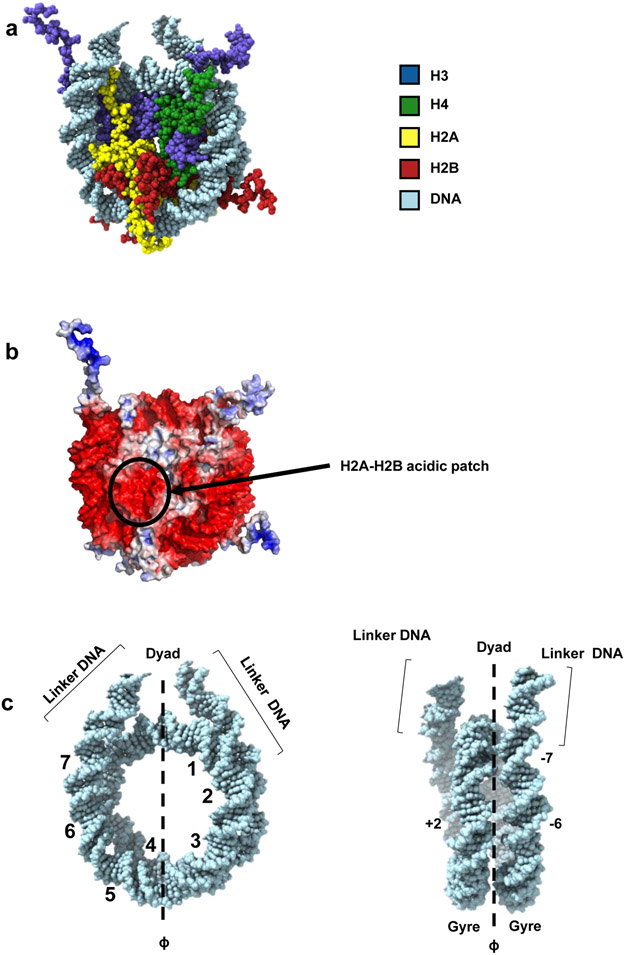
A: Nucleosome disc view, model derived from PDB entries 1KX5 and 1ZBB (DNA from 1ZBB, histone octamer core from 1KX5). B: Electrostatic potential of the nucleosome surface (electrostatic potential calculated from PDB 1KX5, using APBS within PyMOL version 2.2.0). C: Nucleosomal DNA and linker DNA (from PDB entry 1ZBB). Along the 2-fold axis, nucleosomal DNA (145-147 bp) can be divided into two “gyres” (about 72 bp each). The super-helical location (SHL) designation represents the position of each major groove facing inward. The dyad (center of the nucleosomal DNA) is defined as position 0. The numbers “1-7” highlight the SHL on DNA. Linker DNA is the extra-nucleosomal DNA which locates next to the entry/exit site of nucleosomal DNA.
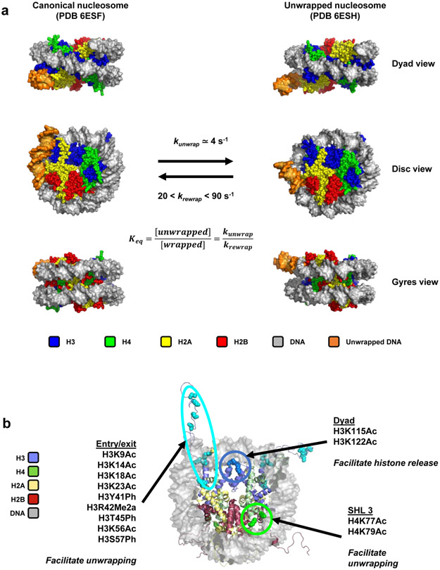
A: CryoEM structures of a canonical nucleosome (PDB entry 6ESF) and a partially unwrapped nucleosome (PDB entry 6ESH). Rate constants of spontaneous unwrapping and rewrapping, determined by stopped-flow spectroscopy, are indicated. B: Location of post-translational modifications that have been studied for their effect on nucleosome unwrapping: at the DNA entry/exit site, at SHL +/−3 (about 35 bp into the nucleosome) and at the dyad. The effects of these post-translational modifications on nucleosome dynamics are indicated. From PDB entry 1KX5.
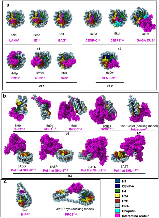
PDB entries of the structures shown in the figure. “1” indicates structures obtained by crystallography. “2” indicates structural models built from single-particle cryoEM maps. “2*” represents docking models generated to interpret cryoEM maps. A: Proteins targeting the surface of nucleosome: a1, small protein fragments or polypeptides recognizing the acidic patch on the nucleosome surface; a2, proteins recognizing both the acidic patch and epigenetic marks on the nucleosome surface; a3, proteins binding to both histones and nucleosomal DNA on the nucleosome surface (the acidic patch also plays an important role in complex a3.1 but not in a3.2). B: Proteins invading nucleosomal DNA gyres. C: Proteins interacting with linker DNA.
References
-
- Woodcock CLF, Safer JP & Stanchfield JE Structural repeating units in chromatin. I. Evidence for their general occurrence. Exp. Cell Res 97, 101–110 (1976). - PubMed
-
- Kornberg RD Chromatin Structure: A Repeating Unit of Histones and DNA. Science (80-. ). 184, 868–871 (1974). - PubMed
-
- Richmond TJ, Finch JT, Rushton B, Rhodes D & Klug A Structure of the nucleosome core particle at 7 resolution. Nature 311, 532–537 (1984). - PubMed
-
-
Luger K, Mäder AW, Richmond RK, Sargent DF & Richmond TJ Crystal structure of the nucleosome core particle at 2.8 Å resolution. Nature 389, 251–260 (1997).
First high-resolution structure of the nucleosome.
-
Publication types
MeSH terms
Substances
Grants and funding
LinkOut - more resources
Full Text Sources
