. 2018 Dec 10;14(12):e1007471.
doi: 10.1371/journal.ppat.1007471. eCollection 2018 Dec.

Palmitoylation mediates membrane association of hepatitis E virus ORF3 protein and is required for infectious particle secretion

Affiliations

Palmitoylation mediates membrane association of hepatitis E virus ORF3 protein and is required for infectious particle secretion

Jérôme Gouttenoire et al. PLoS Pathog. .

Abstract

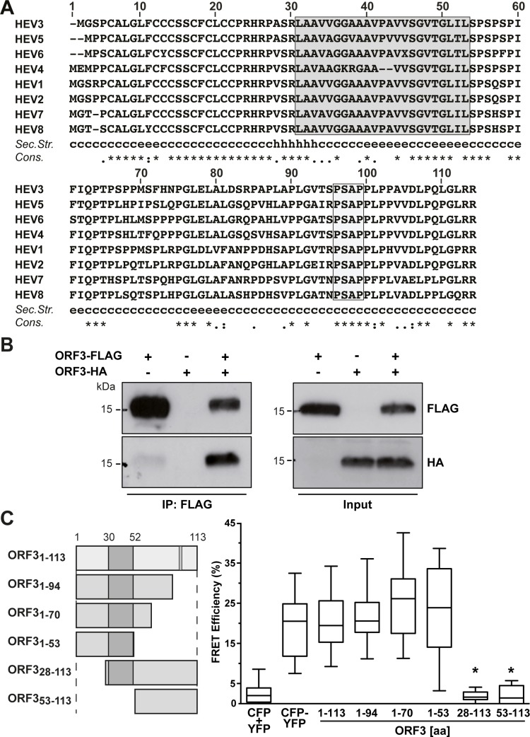
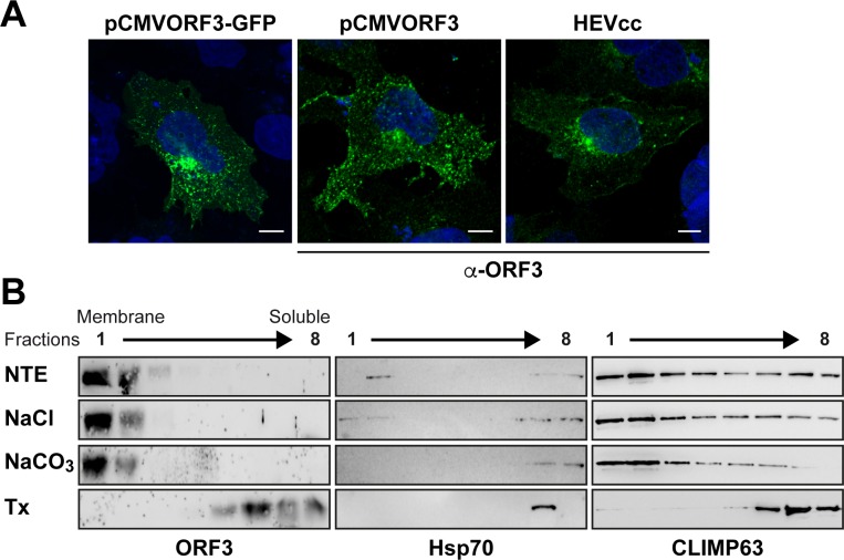
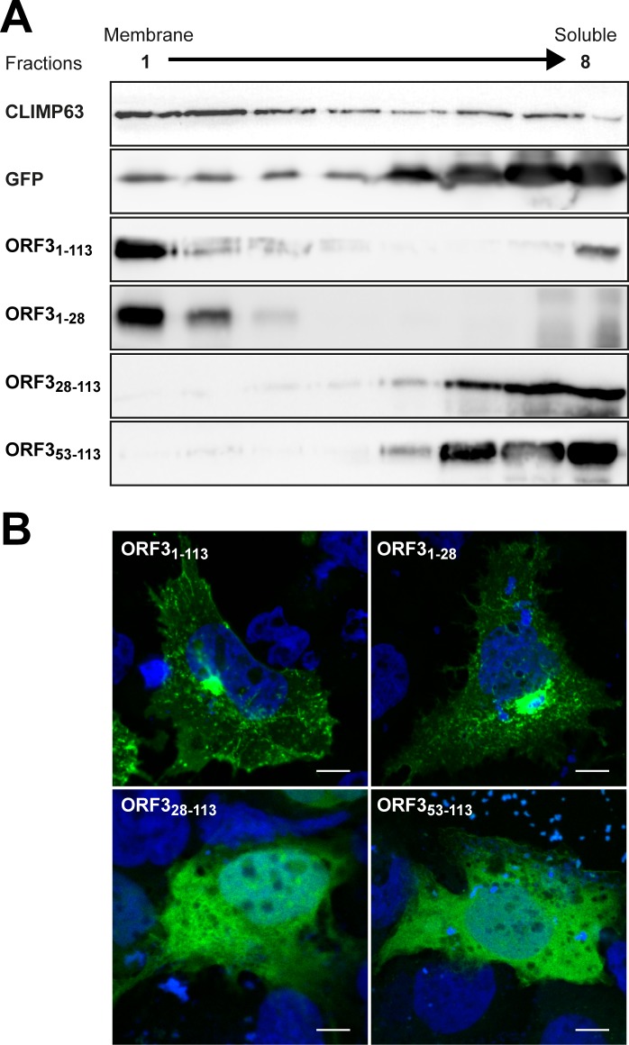
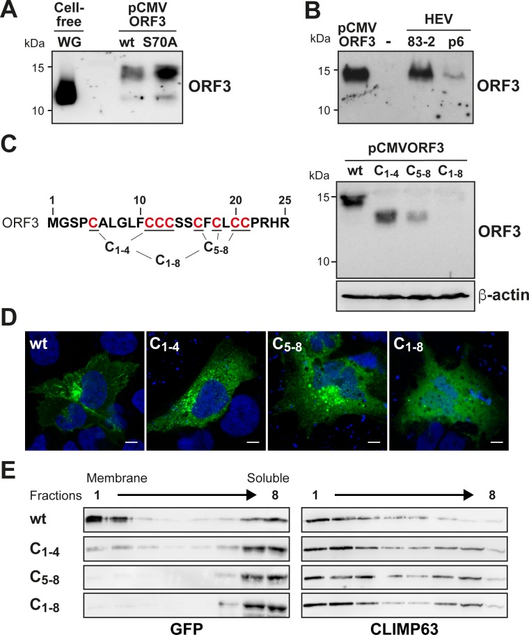
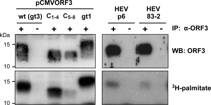
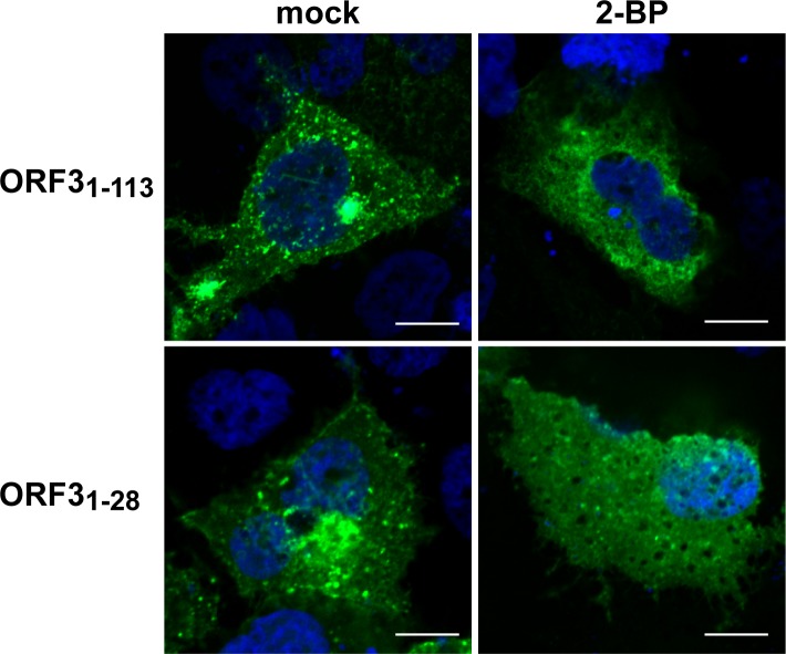
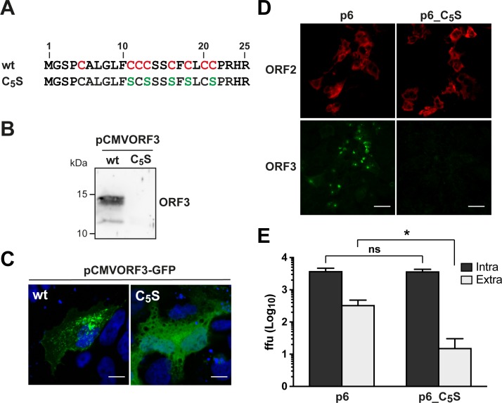
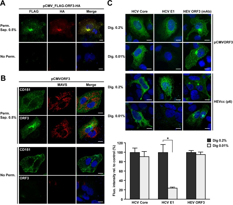
Hepatitis E virus (HEV) is a positive-strand RNA virus encoding 3 open reading frames (ORF). HEV ORF3 protein is a small, hitherto poorly characterized protein involved in viral particle secretion and possibly other functions. Here, we show that HEV ORF3 protein forms membrane-associated oligomers. Immunoblot analyses of ORF3 protein expressed in cell-free vs. cellular systems suggested a posttranslational modification. Further analyses revealed that HEV ORF3 protein is palmitoylated at cysteine residues in its N-terminal region, as corroborated by 3H-palmitate labeling, the investigation of cysteine-to-alanine substitution mutants and treatment with the palmitoylation inhibitor 2-bromopalmitate (2-BP). Abrogation of palmitoylation by site-directed mutagenesis or 2-BP treatment altered the subcellular localization of ORF3 protein, reduced the stability of the protein and strongly impaired the secretion of infectious particles. Moreover, selective membrane permeabilization coupled with immunofluorescence microscopy revealed that HEV ORF3 protein is entirely exposed to the cytosolic side of the membrane, allowing to propose a model for its membrane topology and interactions required in the viral life cycle. In conclusion, palmitoylation determines the subcellular localization, membrane topology and function of HEV ORF3 protein in the HEV life cycle.


Conflict of interest statement

The authors have declared that no competing interests exist.

Figures

Fig 1
Fig 1. HEV ORF3 protein oligomerizes in mammalian cells.
(A) Sequence analysis of ORF3 protein. Amino acid sequences of ORF3 from HEV genotypes 1–8 (GenBank accession numbers AB740232, AF444002, M74506, AJ272108, AB573435, AB856243, KJ496144 and KX387866) were aligned using ClustalW [52]. Segment aa 30–53 predicted as transmembrane passage by TMPred is boxed in grey [53]. A consensus secondary structure was predicted using algorithms MLRC, DSC and PHD (available at https://npsa-prabi.ibcp.fr) and is shown below the alignment (c, random coil; h, α-helix; e, extended strand;?, discrepant prediction). The degree of aa physicochemical conservation at each position is shown on the bottom line and can be inferred with the similarity index according to ClustalW convention (asterisk, invariant; colon, highly similar; dot, similar) [52]. (B) ORF3 protein oligomerization was analyzed by FLAG immunoprecipitation. Lysates (Input) of U-2 OS cells transfected with pCMVORF3-HA and/or pCMVORF3-FLAG as well as immunoprecipitates (IP: FLAG) were subjected to immunoblot with either specific anti-FLAG or anti-HA antibodies. The presence of a strong signal for ORF3-HA after pull-down of ORF3-FLAG indicates oligomerization of ORF3 protein in cells. (C) FRET analyses reveal oligomerization of HEV ORF3. CFP (cyan fluorescent protein) or YFP (yellow fluorescent protein) fused to the C-termini of HEV ORF3 segments aa 1–113, 1–94, 1–70, 1–53, 28–113 or 53–113 were co-expressed in U-2 OS cells. FRET analyses were performed by the acceptor photobleaching method as described in the Materials and Methods section. The CFP-YFP fusion protein and cotransfection of unfused CFP and YFP served as positive and negative controls, respectively. Box-and-whisker plots represent the median FRET efficiency (FRETeff) values of 20 measurements (middle line), the values from the lower to the upper quartile (central box), and the minimum and maximum values (vertical line). The significance of the observed differences was assessed as described in Materials and Methods (*, P<0.0001).
Fig 2
Fig 2. HEV ORF3 protein associates with membranes.
(A) HEV ORF3 protein is expressed in cell membrane compartments. S10-3 cells were transfected with pCMVORF3-GFP, pCMVORF3, or with full-length p6 HEV RNA (HEVcc). Cells were fixed 48 h or 6 d (HEVcc) post-transfection and analyzed by fluorescence microscopy either by direct visualization of GFP or after immunofluorescence staining of HEV ORF3 protein using anti-ORF3 rabbit pAb (α-ORF3). Scale bars indicate 10 μm. (B) Membrane flotation confirms the membrane association of HEV ORF3 protein. S10-3 cells transiently transfected with pCMVORF3 were lysed in a hypotonic buffer and the membrane fraction was obtained by ultracentrifugation as described in the Materials and Methods section. Membrane pellets were resuspended in physiological NTE buffer, 1 M NaCl, 100 mM sodium carbonate (pH 11.5), or 1% Triton X-100 (Tx) and incubated for 20 min at 4°C. Subsequently, membrane flotation analyses were performed as described in the Materials and Methods section. Eight fractions were collected from the top and analyzed by immunoblot using pAb against HEV ORF3. Detection of Hsp70 served as a control for a peripherally membrane-associated protein and CLIMP63 as a control for an integral membrane protein.
Fig 3
Fig 3. The N-terminal region of HEV ORF3 protein determines its membrane-association and plasma membrane localization.
(A) Membrane flotation analyses of ORF3 and deletion constructs. S10-3 cells were transiently transfected with pCMVORF3-GFP, pCMVORF31-53-GFP, pCMVORF31-28-GFP, pCMVORF328-113-GFP, pCMVORF353-113-GFP or pCMV-GFP as indicated. Cell lysates were subjected to membrane flotation assay followed by immunoblot analysis using mAb JL8 against GFP, as described in the Material and Methods section. Endogenous CLIMP63 was used as positive control for an integral membrane protein while GFP served as control for a soluble protein. (B) Subcellular localization of ORF3-GFP fusion constructs. S10-3 cells were transiently transfected with pCMVORF3-GFP, pCMVORF31-53-GFP, pCMVORF31-28-GFP or pCMVORF31-53-GFP as indicated. Nucleus was stained with DAPI. Scale bars indicate 10 μm. Slides were analyzed by confocal laser scanning microscopy.
Fig 4
Fig 4. HEV ORF3 protein is posttranslationally modified at the N-terminal cysteine-rich segment.
(A) S10-3 cells were transfected with pCMVORF3 or pCMVORF3S70A. Protein lysates obtained 48 h post-transfection as well as ORF3 protein expressed using wheat germ extract (WG) were separated by 17% SDS-PAGE and subjected to immunoblot with anti-ORF3 pAb. (B) Protein lysates from pCMVORF3-transfected cells, naïve Hep293TT cells (-) and cells replicating the full-length p6 or 83–2 HEV clone were prepared 6 days post-electroporation. The samples were separated by 17% SDS-PAGE and subjected to immunoblot with anti-ORF3 pAb. (C) Alanine substitution of cysteine residues of HEV ORF3 protein limits its posttranslational modification. S10-3 cells transfected with pCMVORF3, pCMVORF3C1-4, pCMVORF3C45-8 or pCMVORF3C1-8 were harvested 48 h post-transfection and protein lysates were analyzed by immunoblot with anti-ORF3 pAb. (D) S10-3 cells transfected with pCMVORF3-GFP, pCMVORF3C1-4-GFP, pCMVORF3C45-8-GFP or pCMVORF3C1-8-GFP were analyzed by fluorescence microscopy 48 h post-transfection. A representative image is shown for each construct. Scale bars indicate 10 μm. (E) Membrane flotation analyses of wild-type and mutant ORF3-GFP constructs. S10-3 cells were transiently transfected with pCMVORF3-GFP, pCMVORF3C1-4-GFP, pCMVORF3C45-8-GFP or pCMVORF3C1-8-GFP, as indicated. Cell lysates were subjected to membrane flotation assay followed by immunoblot analysis using mAb JL8 against GFP. Endogenous CLIMP63 was used as positive control for an integral membrane protein.
Fig 5
Fig 5. HEV ORF3 protein is palmitoylated.
Protein lysates from S10-3 cells transfected with pCMVORF3, pCMVORF3C1-4, pCMVORF3C45-8 or pCMVORF3_gt1 and from Hep293TT cells replicating the full-length p6 or 83–2 HEV clone were prepared 1 or 6 days post-transfection, respectively, and subjected to immunoprecipitation with either anti-ORF3 pAb (+) or non-relevant rabbit serum (-). After immunoprecipitation, the elution samples were separated by 17% SDS-PAGE and subjected to either immunoblot with anti-ORF3 pAb followed by chemiluminescence revelation or autoradiography (40 d of exposure).
Fig 6
Fig 6. Palmitoylation inhibition induces changes in the subcellular localization of HEV ORF3 protein.
S10-3 cells were transfected with either pCMVORF3-GFP (ORF31-113) or pCMVORF31-28-GFP (ORF31-28). One d post-transfection, cells were treated for 24 h with 25 μM 2-bromopalmitate (2-BP) and analyzed by confocal laser scanning microscopy after staining of the nuclei by DAPI. Scale bars represent 10 μm.
Fig 7
Fig 7. N-terminal cysteine residues of HEV ORF3 protein are essential for infectious particle secretion.
(A) Sequence alignment of the N-terminal 25 aa of wild-type (wt) and cysteine to serine mutant (C5S) HEV ORF3 protein. The conserved cysteine residues in the wt sequence are highlighted in red and the positions bearing a serine substitution in the C5S mutant sequence are indicated in green. (B) S10-3 cells were transfected with pCMVORF3 or pCMVORF3C5S and cell lysates were prepared 48 h post-transfection, followed by SDS-PAGE and immunoblot analysis using anti-ORF3 pAb. (C) S10-3 cells were transiently transfected with pCMVORF3-GFP or pCMVORF3C5S-GFP and analyzed by confocal laser scanning microscopy. Nuclei were stained with DAPI. Scale bars indicate 20 μm. (D) Indirect immunofluorescence detection of HEV ORF2 and ORF3 proteins was carried out 5 d post-transfection with HEV ORF2 pAb (red) and anti-ORF3 mAb MRB198 (green). Scale bars indicate 50 μm. (E) Five d after transfection of p6 or p6_C5S HEV RNA, culture supernatants were harvested and cell lysates were prepared by three freeze-and-thaw cycles followed by clarification by centrifugation at 2,000 g for 15 min. Intracellular (Intra) and extracellular (Extra) infectivities were determined by foci forming assay on HepG2/C3A cells using the cell lysates and the culture supernatants, respectively, as inoculum. Immunofluorescence detection of the capsid protein was performed with rabbit antiserum against HEV ORF2. The asterisk (*) indicates statistically significant results with p<0.0001 while “ns” indicates nonsignificant results. Each bar represents infectivity obtained from 10 independent RNA transfections. ffu: focus forming unit.
Fig 8
Fig 8. Membrane topology of HEV ORF3 protein.
(A) N- and C-terminal ends of HEV ORF3 protein are intracellularly exposed. S10-3 cells were transfected with pCMV_FLAG-ORF3-HA and subjected to immunfluorescence detection of HA and FLAG tags, using rabbit pAb anti-HA (Y-11) and mouse mAb anti-FLAG M2, respectively, after permeabilization with 0.5% saponin (Perm. Sap. 0.5%) or in the absence of permeabilization (No Perm.). (B) Similarly, S10-3 cells transfected with pCMVORF3 were subjected to immunofluorescence to detect the plasma membrane tetraspanin CD151 (mouse mAb 11G5a), the cytoplasmic protein MAVS (rabbit pAb anti-MAVS) or HEV ORF3 protein using mAb MRB198. Nuclei were stained by DAPI. (C) Selective membrane permeabilization. S10-3 cells were transfected with pCMVORF3 or co-transfected with pUHD15-1 and pUHD-Cp7 allowing the expression of the hepatitis C virus (HCV) core-p7 region (top panel) and cultured for 24 h. S10-3 cells were transfected with the HEV p6 infectious clone (middle panel) and cultured for 5 d. All cells were fixed and permeabilized with either 0.2% or 0.01% digitonin. Immunofluorescence detection of the cytoplasmic HCV core with mouse mAb C7-50 or the endoplasmic reticulum luminal HCV E1 glycoprotein with mouse mAb A4 served as controls for selective permeabilization of intracellular membranes. HEV ORF3 protein is detected using anti-ORF3 mAb MRB198. The lower panel shows histograms summarizing fluorescence intensities, as determined by using ImageJ software in 10 to 35 cells per condition, obtained after immunofluorescence with total (Dig. 0.2%) or selective (Dig. 0.01%) membrane permeabilization of S10-3 cells replicating the HEV p6 infectious clone. The asterisk (*) indicates statistically significant results with p<0.001. Scale bars indicate 10 μm.

References

    1. Hoofnagle JH, Nelson KE, Purcell RH. Hepatitis E. N Engl J Med. 2012;367:1237–44. 10.1056/NEJMra1204512 - DOI - PubMed
    1. Kamar N, Izopet J, Pavio N, Aggarwal R, Labrique A, Wedemeyer H, et al. Hepatitis E virus infection. Nat Rev Dis Primers. 2017;3:17086 10.1038/nrdp.2017.86 - DOI - PubMed
    1. Debing Y, Moradpour D, Neyts J, Gouttenoire J. Update on hepatitis E virology: Implications for clinical practice. J Hepatol. 2016;65:200–12. 10.1016/j.jhep.2016.02.045 - DOI - PubMed
    1. Nimgaonkar I, Ding Q, Schwartz RE, Ploss A. Hepatitis E virus: advances and challenges. Nat Rev Gastroenterol Hepatol. 2018;15:96–110. 10.1038/nrgastro.2017.150 - DOI - PMC - PubMed
    1. Smith DB, Simmonds P, Jameel S, Emerson SU, Harrison TJ, Meng XJ, et al. Consensus proposals for classification of the family hepeviridae. J Gen Virol. 2014;95:2223–32. 10.1099/vir.0.068429-0 - DOI - PMC - PubMed

Publication types

Substances