

Utility of Various Ultrafast Magnetic Resonance Sequences in the Detection of Fetal Intracranial Hemorrhage
- PMID: 30532356
- PMCID: PMC6238567
- DOI: 10.4103/aian.AIAN_431_17
Utility of Various Ultrafast Magnetic Resonance Sequences in the Detection of Fetal Intracranial Hemorrhage
Abstract
Objective: The aim of this study is to compare the images obtained from standard ultrafast magnetic resonance (MR) imaging sequences with gradient (GRE) sequence images in identifying fetal intracranial hemorrhage (ICH).
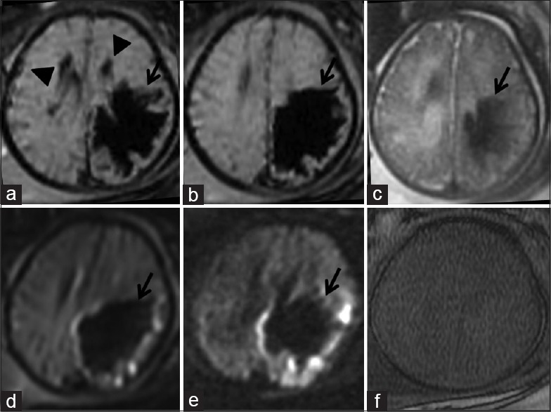
Materials and methods: MR images of fetal brains with ICH done between October 2012 and September 2015 were reviewed. The images obtained from four ultrafast MR sequences- Turbo Fast Low Angle Shot (Turbo FLASH) T1-weighted images, Half Fourier Acquisition single-shot turbo spin echo (HASTE) T2-weighted images, b0 images of diffusion-weighted imaging (DWI) and b800 images of DWI were compared with images obtained from GRE sequence in depicting fetal ICH.
Results: Out of the 212 fetuses during the study period, 15 fetuses had ICH. In the 15 fetuses with ICH as detected on GRE, Grade1 germinal matrix hemorrhage was seen in 5 fetuses, Grade 2 in 4 fetuses, Grade 3 in 3 fetuses, and Grade 4 in two fetuses. Subdural hemorrhage was seen in 1 fetus. In comparison to GRE sequence, b0 of DWI sequence was almost equal in the depiction of ICH. T2 HASTE sequence also delineated hemorrhage, although not as effectively as GRE and b0 images of images DWI. T1 Turbo FLASH and b800 images of DWI were less reliable in the depiction of fetal ICH but were useful in predicting the stage of hemorrhage.
Conclusion: As compared to GRE sequence, b0 images of DWI followed by HASTE are the two preferred ultrafast sequences in the diagnosis of fetal ICH.
Keywords: Fetus; germinal matrix hemorrhage; magnetic resonance imaging; ultrasound.
Conflict of interest statement
There are no conflicts of interest.
Figures


Similar articles
-
Comparison of diffusion weighted imaging b0 with T2*-weighted gradient echo or susceptibility weighted imaging for intracranial hemorrhage detection after reperfusion therapy for ischemic stroke.Neuroradiology. 2023 Nov;65(11):1649-1655. doi: 10.1007/s00234-023-03180-3. Epub 2023 Jun 29. Neuroradiology. 2023. PMID: 37380891 Free PMC article.
-
Detection of intracranial hemorrhage: comparison between gradient-echo images and b(0) images obtained from diffusion-weighted echo-planar sequences.AJNR Am J Neuroradiol. 2001 Aug;22(7):1275-81. AJNR Am J Neuroradiol. 2001. PMID: 11498414 Free PMC article.
-
Detection of intracranial hemorrhage with susceptibility-weighted MR sequences.AJNR Am J Neuroradiol. 1999 Sep;20(8):1527-34. AJNR Am J Neuroradiol. 1999. PMID: 10512241 Free PMC article.
-
Ultrafast MR imaging of the pelvis.Eur J Radiol. 1999 Mar;29(3):233-44. doi: 10.1016/s0720-048x(98)00165-x. Eur J Radiol. 1999. PMID: 10399609 Review.
-
Fetal intracranial hemorrhage and infarct: Main sonographic and MRI characteristics: A review article.Eur J Obstet Gynecol Reprod Biol X. 2024 Nov 6;24:100351. doi: 10.1016/j.eurox.2024.100351. eCollection 2024 Dec. Eur J Obstet Gynecol Reprod Biol X. 2024. PMID: 39610469 Free PMC article. Review.
Cited by
-
Role of Ultrafast MR Imaging in Stroke Patients.J Neurosci Rural Pract. 2020 Jul;11(3):436-441. doi: 10.1055/s-0040-1712716. Epub 2020 Jun 1. J Neurosci Rural Pract. 2020. PMID: 32753809 Free PMC article.
-
Prenatal sonographic diagnosis and postnatal outcomes of fetal intracranial hemorrhage: Two case report.Radiol Case Rep. 2024 Jul 13;19(9):4066-4072. doi: 10.1016/j.radcr.2024.06.035. eCollection 2024 Sep. Radiol Case Rep. 2024. Retraction in: Radiol Case Rep. 2025 Apr 11;20(6):3160. doi: 10.1016/j.radcr.2024.12.028. PMID: 39076884 Free PMC article. Retracted.
References
-
- Reiss I, Gortner L, Moller J, Gehl HB, Baschat AA, Gembruch U, et al. Fetal intracerebral hemorrhage in the second trimester: Diagnosis by sonography and magnetic resonance imaging. Ultrasound Obstet Gynecol. 1996;7:49–51. - PubMed
-
- Papile LA, Burstein J, Burstein R, Koffler H. Incidence and evolution of subependymal and intraventricular hemorrhage: A study of infants with birth weights less than 1,500 gm. J Pediatr. 1978;92:529–34. - PubMed
-
- Brugger PC, Stuhr F, Lindner C, Prayer D. Methods of fetal MR: Beyond T2-weighted imaging. Eur J Radiol. 2006;57:172–81. - PubMed
-
- Gomori JM, Grossman RI. Mechanisms responsible for the MR appearance and evolution of intracranial hemorrhage. Radiographics. 1988;8:427–40. - PubMed
-
- Kothari RU, Brott T, Broderick JP, Barsan WG, Sauerbeck LR, Zuccarello M, et al. The ABCs of measuring intracerebral hemorrhage volumes. Stroke. 1996;27:1304–5. - PubMed
LinkOut - more resources
Full Text Sources
Research Materials
