

Acute inferolateral ST-elevation myopericarditis diagnosed by delayed enhancement cardiac computed tomography
- PMID: 30532846
- PMCID: PMC6265109
- DOI: 10.1016/j.jccase.2010.11.003
Acute inferolateral ST-elevation myopericarditis diagnosed by delayed enhancement cardiac computed tomography
Abstract
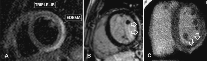
A 20-year-old man with no previous medical history presented to the Emergency Department (ED) complaining of 3 h of chest pressure. He denied drug abuse or risk factors for coronary artery disease. He had no symptoms of viral infection. Physical examination was unremarkable. The first electrocardiogram (ECG) showed a 4 mm ST-segment elevation in the inferior leads and no PR depression. His troponin and CK-MB levels were abnormal. Urgent coronary angiography showed no lesions. Echocardiography was normal. The patient was investigated with cardiac computed tomography (CT) and late enhancement imaging. Cardiac anatomy and coronary arteries were normal in the first pass images. Later image acquisition showed an inferolateral enhancement. Since cardiac magnetic resonance (CMR) is the gold standard for myocarditis evaluation, the patient was transferred for CMR evaluation which showed edema and late enhancement in the same myocardial territory diagnosed by CT. The patient was discharged with a diagnosis of myocarditis and presented asymptomatic at 1 month follow-up. This is the first report to show the topographic correlation of the ECG ST elevation with the myocarditis diagnosed by CT and CMR. Since CT is more widely available, its use in myocarditis diagnosis might become part of its routine work up.
Keywords: Cardiac computed tomography; Myocarditis.
Figures


References
-
- Nieman K., Shapiro M.D., Ferencik M., Nomura C.H., Abbara S., Hoffmann U., Gold H.K., Jang I.K., Brady T.J., Cury R.C. Reperfused myocardial infarction: contrast-enhanced 64-Section CT in comparison to MR imaging. Radiology. 2008;247:49–56. - PubMed
-
- Brooks M.A., Sane D.C. CT Findings in acute myocarditis – 2 cases. J Thorac Imaging. 2007;22:277–279. - PubMed
-
- Imazio M., Trinchero R. The spectrum of inflammatory myopericardial diseases. Int J Cardiol. 2008;127:17–26. - PubMed
LinkOut - more resources
Full Text Sources
Research Materials
Miscellaneous
